at dilution of 1:3000 incubated at room temperature for 1.5 hours.")
 at dilution of 1:20000 incubated at room temperature for 1.5 hours.")
 at dilution of 1:5000 incubated at room temperature for 1.5 hours.")
 at dilution of 1:3000 incubated at room temperature for 1.5 hours.")
 at dilution of 1:2000 incubated at room temperature for 1.5 hours.")
 at dilution of 1:2000 incubated at room temperature for 1.5 hours.")
 at dilution of 1:3000 incubated at room temperature for 1.5 hours.")
Tested Applications
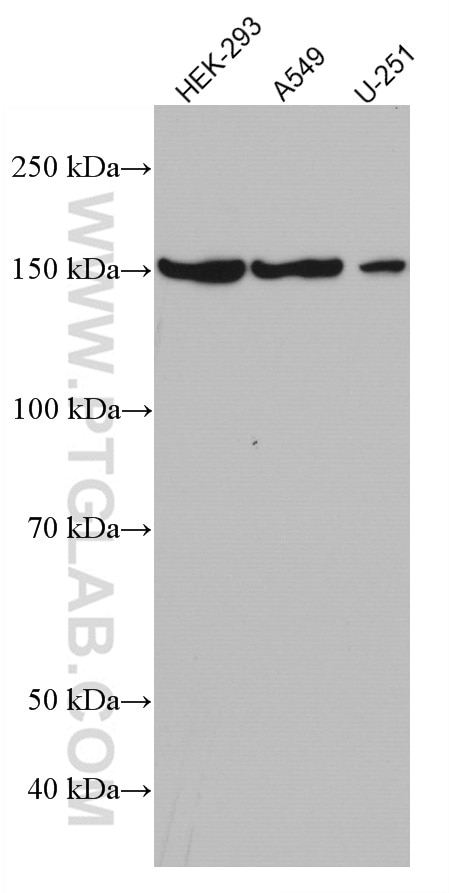
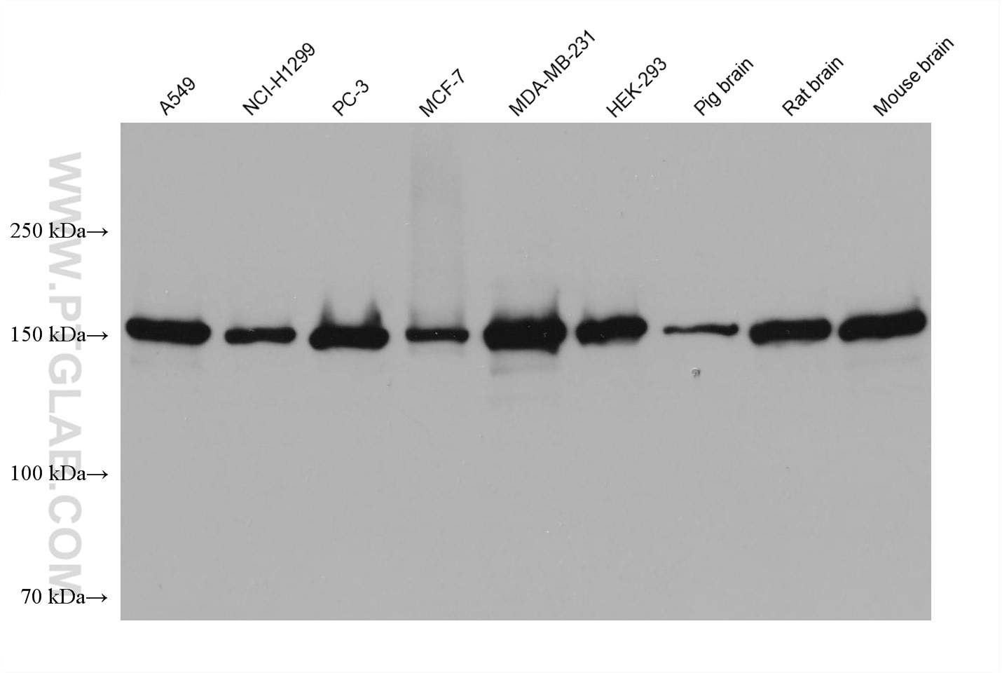
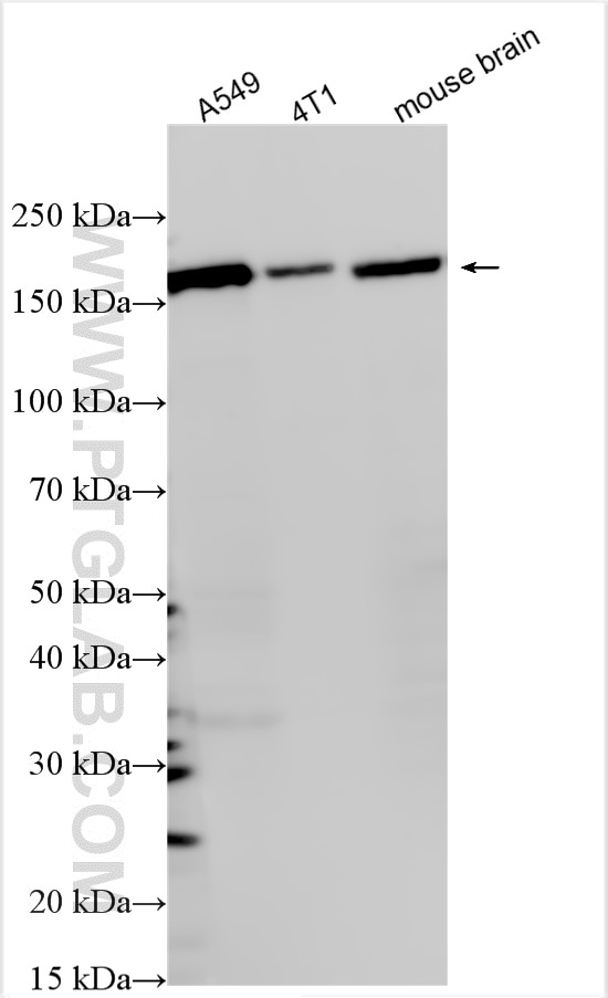
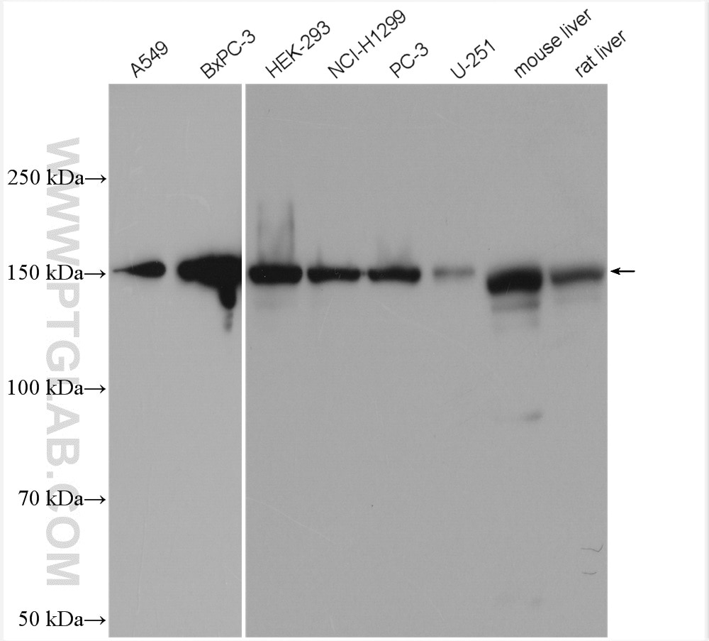
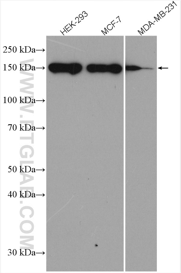
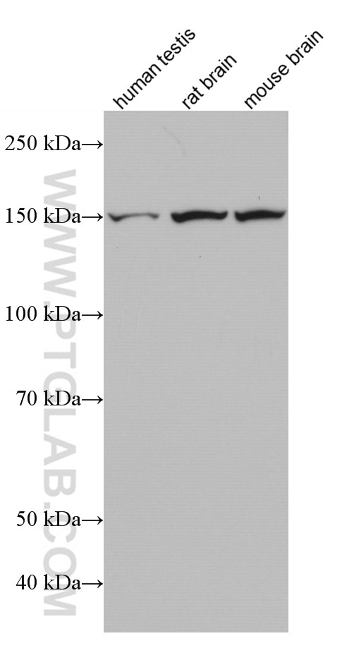
| Positive WB detected in | A549 cells, C6 cells, HEK-293 cells, human testis tissue, 4T1 cells, mouse brain tissue, NCI-H1299 cells, PC-3 cells, MCF-7 cells, MDA-MB-231 cells, pig brain tissue, rat brain tissue, BxPC-3 cells, U-251 cells, mouse liver tissue, rat liver tissue |
Recommended dilution
| Application | Dilution |
|---|---|
| Western Blot (WB) | WB : 1:2000-1:20000 |
| It is recommended that this reagent should be titrated in each testing system to obtain optimal results. | |
| Sample-dependent, Check data in validation data gallery. | |
Published Applications
| KD/KO | See 5 publications below |
| WB | See 85 publications below |
| IHC | See 17 publications below |
| IF | See 11 publications below |
| IP | See 1 publications below |
| CoIP | See 1 publications below |
Product Information
66905-1-Ig targets GLI1 in WB, IHC, IF, IP, CoIP, ELISA applications and shows reactivity with human, mouse, rat, pig samples.
| Tested Reactivity | human, mouse, rat, pig |
| Cited Reactivity | human, mouse, rat |
| Host / Isotype | Mouse / IgG1 |
| Class | Monoclonal |
| Type | Antibody |
| Immunogen |
CatNo: Ag27832 Product name: Recombinant human GLI1 protein Source: e coli.-derived, PET28a Tag: 6*His Domain: 470-659 aa of BC013000 Sequence: NAGGSTEDLSSLDEGPCIAGTGLSTLRRLENLRLDQLHQLRPIGTRGLKLPSLSHTGTTVSRRVGPPVSLERRSSSSSSISSAYTVSRRSSLASPFPPGSPPENGASSLPGLMPAQHYLLRARYASARGGGTSPTAASSLDRIGGLPMPPWRSRAEYPGYNPNAGVTRRASDPAQAADRPAPARVQRFKS Predict reactive species |
| Full Name | GLI family zinc finger 1 |
| Calculated Molecular Weight | 1106 aa, 118 kDa |
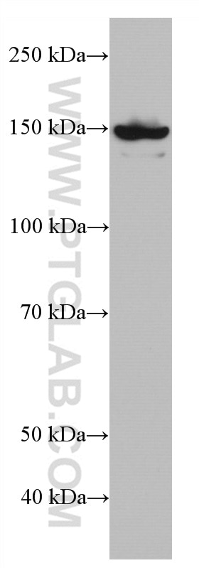
| Observed Molecular Weight | 150 kDa |
| GenBank Accession Number | BC013000 |
| Gene Symbol | GLI1 |
| Gene ID (NCBI) | 2735 |
| RRID | AB_2882232 |
| Conjugate | Unconjugated |
| Form | Liquid |
| Purification Method | Protein G purification |
| UNIPROT ID | P08151 |
| Storage Buffer | PBS with 0.02% sodium azide and 50% glycerol, pH 7.3. |
| Storage Conditions | Store at -20°C. Stable for one year after shipment. Aliquoting is unnecessary for -20oC storage. 20ul sizes contain 0.1% BSA. |
Background Information
GLI1, also known as Glioma-associated oncogene, is a 1106 amino acid protein, which belongs to the GLI C2H2-type zinc-finger protein family. GLI1 is detected in testis and Also expressed in the brain with highest expression in the cerebellum, optic nerve and olfactory tract. GLI1 is tethered in the cytoplasm by binding to SUFU (PubMed:10806483). GLI1 is activated and translocated to the nucleus promoted by interaction with STK36 (PubMed:10806483). Phosphorylation of GLI1 by ULK3 may promote nuclear localization (PubMed:19878745). GLI1 as a transcriptional activator may regulate the transcription of specific genes during normal development (PubMed:19706761). GLI1 plays a role in craniofacial development and digital development, as well as development of the central nervous system and gastrointestinal tract and Mediates SHH signaling (PubMed:19706761). GLI1 plays a role in cell proliferation and differentiation via its role in SHH signaling.
Protocols
| Product Specific Protocols | |
|---|---|
| WB protocol for GLI1 antibody 66905-1-Ig | Download protocol |
| Standard Protocols | |
|---|---|
| Click here to view our Standard Protocols |
Publications
| Species | Application | Title |
|---|---|---|
Mol Cancer Circular RNA circIPO11 drives self-renewal of liver cancer initiating cells via Hedgehog signaling. | ||
Nat Commun TSPAN8 promotes cancer cell stemness via activation of sonic Hedgehog signaling. | ||
J Exp Clin Cancer Res ACTN1 promotes HNSCC tumorigenesis and cisplatin resistance by enhancing MYH9-dependent degradation of GSK-3β and integrin β1-mediated phosphorylation of FAK | ||
J Nanobiotechnology NaHS@Cy5@MS@SP nanoparticles improve rheumatoid arthritis by inactivating the Hedgehog signaling pathway through sustained and targeted release of H2S into the synovium | ||
JCI Insight Mutations in OSBPL2 cause hearing loss associated with primary cilia defects via Sonic Hedgehog signaling. | ||
Cancer Immunol Res Remodeling Chondroitin-6-Sulfate-Mediated Immune Exclusion Enhances Anti-PD-1 Response in Colorectal Cancer with Microsatellite Stability. |
Reviews
The reviews below have been submitted by verified Proteintech customers who received an incentive for providing their feedback.
FH MALLIKARJUNA (Verified Customer) (10-17-2025) | work best for IHC
|
FH Bárbara (Verified Customer) (07-31-2025) | We tested the GLI1 antibody (Proteintech) for Western blot in human PBMC lysates at 1:1000 dilution. The expected ~140 kDa band low, but with one band
|
FH Boyan (Verified Customer) (07-23-2019) | In my experiment, I used the untreated and Shh ligand-treated cell lysates, in which the protein level of Gli1 should be increased after Shh treatment.With these two samples, I could detect a significant induction of the Gli1 protein using another Gli1 antibody. But, I could not see this induction with this antibody (66905-1-lg). A tricky thing is that, comparing with the real Gli1 band shown by the other antibody, this antibody (66905-1-lg) could recognise a protein at a similar molecular weight (but a little higher than that real Gli1). I think this is misleading, because although being shown on the website, that band is not the real band of Gli1.
|
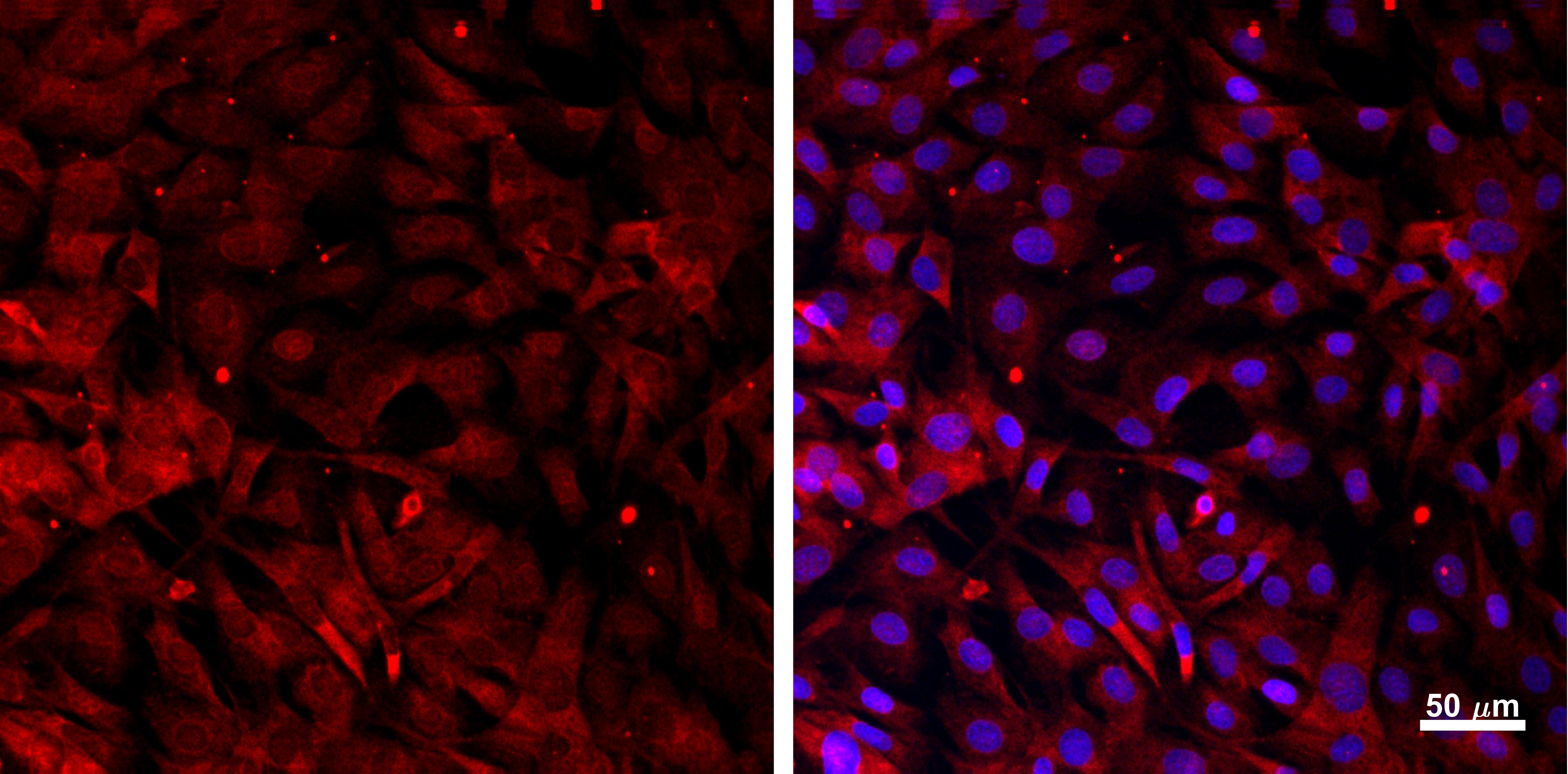
FH Joshua (Verified Customer) (04-28-2019) | Cells paraformaldehyde fixed and stained for GLI1 (red). Counterstained with DRAQ7 (blue). Observed mixed nuclear/cytosolic staining as expected for GLI1.
 |