


Tested Applications
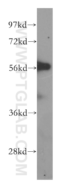
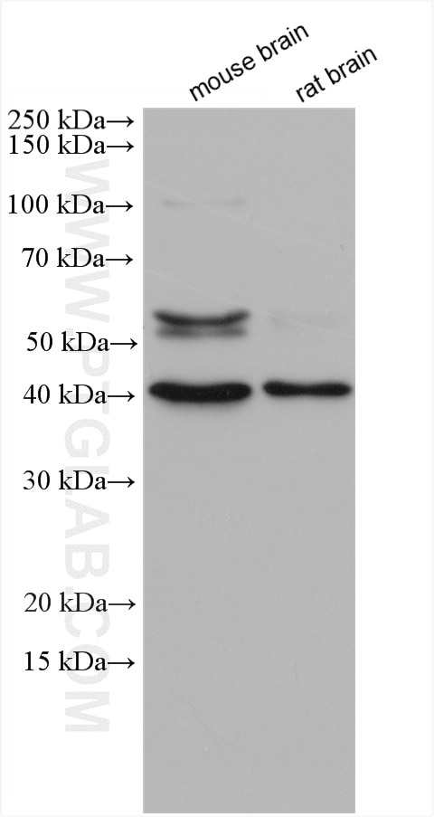
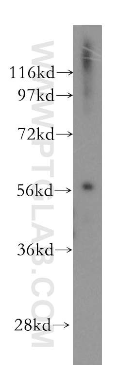
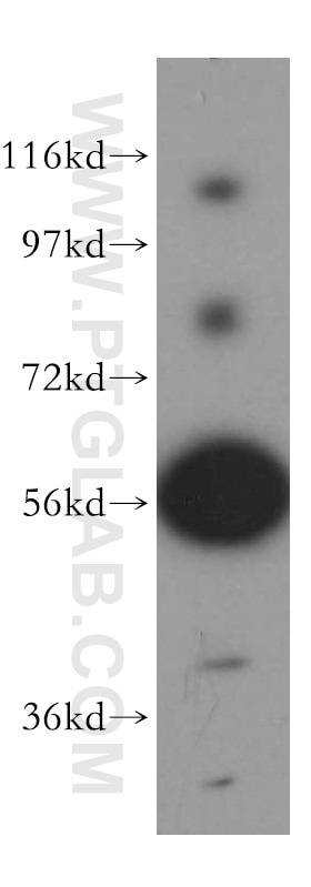
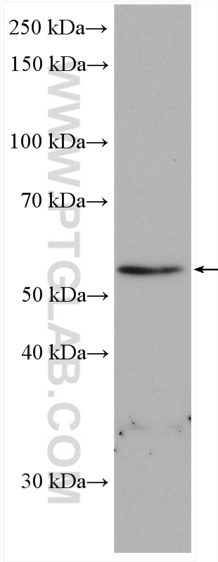
| Positive WB detected in | mouse brain tissue, human heart tissue, mouse lung tissue, human brain tissue, mouse heart tissue, rat brain tissue |
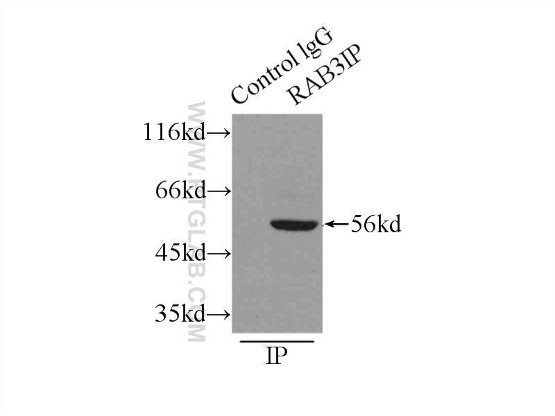
| Positive IP detected in | mouse lung tissue |
| Positive IHC detected in | human stomach cancer tissue Note: suggested antigen retrieval with TE buffer pH 9.0; (*) Alternatively, antigen retrieval may be performed with citrate buffer pH 6.0 |
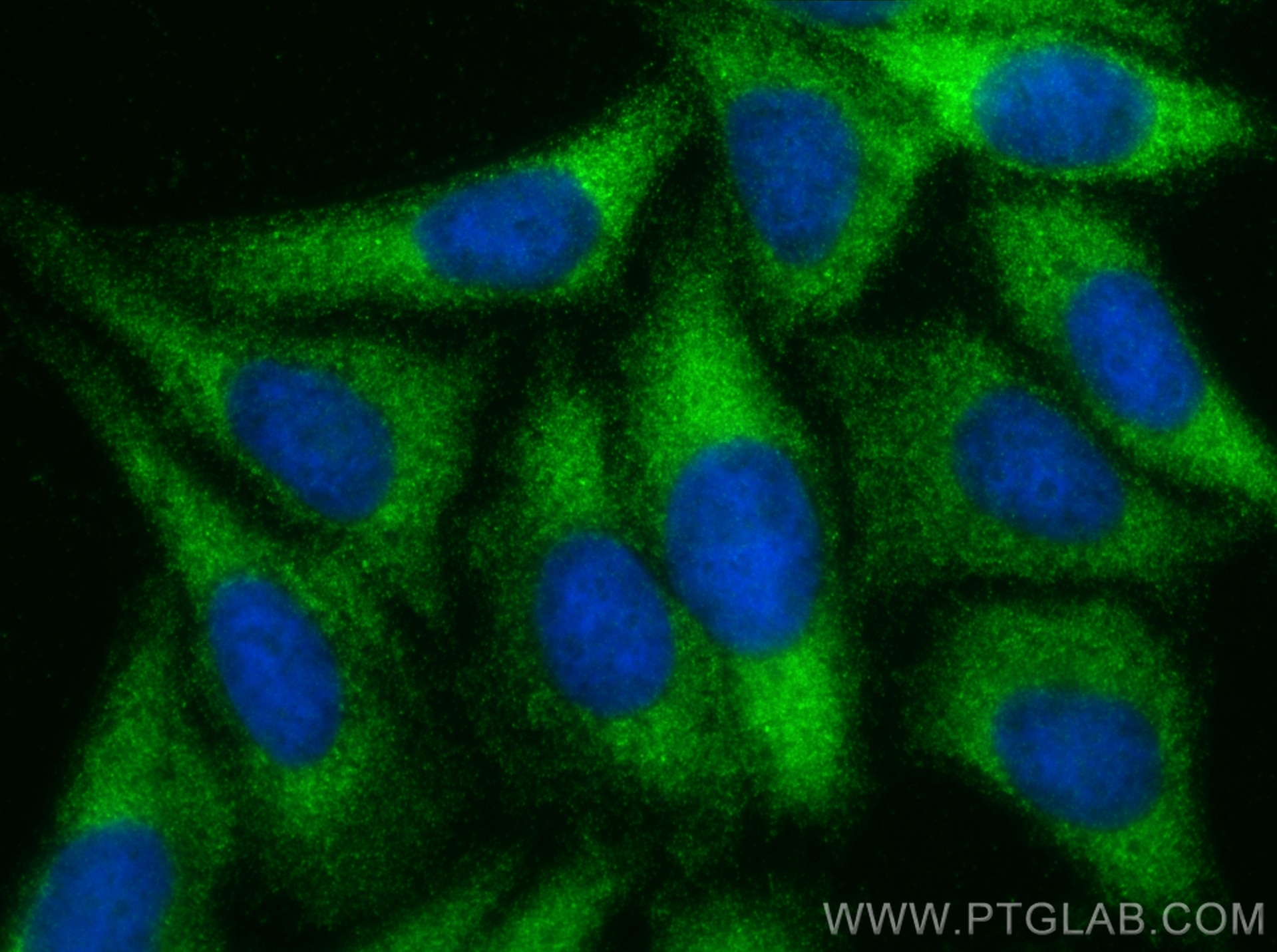
| Positive IF/ICC detected in | HepG2 cells |
Recommended dilution
| Application | Dilution |
|---|---|
| Western Blot (WB) | WB : 1:500-1:3000 |
| Immunoprecipitation (IP) | IP : 0.5-4.0 ug for 1.0-3.0 mg of total protein lysate |
| Immunohistochemistry (IHC) | IHC : 1:400-1:1600 |
| Immunofluorescence (IF)/ICC | IF/ICC : 1:50-1:500 |
| It is recommended that this reagent should be titrated in each testing system to obtain optimal results. | |
| Sample-dependent, Check data in validation data gallery. | |
Product Information
12321-1-AP targets RAB3IP/Rabin8 in WB, IHC, IF/ICC, IP, ELISA applications and shows reactivity with human, mouse, rat samples.
| Tested Reactivity | human, mouse, rat |
| Cited Reactivity | human, mouse, rat, canine |
| Host / Isotype | Rabbit / IgG |
| Class | Polyclonal |
| Type | Antibody |
| Immunogen |
CatNo: Ag2974 Product name: Recombinant human RABIN8 protein Source: e coli.-derived, PGEX-4T Tag: GST Domain: 21-254 aa of BC015548 Sequence: GKIDVLQAEVAALKTLVLSSSPTSPTQEPLPGGKTPFKKGHTRNKSTSSAMSGSHQDLSVIQPIVKDCKEADLSLYNEFRLWKDEPTMDRTCPFLDKIYQEDIFPCLTFSKSELASAVLEAVENNTLSIEPVGLQPIRFVKASAVECGGPKKCALTGQSKSCKHRIKLGDSSNYYYISPFCRYRITSVCNFFTYIRYIQQGLVKQQDVDQMFWEVMQLRKEMSLAKLGYFKEEL Predict reactive species |
| Full Name | RAB3A interacting protein (rabin3) |
| Calculated Molecular Weight | 476 aa, 53 kDa |
| Observed Molecular Weight | 53/51/43 kDa |
| GenBank Accession Number | BC015548 |
| Gene Symbol | RAB3IP |
| Gene ID (NCBI) | 117177 |
| RRID | AB_2177510 |
| Conjugate | Unconjugated |
| Form | Liquid |
| Purification Method | Antigen affinity purification |
| UNIPROT ID | Q96QF0 |
| Storage Buffer | PBS with 0.02% sodium azide and 50% glycerol, pH 7.3. |
| Storage Conditions | Store at -20°C. Stable for one year after shipment. Aliquoting is unnecessary for -20oC storage. 20ul sizes contain 0.1% BSA. |
Background Information
RAB3IP, also hnown as Rabin8, is a deduced 476-amino acid protein containing a central coiled-coil domain, a bipartite nuclear localization signal, and a C-terminal endoplasmic reticulum retention motif. Rabin8 has several sites for N-glycosylation and phosphorylation. Rabin8 plays important roles in various cellular processes, including ciliogenesis.The GEF activity of Rabin8 towards Rab8 is essential for ciliogenesis and cyst apical membrane transport,but not for exocytosis of discoidal/fusiform-shaped vesicles. This antibody can recognize isoforms with predicted MW's of 53 kDa, 51 kDa, 43 kDa, 45 kDa, 33 kDa and 35 kDa, 29 kDa respectively.
Protocols
| Product Specific Protocols | |
|---|---|
| IF protocol for RAB3IP/Rabin8 antibody 12321-1-AP | Download protocol |
| IHC protocol for RAB3IP/Rabin8 antibody 12321-1-AP | Download protocol |
| IP protocol for RAB3IP/Rabin8 antibody 12321-1-AP | Download protocol |
| WB protocol for RAB3IP/Rabin8 antibody 12321-1-AP | Download protocol |
| Standard Protocols | |
|---|---|
| Click here to view our Standard Protocols |
Publications
What published studies show
RAB8A antibody (Cat# 12321-1-AP) has 21 citations across 16 journals including Nature Cell Biology, Neuron, PNAS, EMBO Journal, Cell Reports, PLoS Biology, Journal of Cell Biology, iScience, and FEBS Letters. Research fields span ciliogenesis, membrane trafficking, Rab GTPase signaling, exosome-mediated tumor growth, lipid metabolism and obesity, Parkinson's disease, endocytosis, neurite outgrowth, immune signaling, and protein quality control.
| Species | Application | Title |
|---|---|---|
Nat Cell Biol Stiff matrix induces exosome secretion to promote tumour growth
| ||
Nat Cell Biol A molecular network for de novo generation of the apical surface and lumen.
| ||
Neuron Chemical genetic identification of NDR1/2 kinase substrates AAK1 and Rabin8 Uncovers their roles in dendrite arborization and spine development. | ||
Proc Natl Acad Sci U S A Disruption of the AMPK-TBC1D1 nexus increases lipogenic gene expression and causes obesity in mice via promoting IGF1 secretion.
| ||
EMBO J Phosphoproteomic screening identifies Rab GTPases as novel downstream targets of PINK1. |
Reviews
What customers say
Both reviewers used this antibody for immunofluorescence and reported positive results. One user (5/5) found that a 1:200 dilution worked well on NPC tissue. The other (4/5) used a 1:100 dilution on spleen B cells and observed staining similar to the datasheet example, though noting it was not very sharp with some diffuse signal across the cell. Overall, users find the antibody functional for IF applications, with one planning to test it further in Western blot.
The reviews below have been submitted by verified Proteintech customers who received an incentive for providing their feedback.
Silvia (Verified Customer) (09-30-2021) | Immunofluorescence (1:200) works well on NPC
|
Sara (Verified Customer) (04-07-2020) | I got a quite similar staining to the one shown in the example figure of this product data sheet. Not very sharp staining (a bit all over the cell), but working. Next I will try in WB.
|









