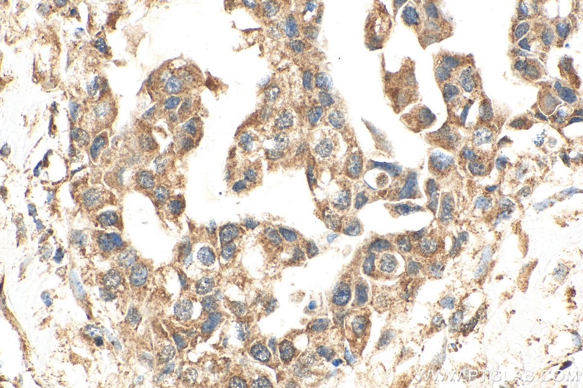
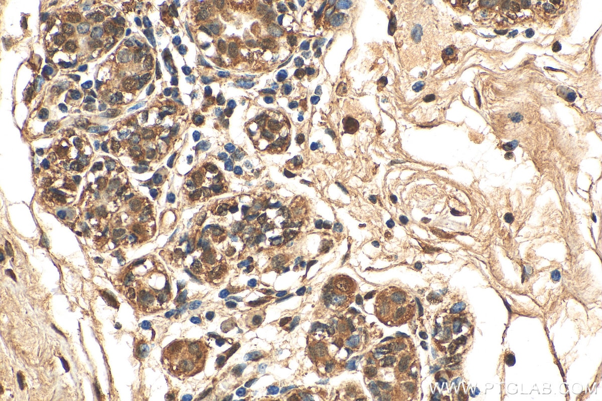
Tested Applications
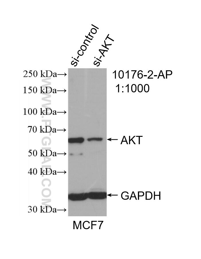
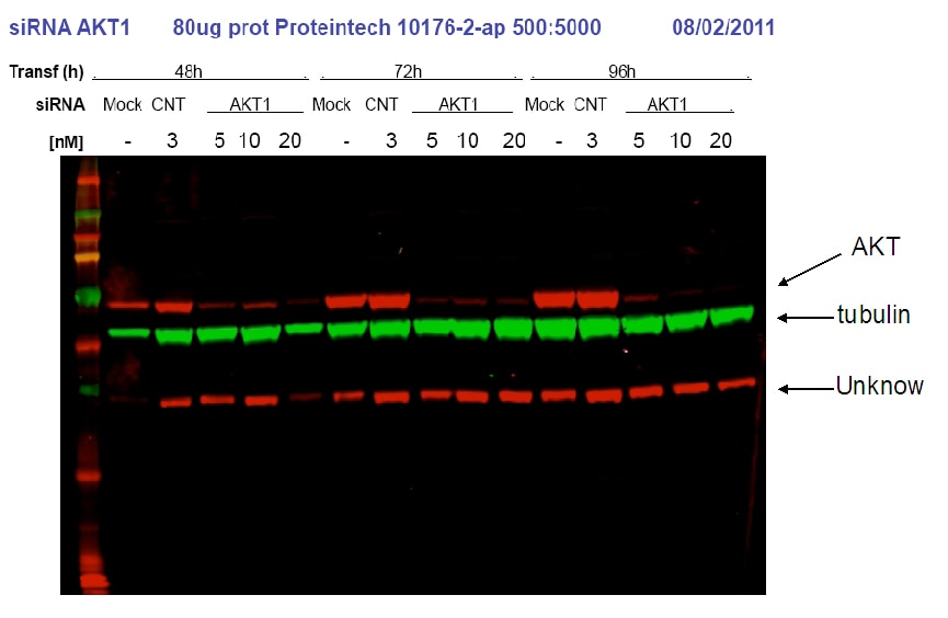
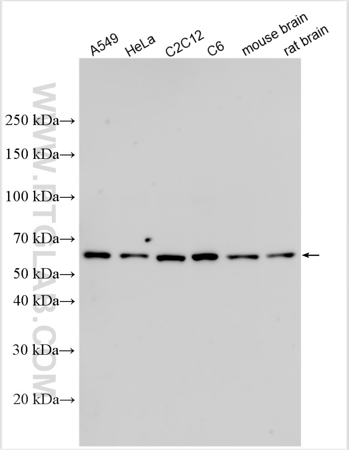
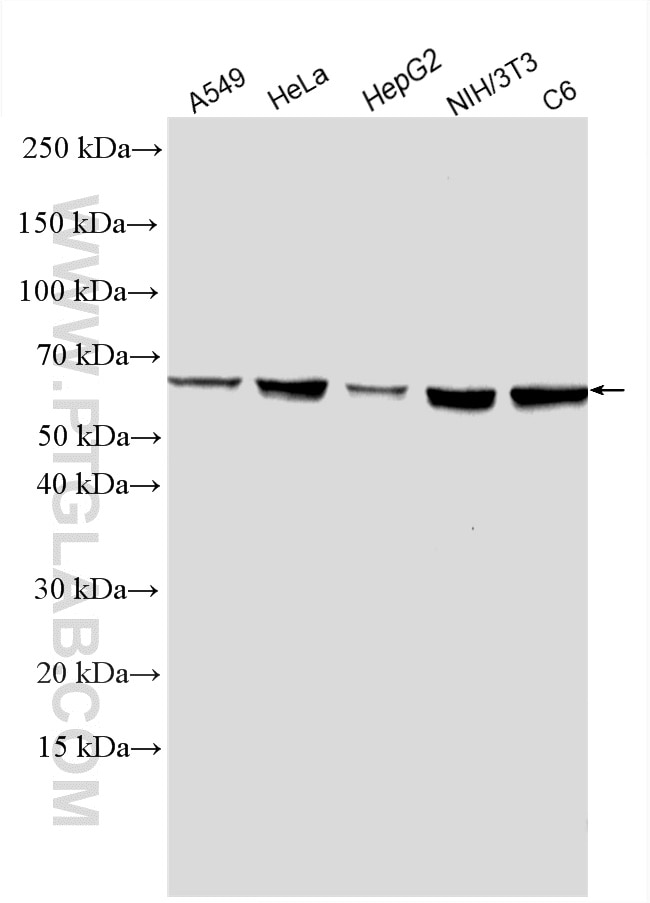
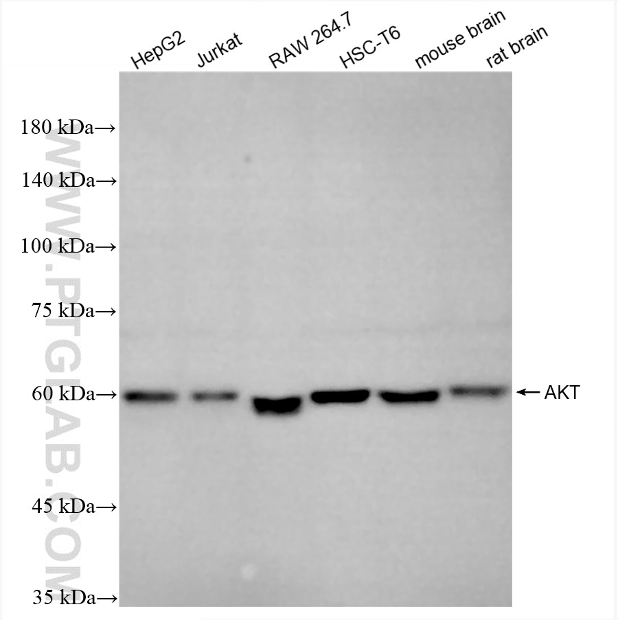
| Positive WB detected in | A549 cells, HeLa cells, HepG2 cells, MCF-7 cells, NIH/3T3 cells, C6 cells, C2C12 cells, mouse brain tissue, rat brain tissue, Jurkat cells, RAW 264, HSC-T6 cells |
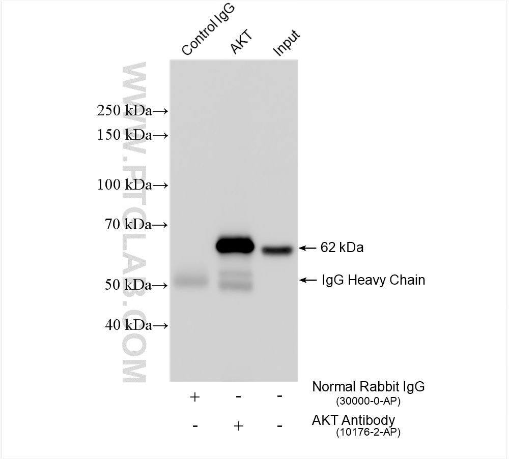
| Positive IP detected in | HeLa cells |
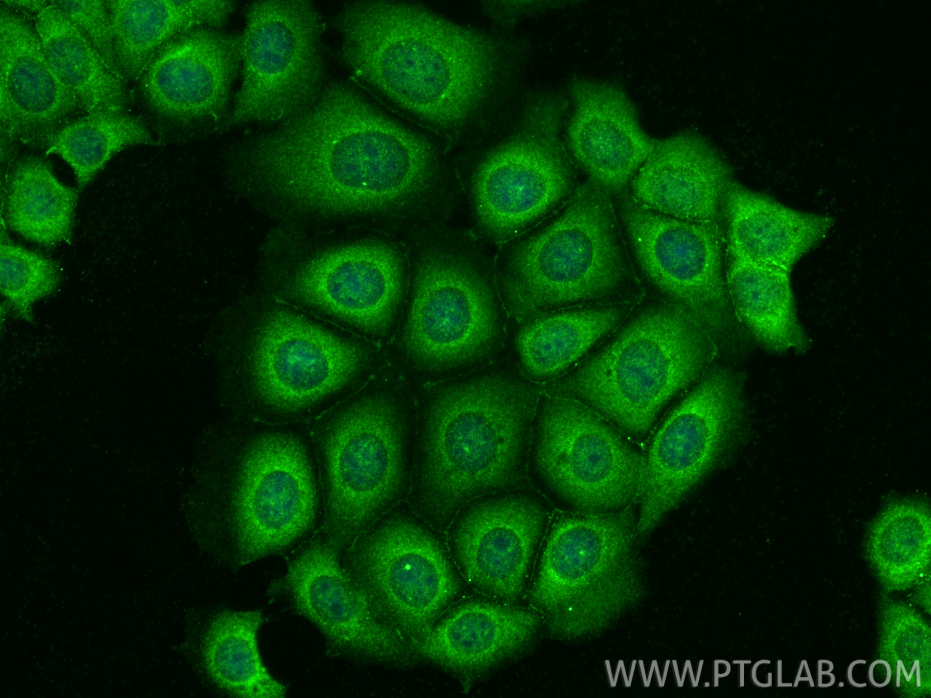
| Positive IF/ICC detected in | MCF-7 cells |
Recommended dilution
| Application | Dilution |
|---|---|
| Western Blot (WB) | WB : 1:2000-1:12000 |
| Immunoprecipitation (IP) | IP : 0.5-4.0 ug for 1.0-3.0 mg of total protein lysate |
| Immunofluorescence (IF)/ICC | IF/ICC : 1:200-1:800 |
| It is recommended that this reagent should be titrated in each testing system to obtain optimal results. | |
| Sample-dependent, Check data in validation data gallery. | |
Published Applications
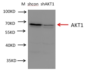
| KD/KO | See 10 publications below |
| WB | See 1904 publications below |
| IHC | See 58 publications below |
| IF | See 33 publications below |
| IP | See 12 publications below |
| ELISA | See 1 publications below |
| CoIP | See 5 publications below |
Product Information
10176-2-AP targets AKT in WB, IHC, IF/ICC, IP, CoIP, ELISA applications and shows reactivity with human, mouse, rat samples.
| Tested Reactivity | human, mouse, rat |
| Cited Reactivity | human, mouse, rat, rabbit, monkey, chicken, sheep, goat, zebra finches |
| Host / Isotype | Rabbit / IgG |
| Class | Polyclonal |
| Type | Antibody |
| Immunogen |
CatNo: Ag0213 Product name: Recombinant human AKT protein Source: e coli.-derived, PGEX-4T Tag: GST Domain: 1-224 aa of BC000479 Sequence: MSDVAIVKEGWLHKRGEYIKTWRPRYFLLKNDGTFIGYKERPQDVDQREAPLNNFSVAQCQLMKTERPRPNTFIIRCLQWTTVIERTFHVETPEEREEWTTAIQTVADGLKKQEEEEMDFRSGSPSDNSGAEEMEVSLAKPKHRVTMNEFEYLKLLGKGTFGKVILVKEKATGRYYAMKILKKEVIVAKDEVAHTLTENRVLQNSRHPFLTALKYSFQTHDRLC Predict reactive species |
| Full Name | v-akt murine thymoma viral oncogene homolog 1 |
| Calculated Molecular Weight | 56 kDa |
| Observed Molecular Weight | 56-62 kDa |
| GenBank Accession Number | BC000479 |
| Gene Symbol | AKT1 |
| Gene ID (NCBI) | 207 |
| RRID | AB_2224574 |
| Conjugate | Unconjugated |
| Form | Liquid |
| Purification Method | Antigen affinity purification |
| UNIPROT ID | P31749 |
| Storage Buffer | PBS with 0.02% sodium azide and 50% glycerol, pH 7.3. |
| Storage Conditions | Store at -20°C. Stable for one year after shipment. Aliquoting is unnecessary for -20oC storage. 20ul sizes contain 0.1% BSA. |
Background Information
The serine-threonine protein kinase AKT1 is catalytically inactive in serum-starved primary and immortalized fibroblasts. AKT1 and the related AKT2 are activated by platelet-derived growth factor. The activation is rapid and specific, and it is abrogated by mutations in the pleckstrin homology domain of AKT1. It was shown that the activation occurs through phosphatidylinositol 3-kinase. In the developing nervous system AKT is a critical mediator of growth factor-induced neuronal survival. Survival factors can suppress apoptosis in a transcription-independent manner by activating the serine/threonine kinase AKT1, which then phosphorylates and inactivates components of the apoptotic machinery.
Protocols
| Product Specific Protocols | |
|---|---|
| IF protocol for AKT antibody 10176-2-AP | Download protocol |
| IHC protocol for AKT antibody 10176-2-AP | Download protocol |
| IP protocol for AKT antibody 10176-2-AP | Download protocol |
| WB protocol for AKT antibody 10176-2-AP | Download protocol |
| Standard Protocols | |
|---|---|
| Click here to view our Standard Protocols |
Publications
| Species | Application | Title |
|---|---|---|
Nat Commun Genome-wide enhancer-gene regulatory maps link causal variants to target genes underlying human cancer risk | ||
Adv Sci (Weinh) Cis-Regulation of an m6A Eraser by an Insertion Variant Associated with Survival of Patients With Non-Small Cell Lung Carcinoma | ||
Neuron C5aR1+ microglia exacerbate neuroinflammation and cerebral edema in acute brain injury | ||
Sci Adv Platelet P-selectin initiates cross-presentation and dendritic cell differentiation in blood monocytes. |
Reviews
The reviews below have been submitted by verified Proteintech customers who received an incentive for providing their feedback.
FH Ana (Verified Customer) (06-17-2025) | The staining looks very good. It might be better to dilute a bit more than 1:2000 to reduce background noise.
|
FH Hyopil (Verified Customer) (08-23-2021) | Worked well with western blotting
|
FH Azita (Verified Customer) (06-02-2021) | Western blot analysis using AKT polyclonal antibody in NSC34 cell line at dilution of 1:500.
|