Anti-IgG VHH Beads for Immunoprecipitation
A Smarter Alternative to Protein A/G
While Protein A and G remain the most common choices for antibody capture, their broad and unspecific Fc binding and potential for leaching from beads can compromise specificity and lead to interfering background in sensitive assays. Our VHH beads are optimized for targeted precision, clean results, and redefine specificity and purity in immunoprecipitation.
-
 Excellent tissue and cell penetration
-
Minimal epitope-label displacement
-
Recombinant production
Available anti-IgG VHH Beads
Choose from broad-specificity or species-specific anti-IgG VHH beads in agarose and magnetic agarose formats for immunoprecipitation workflows.
Anti-Rabbit / Anti-Mouse VHH Beads
Our anti-rabbit / anti-mouse VHH beads are conjugated with a mix of three high-affinity VHHs targeting rabbit IgG, mouse IgG1, and mouse kappa light chains. This combination ensures broad compatibility with the most commonly used rabbit and mouse antibodies. Whether you're performing routine IPs across various antibody subtypes or working with antibodies of unknown isotype, these beads offer a powerful, reliable alternative to Protein A or Protein G, delivering clean, efficient pulldowns with consistent performance.
| Target | Formats | Available Sizes | Specificity | |
|---|---|---|---|---|
| Rabbit / Mouse | Agarose | Magnetic agarose | 1ml, 2ml, 5ml | Rabbit IgG, Mouse IgG1, 2a*, 2b*, 3* *kappa light chain |
Species specific VHH Beads
Our species specific VHH beads offer excellent specificity to only one IgG subtype, enabling you to perform precise pulldowns with maximum efficiency when you know the subtype you’re working with. If you regularly perform IPs with the same antibody or antibodies of the same subtype, this solution is the best pick for you.
| Target | Formats | Available Sizes | Specificity | |
|---|---|---|---|---|
| Rabbit | Agarose | Magnetic agarose | 1ml, 2ml, 5ml | Rabbit IgG |
| Mouse | Agarose | Magnetic agarose | 1ml, 2ml, 5ml | Mouse (All Isotypes) |
| Mouse IgG1 | Agarose | Magnetic agarose | 1ml, 2ml, 5ml | Mouse IgG1 |
| Mouse IgG2a | Agarose | Magnetic agarose | 1ml, 2ml, 5ml | Mouse IgG2a |
| Mouse IgG2b | Agarose | Magnetic agarose | 1ml, 2ml, 5ml | Mouse IgG2b |
| Mouse IgG3 | Agarose | Magnetic agarose | 1ml, 2ml, 5ml | Mouse IgG3 |
Anti-IgG VHH Bead Performance Data
Explore validation data for Anti-IgG VHH Beads across immunoprecipitation workflows, benchmarking against Protein A/G, binding capacity, species specificity, and background performance in common sample types.
Co-IP of whole MCM-Complex via specific anti-MCM6 antibody
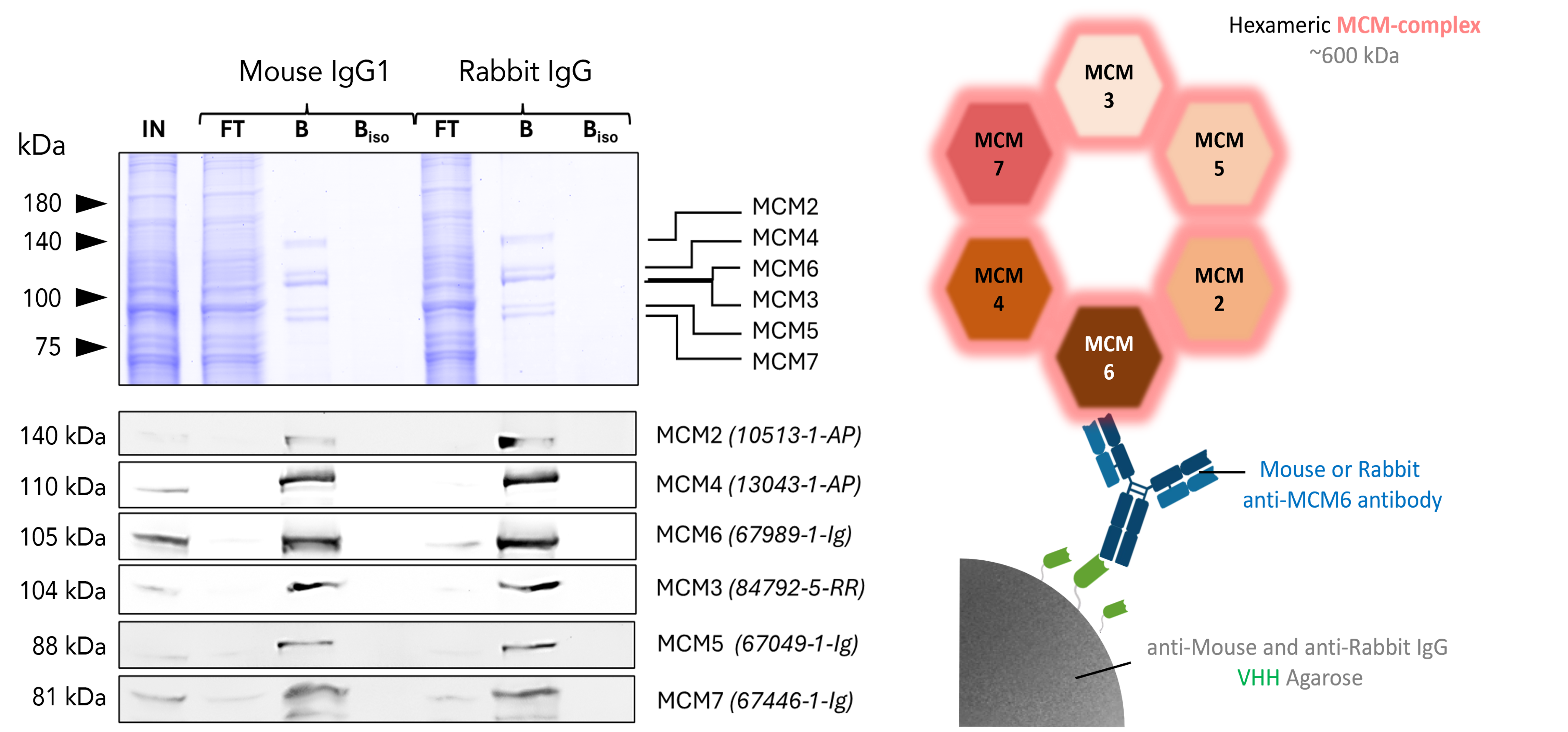
Co-IP of MCM complex via pulldown of MCM6 using 5 µg of anti-MCM6 antibodies and anti-rabbit IgG / anti-mouse IgG VHH agarose. All subunits of the 600 kDa hetero-hexameric complex are successfully precipitated using both a mouse IgG1 (67989-1-Ig) and a rabbit (13347-2-AP) MCM6 antibody, as shown by western blot analysis using subunit specific antibodies. Apparent molecular weights are provided. For input (IN) and flowthrough (FT) fractions, 1% was loaded. For bound (B) fraction and bound fraction of isotype control antibody (BISO), 20% was loaded. Product codes for PTG-antibodies are provided in parentheses. BISO is an isotype control used as negative control (ms1: 66260-1-Ig; rb: 30000-0-AP). Results were reproducible for anti-rabbit IgG/ anti-mouse IgG Magnetic VHH Agarose.
Species specificity of anti-rabbit IgG / anti-mouse IgG VHH agarose
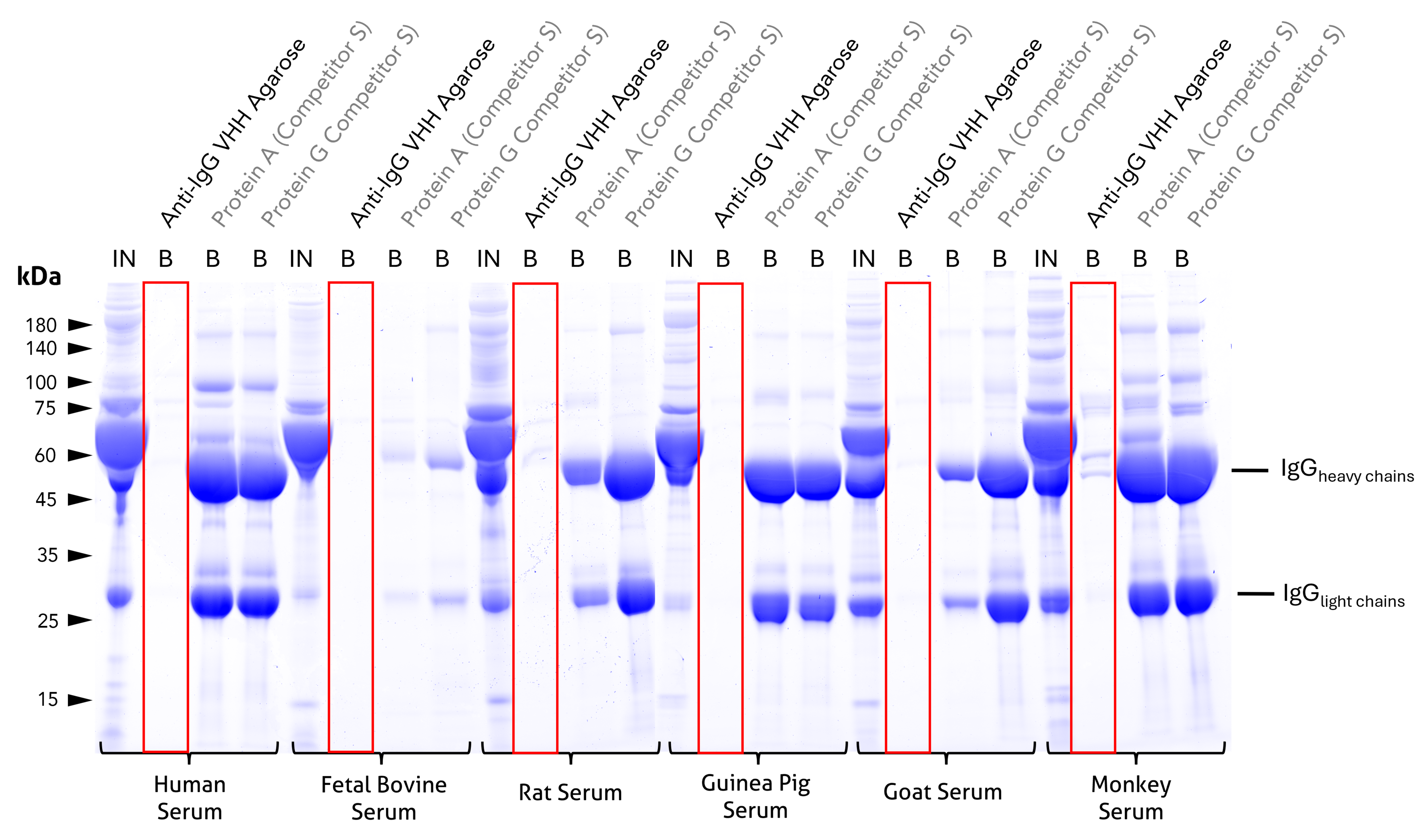
Cross-reactivity test and comparison to competitor Protein A and Protein G beads. Coomassie-stained SDS-PAGE gel showing input (IN) and bound (B) fractions for different beads incubated with 1:10 diluted sera of relevant model species. Unlike the tested protein A or G beads, bound fractions of Proteintech anti-rabbit IgG / anti-mouse IgG VHH agarose reveal no cross-reactivity to human, bovine, rat, guinea pig, or goat IgGs and only minor cross-reactivity to cyno monkey IgGs. For IN and B fractions 1% and 25% were loaded, respectively. Lanes of anti-IgG VHH beads are highlighted red. Results were reproducible for anti-rabbit IgG/ anti-mouse IgG Magnetic VHH Agarose.
Clean precipitation and detection of PCNA with anti-rabbit IgG / anti-mouse IgG VHH beads using anti-PCNA antibody
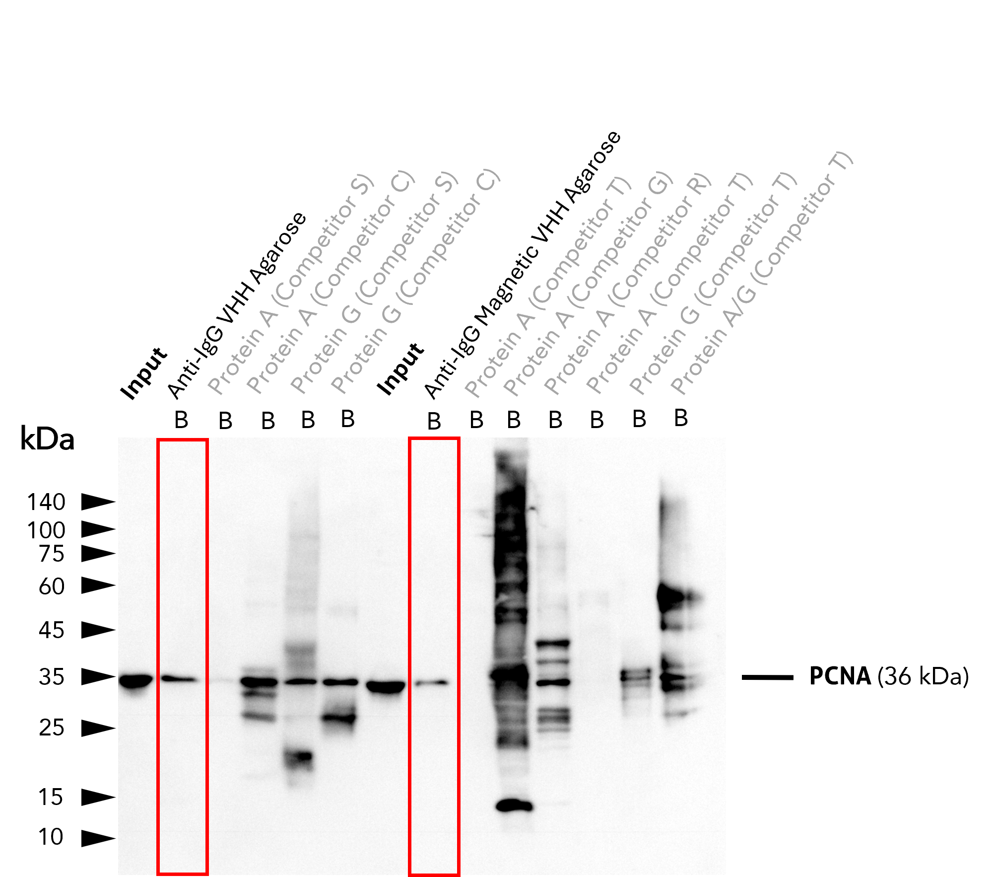
IP of PCNA using 5 µg of anti-PCNA antibody (IgG1, 60097-1-Ig) by anti-rabbit IgG / anti-mouse IgG VHH beads. For Western blot analysis, the PCNA polyclonal antibody (10205-2-AP, 1:2000) was labeled using a conformation-specific HRP-conjugated secondary to avoid staining of heavy and light chain of the IP-antibody. In contrast to many competitors' protein A and G beads, clean pull-down of PCNA by Proteintech anti-rabbit IgG/ anti-mouse IgG VHH Beads (Ag. = Agarose; M.Ag. = Magnetic Agarose) facilitates unambiguous identification of the target protein. Other beads show leaching of protein A or protein G fragments into the final IP fractions, which can lead to binding of the detection antibody and unspecific signals, as reported previously (Grant et al., 2019, Biol Proced Online, doi: 10.1186/s12575-019-0095-z). For input (IN) and bound (B) fractions, 1% and 30% were loaded, respectively. Lanes of anti-IgG VHH beads are highlighted in red.
VHH Agarose
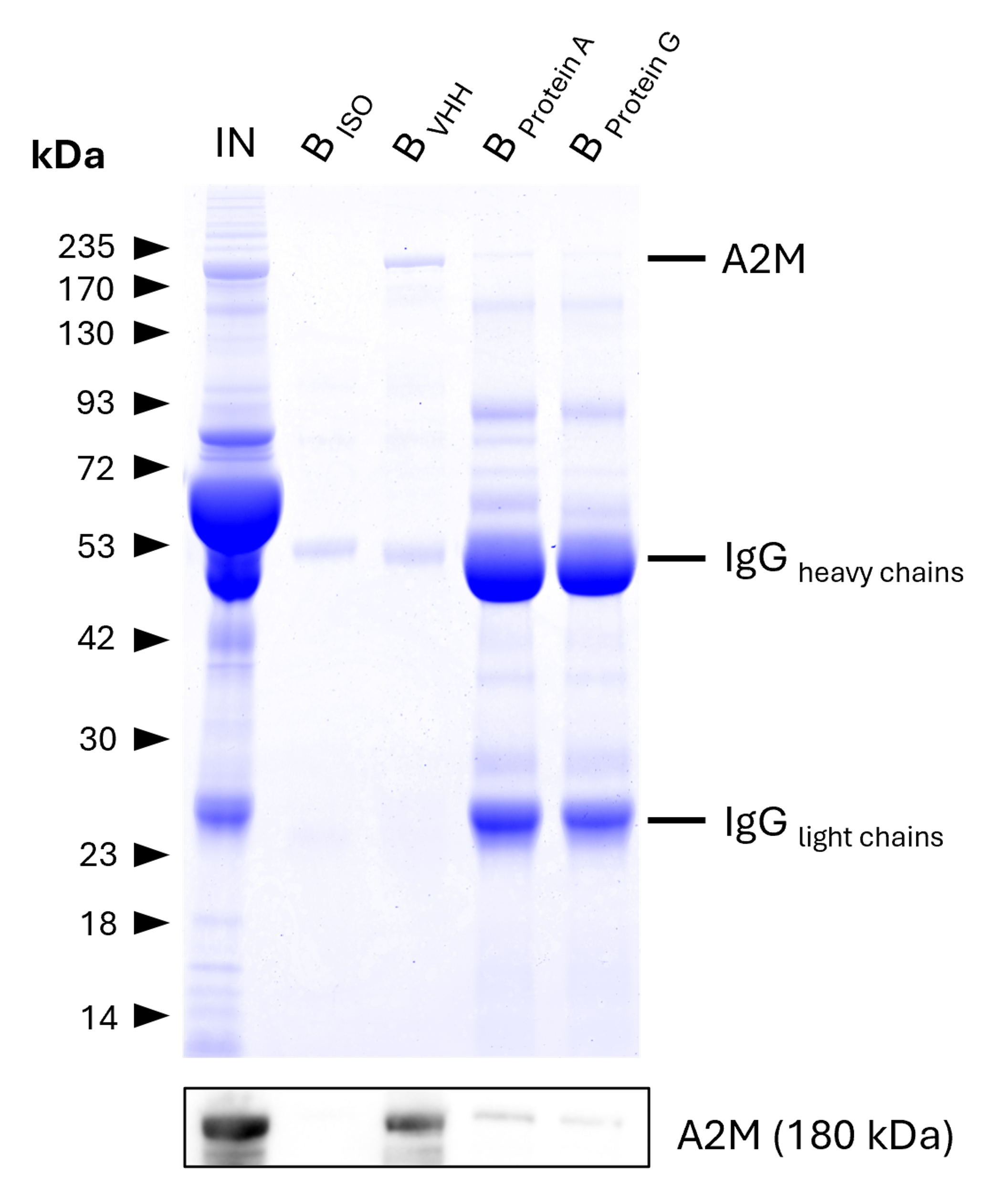
IP of Alpha-2-Macroglobulin (A2M) from human serum using anti-rabbit IgG / anti-mouse IgG VHH Agarose and comparison to Protein A and G beads. IP was performed using 5 μg of rabbit anti-A2M IgG (13545-1-AP) in 1:10 diluted human serum. 0.5% and 6.25% of input (IN) and bound (B) fractions were analyzed by SDS-PAGE, respectively. 5 μg of rabbit isotype control antibody (BISO, 98136-1-RR) was used as negative control. The Coomassie-stained gel shows precipitation of A2M by anti-rabbit IgG / anti-mouse IgG VHH agarose and reveals its absence for ´protein A and G beads. This is likely due to human IgGs competing with the spiked IP-antibody for the latter beads. Results were verified using Western blot analysis, which detected A2M at approximately 180 kDa using an anti-A2M primary IgG (13545-1-AP, 1:5000). Data was reproducible for anti-rabbit IgG / anti-mouse IgG magnetic VHH agarose.
Binding efficiency – Agarose
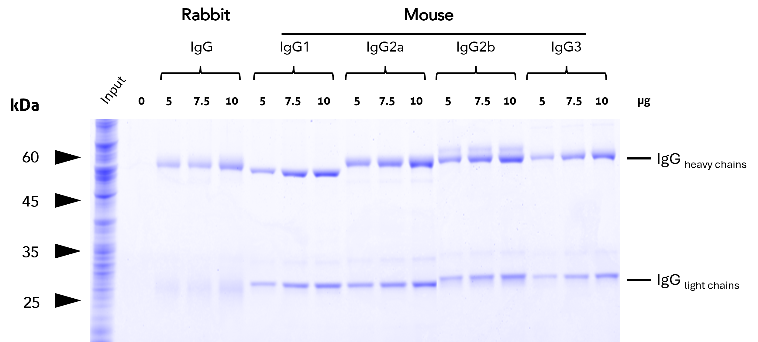
Binding efficiency and specificity of anti-rabbit IgG / anti-mouse IgG VHH agarose for rabbit and mouse IgG subclasses. Binding of antibodies of different subtypes was tested at increasing concentrations from HEK293T cell lysate. The high capacity of the anti-IgG VHH beads allows for efficient binding of all IgGs tested.
Binding efficiency – Magnetic agarose
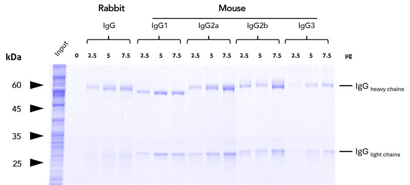
Binding efficiency and specificity of anti-rabbit IgG / anti-mouse IgG magnetic VHH agarose for rabbit and mouse IgG subclasses. Binding of antibodies of different subtypes was tested at increasing concentrations from HEK293T cell lysate. The high capacity of the anti-IgG VHH beads allows for efficient binding of all IgGs tested.
Magnetic VHH agarose
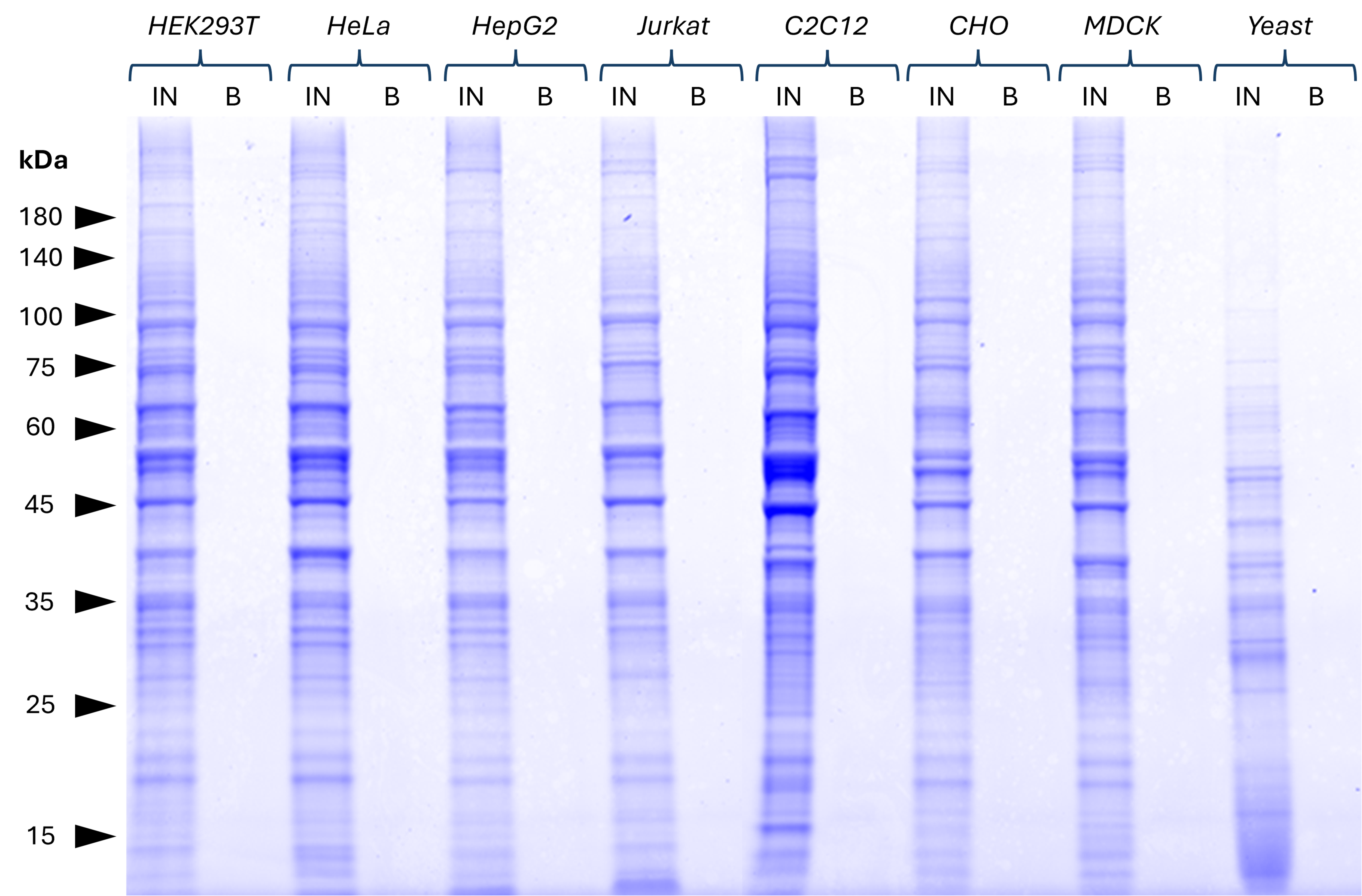
No background binding of anti-rabbit IgG/ anti-mouse IgG magnetic VHH agarose in commonly used cell lines. Various cell lysates (derived from 1x10⁷ cells) were tested for non-specific binding to the beads. Clean backgrounds were observed in bound (B) fractions of the Coomassie-stained SDS-PAGE gel for all lysates. This indicates that no significant non-specific binding occurs. 25% of B fraction and 1% of Input (IN) fractions were loaded. HEK293T, HeLa, HepG2, Jurkat: Human; C2C12: Mouse; CHO: Hamster; MDCK: Canine; Yeast: Fungi. Results were reproducible for anti-rabbit IgG / anti-mouse IgG VHH agarose.
Magnetic VHH agarose
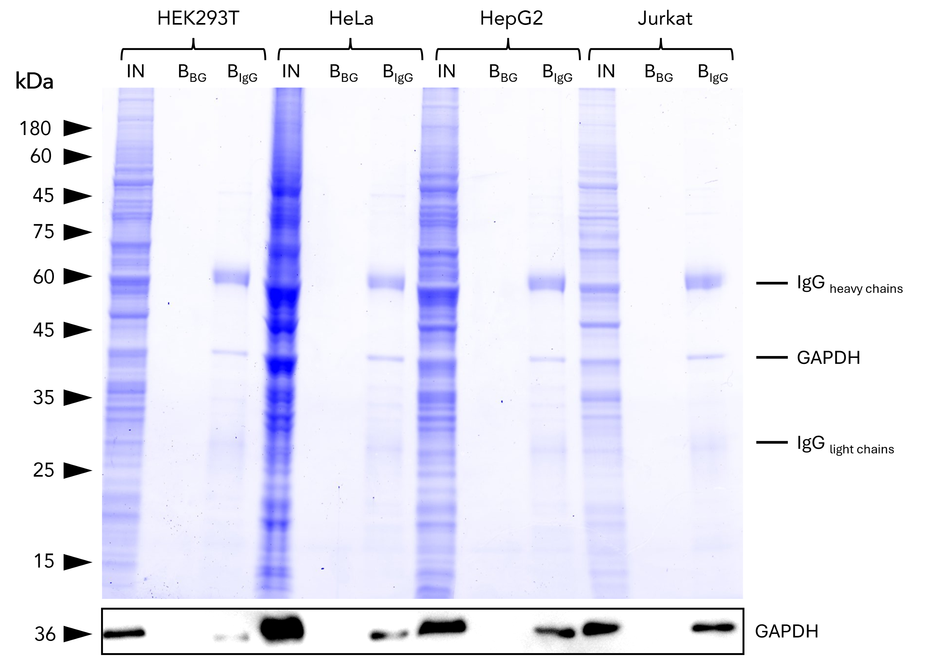
IP of GAPDH from common human cell lines using anti-rabbit IgG/ anti-mouse IgG magnetic VHH agarose. IP was performed using 5 μg of rabbit anti-GAPDH IgG (10494-1-AP) with 1x10⁷ cells used per IP reaction. 1% and 25% of input (IN) and bound (B) fractions were analyzed by SDS-PAGE, respectively. Bound fractions without antibody spiked (BBG) are shown as background control. The Coomassie-stained gel shows precipitation of GAPDH by anti-rabbit IgG / anti-mouse IgG magnetic VHH agarose, what was verified using Western blot analysis using the anti-GAPDH mouse monoclonal antibody (60004-1-Ig). Results are reproducible for anti-rabbit IgG / anti-mouse IgG VHH agarose.
How anti-IgG VHH Beads compare with Nano-Traps and Protein A/G
Compare anti-IgG VHH Beads with Nano-Traps and Protein A/G to see when VHH-based IgG capture offers the best balance of specificity, background reduction, and workflow compatibility.
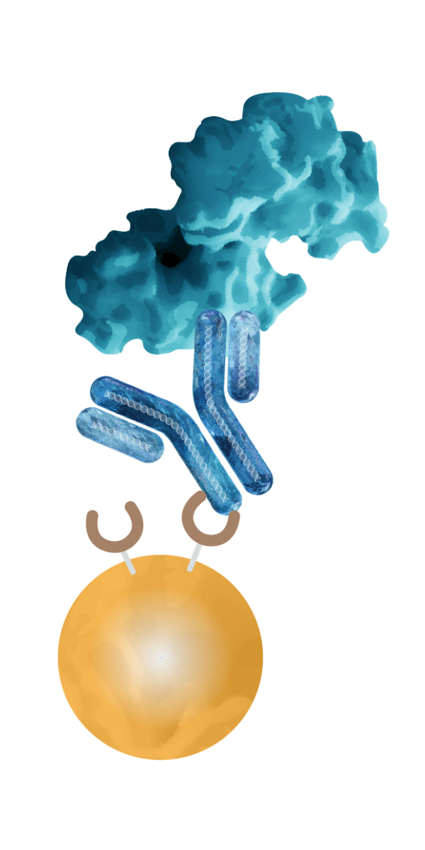
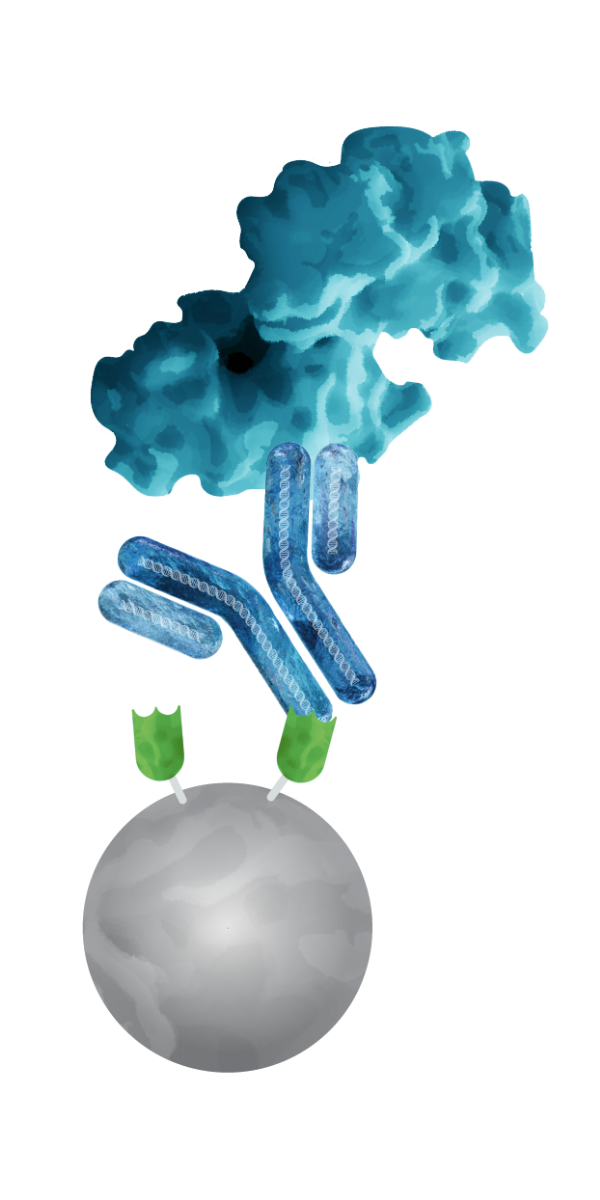
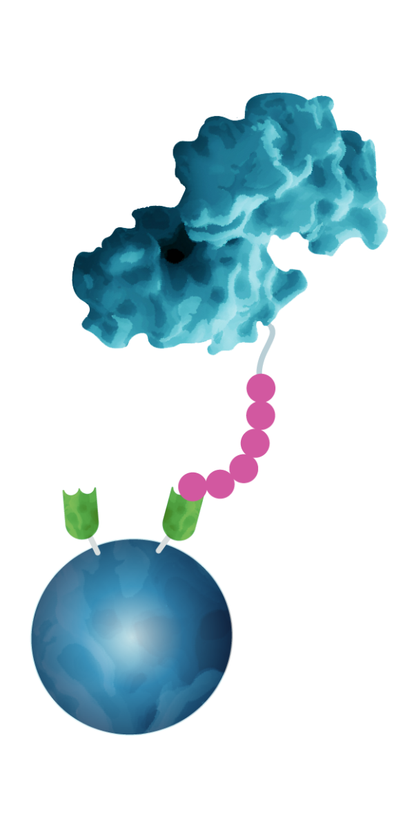
| Protein A/G | Anti-IgG VHH Beads | Nano-Traps | |
|---|---|---|---|
 |
 |
 |
|
| Binding via | Bacterial Protein A or G | Recombinant Secondary VHH | Recombinant Primary VHH |
| Binding target | IgG | IgG | Protein or peptide tag Endogenous proteins |
| Species-specific IgG recognition? | No | Yes | Binds directly to target |
| Background in SDS-PAGE and Western Blot |
High Protein A/G leaching into IP fraction and unspecific binding of detection antibodies; Heavy and light chain antibody bands possible. |
Low Heavy and light chain antibody bands possible. You can use SpeedAb Kits or conformation-specific secondary antibodies to avoid recognition of your pulldown antibody. |
Ultra-low |
| Duration | > 3h | > 3h | < 2h |
| Binding affinity to IgG or tag? |
Very low to high (depending on IgG species) |
High (optimized for the respective IgG subtype) |
High to very high |
| Ready-to-use? | Depends on vendor | Yes | Yes |
| Use for | Standard IPs if background is not critical | Standard IPs where clean, efficient, and consistent performance is required, especially when working with antibody species that have low affinity for Protein A/G. | IPs of tagged proteins, especially for low abundant targets and workflows requiring stringent washing (e.g., IP-MS), or for applications where speed and reproducibility are key. |
| Learn more about Nano-Traps |
FAQs
Need help choosing the right immunoprecipitation reagent? Explore when to use anti-IgG VHH beads for your experiments.
When are anti-IgG VHH Beads a better choice than Protein A/G?
While Protein A and G remain the most common choices for antibody capture, their broad and unspecific Fc binding and potential for leaching from beads can compromise specificity and lead to interfering background in sensitive assays. Our anti-IgG VHH Beads use secondary VHHs coupled to agarose or magnetic agarose beads and are optimized for targeted precision, and clean results. The anti-IgG VHH Beads are particularly useful when your sample contains endogenous antibodies, e.g. serum or plasma.
Do anti-IgG VHH beads work with all mouse and rabbit antibodies?
We offer species-specific anti-IgG VHH beads for rabbit IgG and mouse IgG1, IgG2a, IgG2b, and IgG3, as well as an anti-rabbit/anti-mouse combination resin.
Species-specific VHH beads provide high selectivity for a single IgG subtype, enabling precise and efficient immunoprecipitation when the antibody subtype is known. They are ideal for routine IP workflows using the same antibody or IgG subtype.
Anti-rabbit/anti-mouse VHH combi beads contain a mixture of three high-affinity VHHs that bind specifically to rabbit IgG (Fc fragment), mouse IgG1 (Fc fragment), and mouse Ig κ light chain (Fab fragment), thus covering 100% and 98% of available rabbit and mouse primary antibodies, respectively. They are ideal when you perform routine IPs across various antibody subtypes or work with antibodies of unknown isotypes.
Can I use anti-IgG VHH Beads to perform immunoprecipitation directly from blood serum or samples containing BSA, FCS or HSA?
Yes, you can use anti-Rabbit or anti-Mouse IgG VHH Beads to perform immunoprecipitation directly from complex matrices like blood serum or samples containing BSA, FCS, or HSA. Anti-IgG VHH beads are engineered exclusively for their target host IgG (Rabbit or Mouse) and offer exceptional species and even isotype specificity. They do not cross-react with human, bovine, rat, guinea pig or goat antibodies.
What lysis and wash buffers are compatible with anti-IgG VHH Beads?
ChromoTek’s anti-IgG VHH Beads are coupled with recombinant secondary nanobodies. Because nanobodies are very small and have a compact structure, they show increased thermostability and robustness under stringent washing and lysis buffer conditions. However, unlike one-step Nano-Traps, anti-IgG VHH Beads do not bind the protein of interest directly but require the use of a conventional primary capture antibody. Therefore, the total buffer compatibility of your experiment is ultimately limited by what the primary antibody can tolerate.
Where can I find an immunoprecipitation protocol for my application?
Protocols are available in the following places:
- Standard immunoprecipitation protocol.
- Protocols for anti-IgG VHH Beads can be found on each respective product page under the product information section
- Find out more about our immunoprecipitation tools, workflow guidance, and FAQs.