Tested Applications
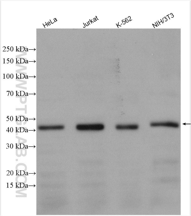
| Positive WB detected in | HeLa cells, Jurkat cells, K-562 cells, NIH/3T3 cells |
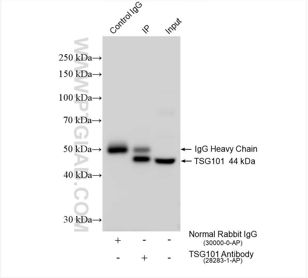
| Positive IP detected in | HeLa cells |
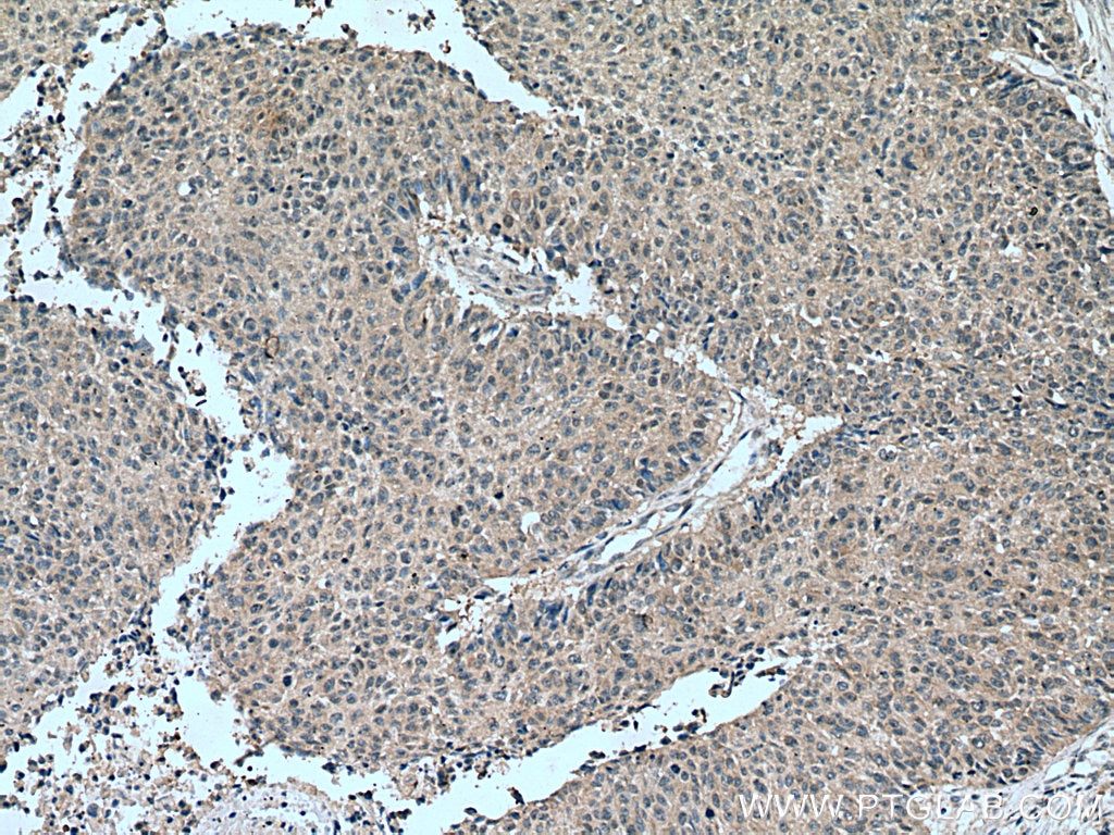
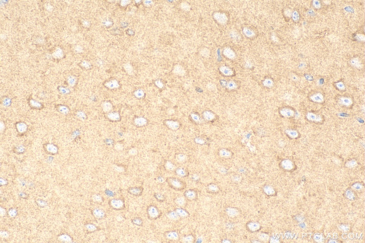
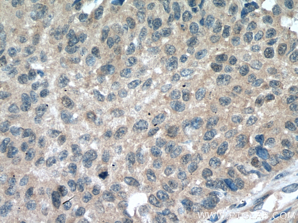
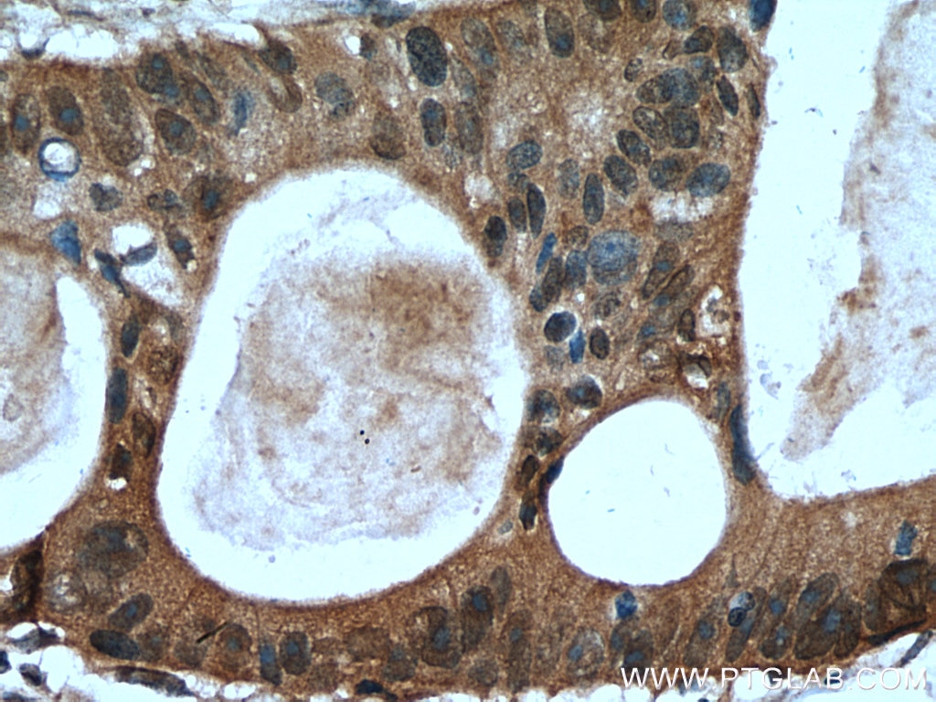
| Positive IHC detected in | human lung cancer tissue, human colon cancer tissue, mouse brain tissue Note: suggested antigen retrieval with TE buffer pH 9.0; (*) Alternatively, antigen retrieval may be performed with citrate buffer pH 6.0 |
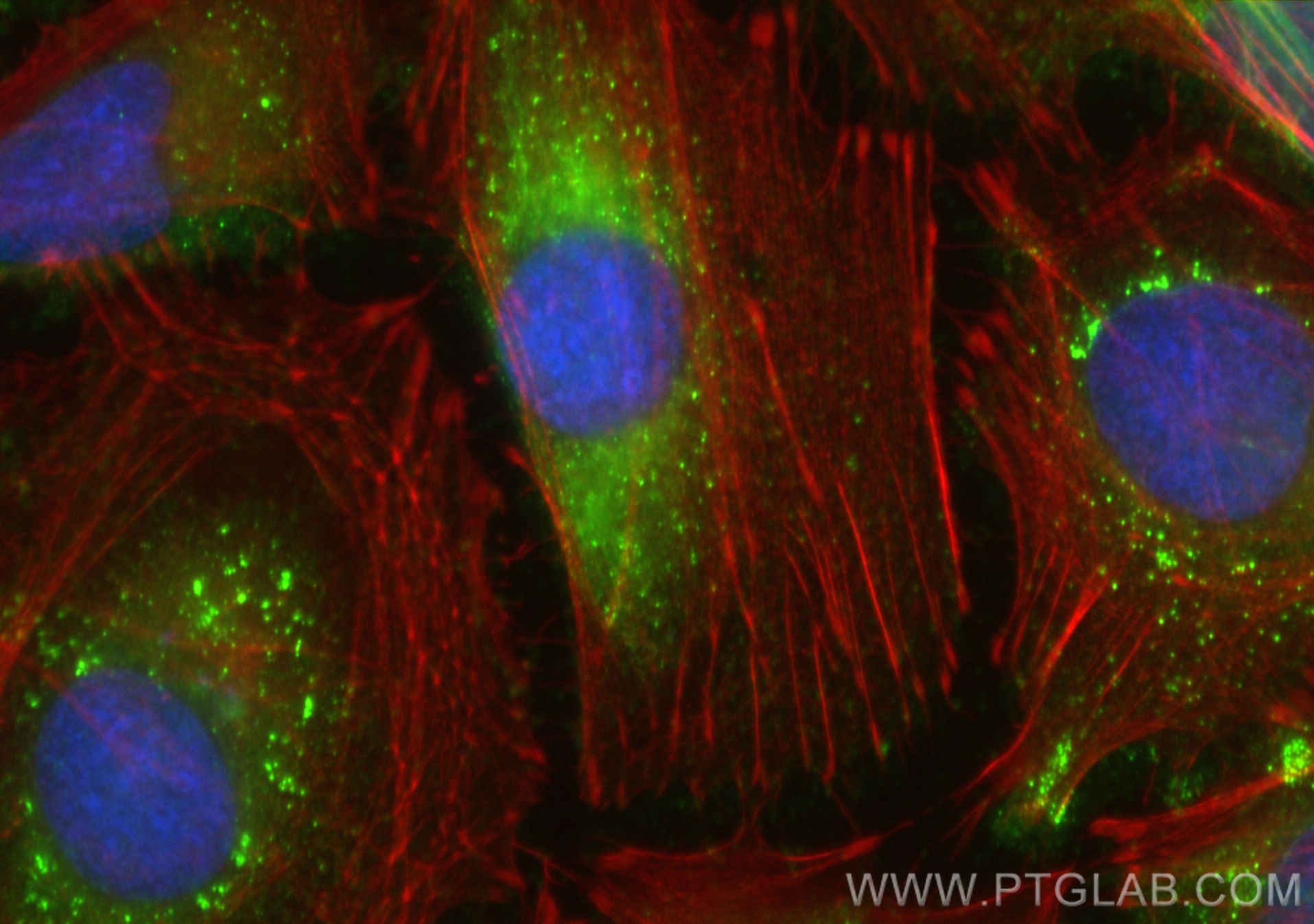
| Positive IF/ICC detected in | HeLa cells |
Recommended dilution
| Application | Dilution |
|---|---|
| Western Blot (WB) | WB : 1:1000-1:10000 |
| Immunoprecipitation (IP) | IP : 0.5-4.0 ug for 1.0-3.0 mg of total protein lysate |
| Immunohistochemistry (IHC) | IHC : 1:50-1:500 |
| Immunofluorescence (IF)/ICC | IF/ICC : 1:50-1:500 |
| It is recommended that this reagent should be titrated in each testing system to obtain optimal results. | |
| Sample-dependent, Check data in validation data gallery. | |
Published Applications
| KD/KO | See 6 publications below |
| WB | See 455 publications below |
| IHC | See 2 publications below |
| IF | See 8 publications below |
| IP | See 5 publications below |
| CoIP | See 2 publications below |
Product Information
28283-1-AP targets TSG101 in WB, IHC, IF/ICC, IP, CoIP, ELISA applications and shows reactivity with human, mouse samples.
| Tested Reactivity | human, mouse |
| Cited Reactivity | human, mouse, rat, pig, rabbit, monkey, chicken, hamster, sheep, goat |
| Host / Isotype | Rabbit / IgG |
| Class | Polyclonal |
| Type | Antibody |
| Immunogen |
CatNo: Ag28569 Product name: Recombinant human TSG101 protein Source: e coli.-derived, PET28a Tag: 6*His Domain: 214-390 aa of BC002487 Sequence: GPSRDGTISEDTIRASLISAVSDKLRWRMKEEMDRAQAELNALKRTEEDLKKGHQKLEEMVTRLDQEVAEVDKNIELLKKKDEELSSALEKMENQSENNDIDEVIIPTAPLYKQILNLYAEENAIEDTILYLGEALRRGVIDLDVFLKHVRLLSRKQFQLRALMQKARKTAGLSDLY Predict reactive species |
| Full Name | tumor susceptibility gene 101 |
| Calculated Molecular Weight | 44 kDa |
| Observed Molecular Weight | 44 kDa |
| GenBank Accession Number | BC002487 |
| Gene Symbol | TSG101 |
| Gene ID (NCBI) | 7251 |
| RRID | AB_2881104 |
| Conjugate | Unconjugated |
| Form | Liquid |
| Purification Method | Antigen affinity purification |
| UNIPROT ID | Q99816 |
| Storage Buffer | PBS with 0.02% sodium azide and 50% glycerol, pH 7.3. |
| Storage Conditions | Store at -20°C. Stable for one year after shipment. Aliquoting is unnecessary for -20oC storage. 20ul sizes contain 0.1% BSA. |
Background Information
TSG101(Tumor susceptibility gene 101 protein) is essential for endosomal sorting, membrane receptor degradation and the final stages of cytokinesis. It plays a crucial role for cell proliferation and cell survival. TSG101 has been identified as a candidate tumor suppressor gene and belongs to the ubiquitin-conjugating enzyme family. TSG101 is a marker for exosome. This protein has 2 isoforms produced by alternative splicing with the molecular mass of 44 and 32 kDa.
Protocols
| Product Specific Protocols | |
|---|---|
| IF protocol for TSG101 antibody 28283-1-AP | Download protocol |
| IHC protocol for TSG101 antibody 28283-1-AP | Download protocol |
| IP protocol for TSG101 antibody 28283-1-AP | Download protocol |
| WB protocol for TSG101 antibody 28283-1-AP | Download protocol |
| Standard Protocols | |
|---|---|
| Click here to view our Standard Protocols |
Publications
| Species | Application | Title |
|---|---|---|
Nat Commun Injectable ECM-mimetic dynamic hydrogels abolish ferroptosis-induced post-discectomy herniation through delivering nucleus pulposus progenitor cell-derived exosomes | ||
J Extracell Vesicles Quantification of urinary podocyte-derived migrasomes for the diagnosis of kidney disease | ||
Nat Commun A functionally tunable magnetic nanochains platform for N-glycoproteomic analysis of extracellular vesicles from ultratrace biofluids | ||
Adv Sci (Weinh) LIMA1 O-GlcNAcylation Promotes Hepatic Lipid Deposition through Inducing β-catenin-Regulated FASn Expression in Metabolic Dysfunction-Associated Steatotic Liver Disease | ||
J Am Chem Soc Efficient Metabolomics Profiling from Plasma Extracellular Vesicles Enables Accurate Diagnosis of Early Gastric Cancer |
Reviews
The reviews below have been submitted by verified Proteintech customers who received an incentive for providing their feedback.
FH Angélique (Verified Customer) (04-28-2026) | western-blot on mouse liver tumors compared to non-tumor tissue
 |
FH Sonam (Verified Customer) (11-17-2025) | highly recommend
|
FH Kamal (Verified Customer) (02-15-2024) | Mouse liver lysates were subjected to SDS PAGE followed by western blot with 28283-1-AP (TSG101 antibody) at dilution of 1:10000 incubated at 4 degree C for 1.5 hours. TSG101 bands appeared at 42 kDa.
|
FH SONAM (Verified Customer) (01-09-2024) | Antibody works well. Ordered three times..
|