Working with green fluorescent proteins: Tools and properties
Overview about the three most commonly used classes of green fluorescent proteins
Jellyfish Green Fluorescent Protein (GFP) and its derivatives are still the most frequently used fluorescent proteins in biomedical research. Recently, additional green fluorescent proteins have been discovered in higher animals such as crustaceans and lancelets. These FPs share a common fold, but diverge widely in their primary sequence. Thus, they require novel, dedicated antibody research tools. Here is an overview about EGFP (the most commonly used GFP derivative), TurboGFP and mNeonGreen.
|
|
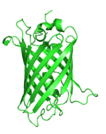
EGFP |
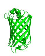
TurboGFP |
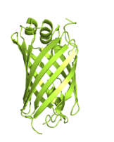
mNeonGreen |
|
Discovery/ |
1995 |
2004 |
2013 |
|
Structure |
|
|
|
|
Origin |
GFP from Jellyfish |
Tetrameric CopGFP from copepod Pontellina plumata |
Tetrameric LanYFP from Lancelet Branchiostoma lanceolatum |
|
Sequence identity to EGFP |
(100%) |
~20% |
20-25% |
|
Derived from GFP? |
Yes |
No |
No |
|
Structure |
Monomer |
Dimer |
Monomer |
|
Length/ molecular weight (MW) |
239 amino acids, |
2x 232 amino acids, |
237 amino acids, |
|
Excitation / Emission maximum |
488/ 509 nm |
482 nm/ 502 nm |
506 nm/ 517 nm |
|
Common variants |
GFP, CFP, YFP, Venus, BFP and many more |
TurboGFP, CopGFP, max GFP, maxFP-Green |
mNeonGreen |
|
Research Tools |
Immunoprecipitation Immunofluorescence Western Blotting |
Immunoprecipitation |
Immunoprecipitation Immunofluorescence Western Blotting |