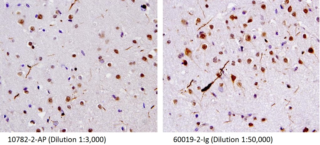
at dilution of 1:60000 incubated at room temperature for 1.5 hours.")
 at dilution of 1:5000 incubated at room temperature for 1.5 hours.")
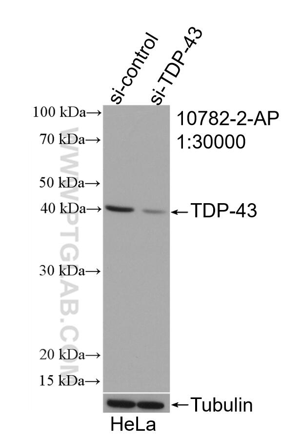
 with sh-Control and sh-TDP-43 transfected HeLa cells.")
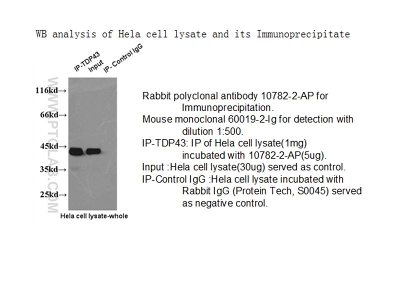
 with HeLa cells lysate 1520 ug.")
.")
.")
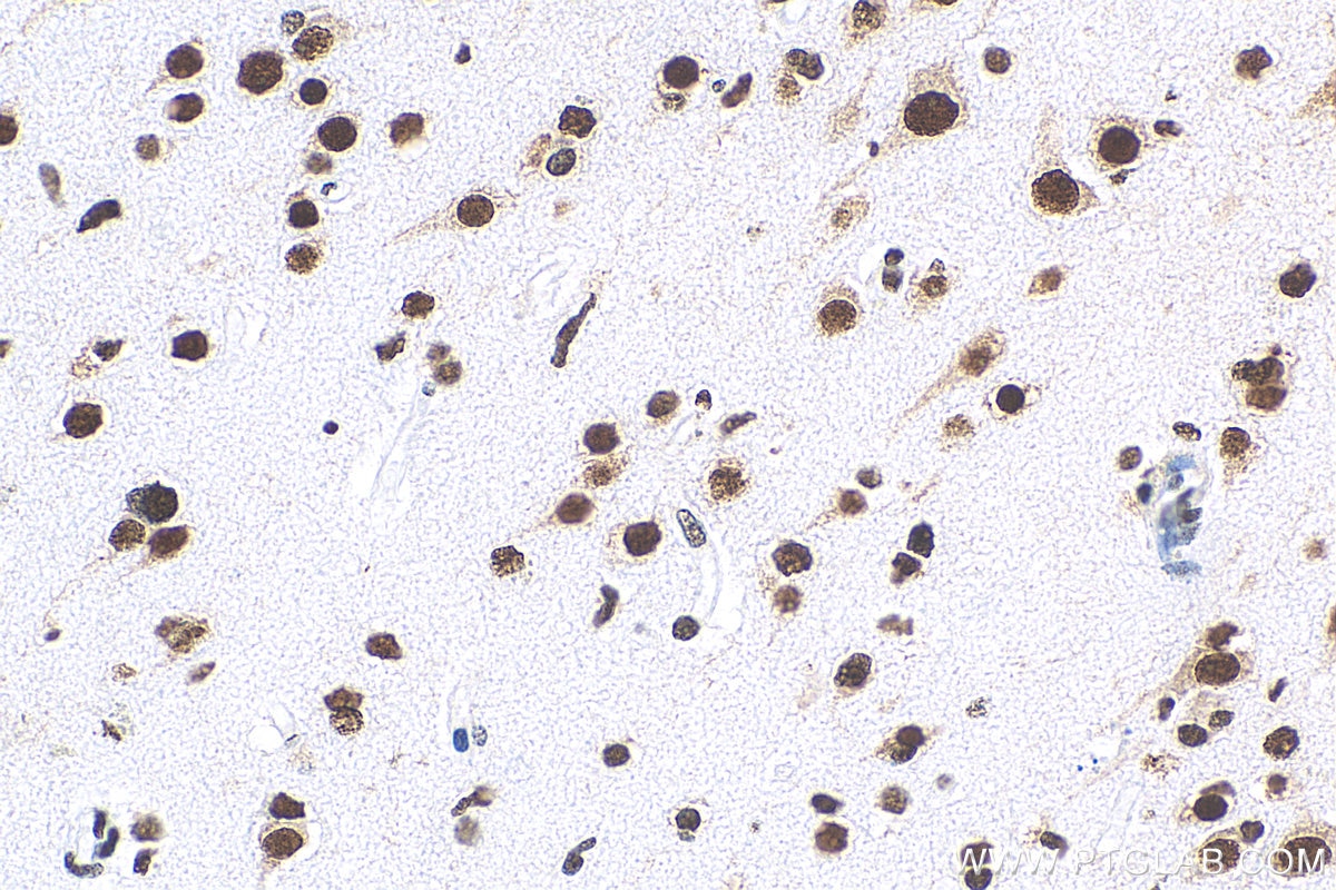
 at dilution of 1:4000 (under 40x lens). Heat mediated antigen retrieval with Tris-EDTA buffer (pH 9.0).")
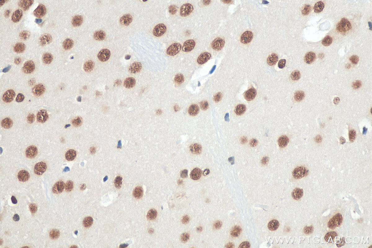
 antibody) at dilution of 1:5000 (under 40x lens). Heat mediated antigen retrieval with Tris-EDTA buffer (pH 9.0).")
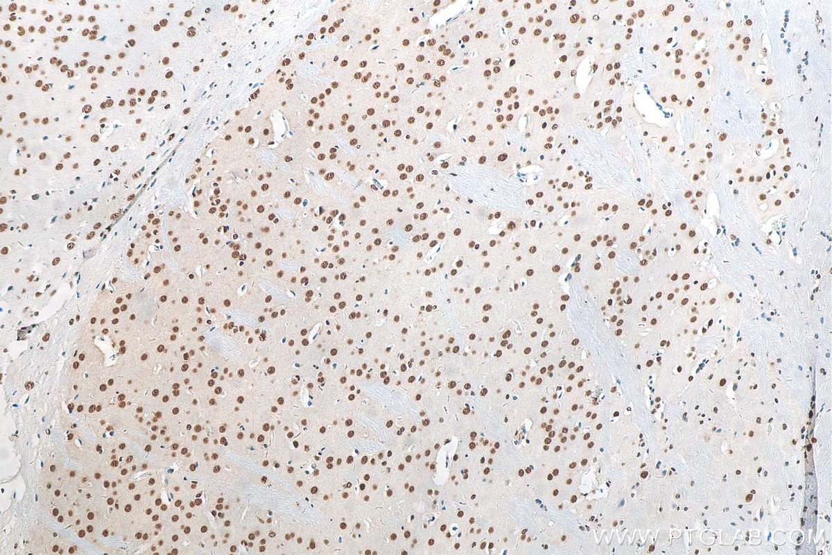
 antibody) at dilution of 1:5000 (under 40x lens). Heat mediated antigen retrieval with Tris-EDTA buffer (pH 9.0).")
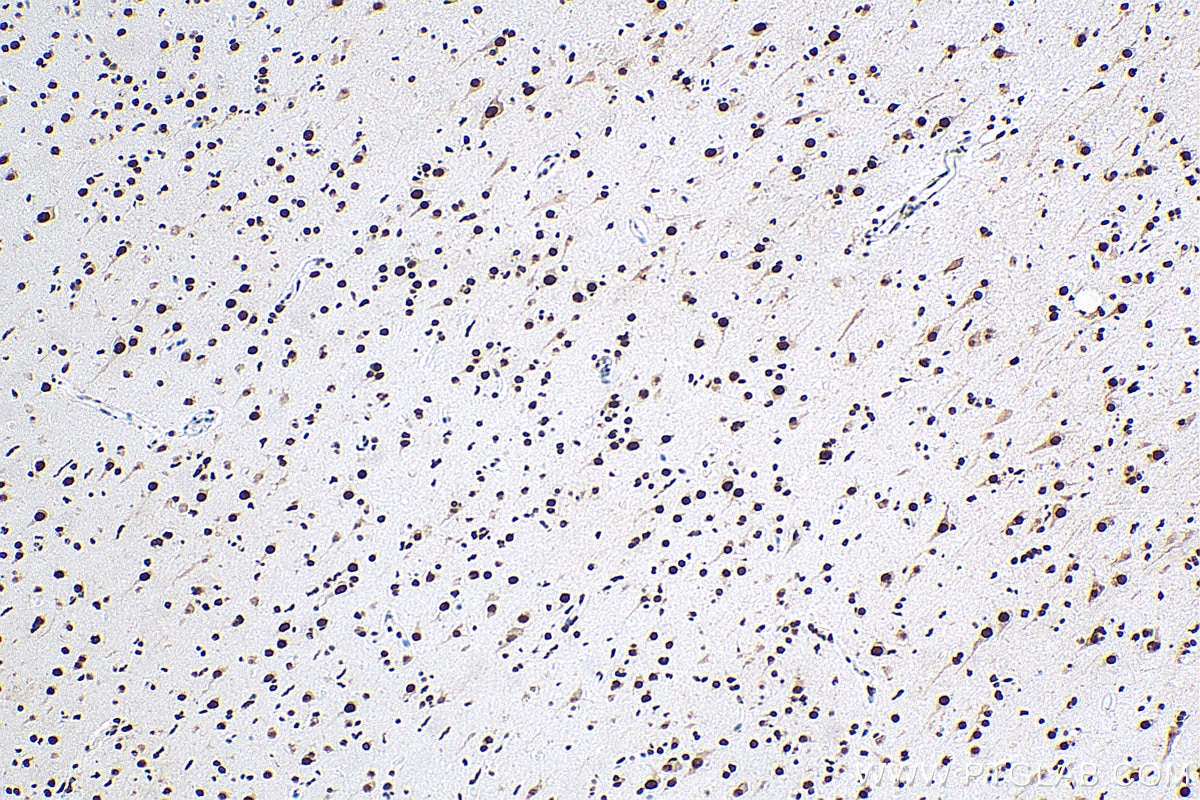
 at dilution of 1:4000 (under 10x lens). Heat mediated antigen retrieval with Tris-EDTA buffer (pH 9.0).")
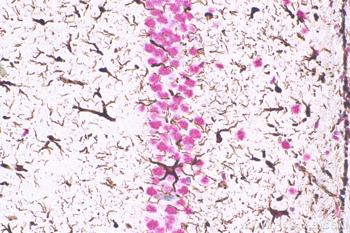
 at dilution of 1:2000 (under 40x lens). Heat mediated antigen retrieval with Tris-EDTA buffer (pH 9.0).")
 at dilution of 1:2000 (under 10x lens). Heat mediated antigen retrieval with Tris-EDTA buffer (pH 9.0).")
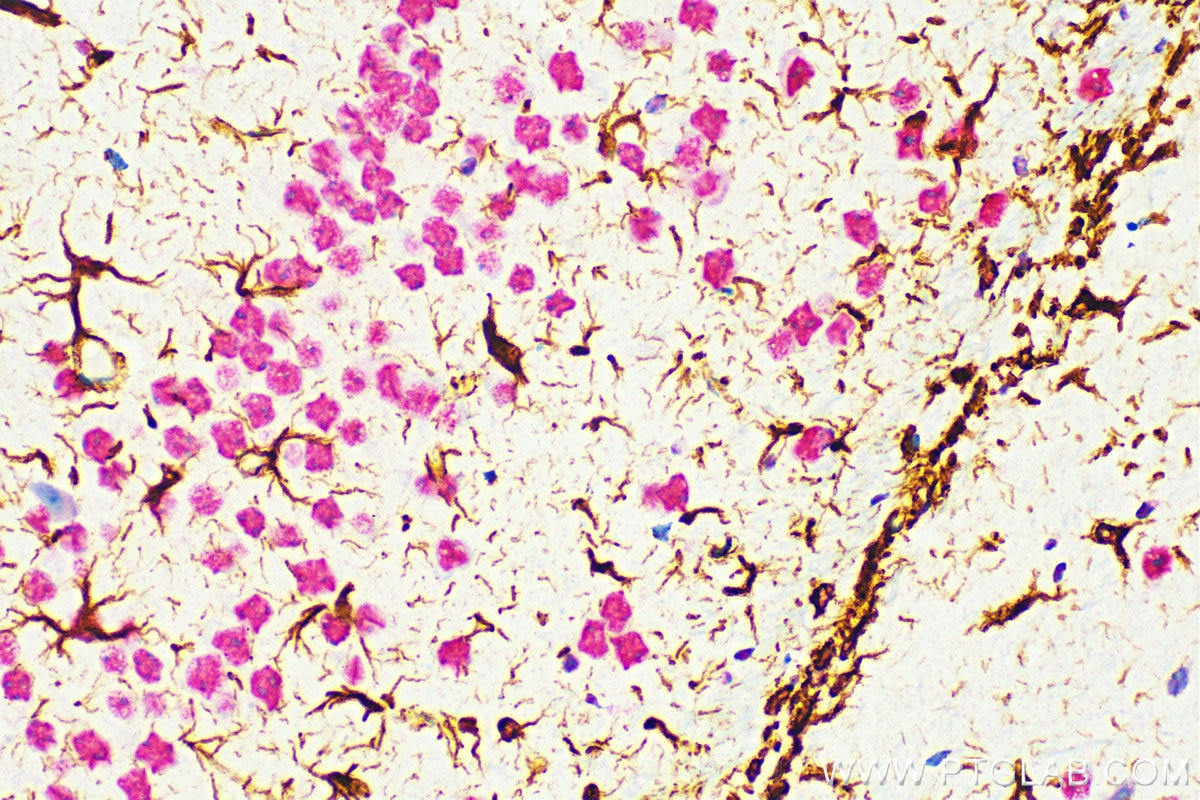
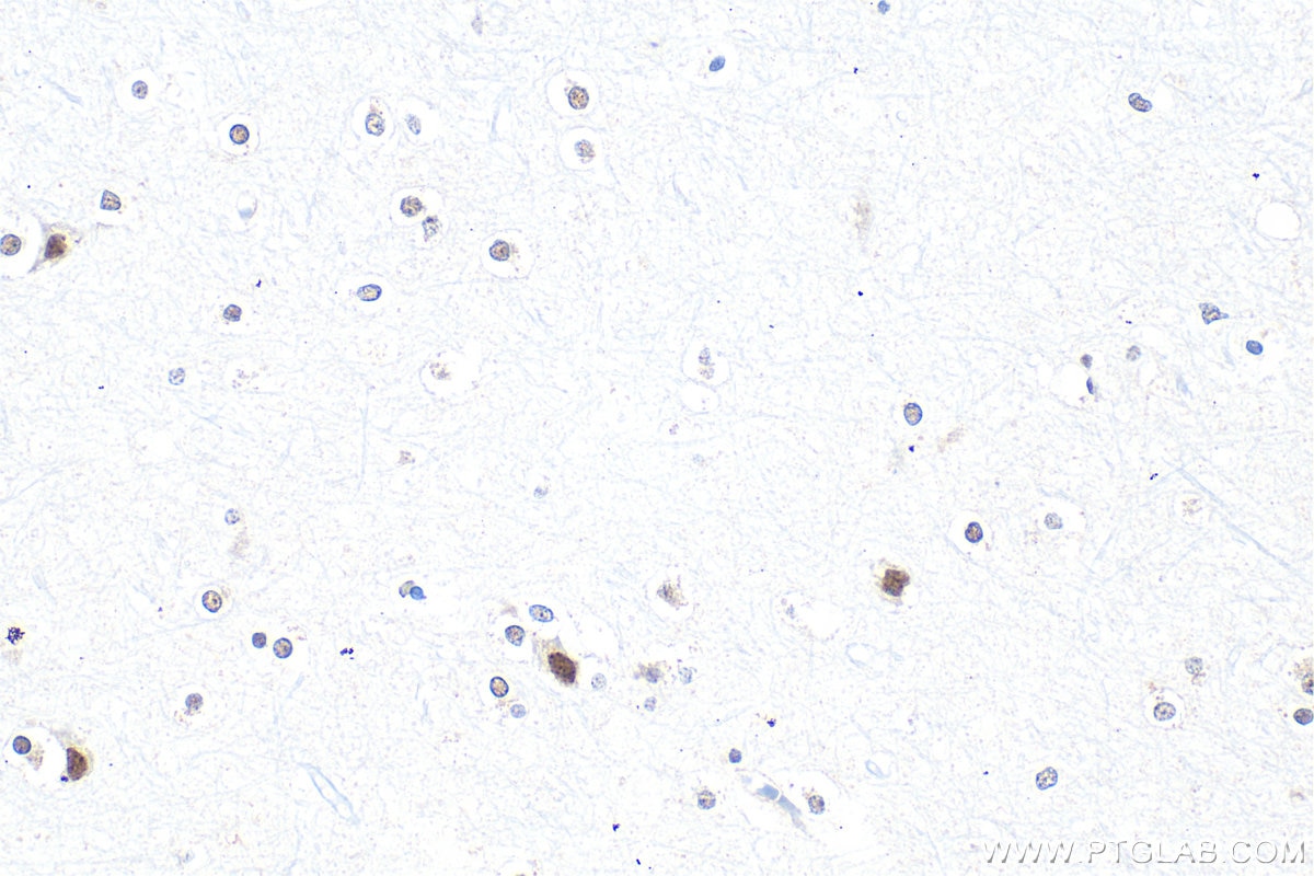
 at dilution of 1:6000 (under 40x lens). Heat mediated antigen retrieval with Tris-EDTA buffer (pH 9.0).")
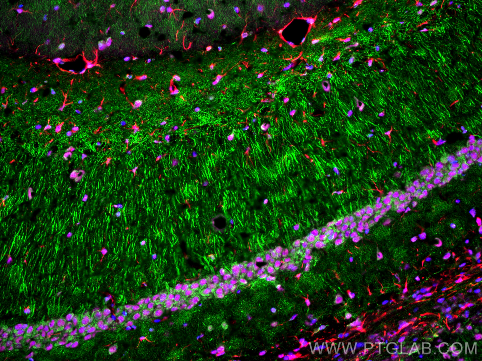
 fixed frozen OCT-embedded rat brain tissue using TDP-43 antibody (10782-2-AP) at dilution of 1:2000 and CoraLite®488-Conjugated Goat Anti-Rabbit IgG(H+L) (SA00013-2), CoraLite®594 GFAP antibody (CL594-16825, red).")
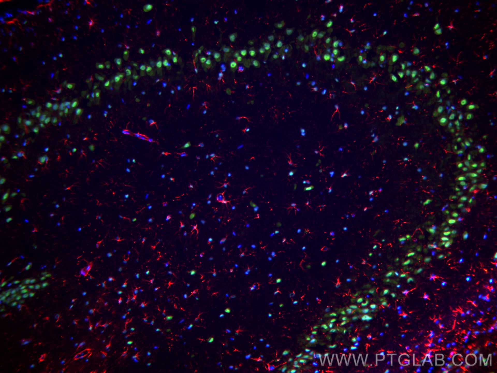
 fixed frozen OCT-embedded mouse brain tissue using TDP-43 antibody (10782-2-AP) at dilution of 1:2000 and CoraLite®647-conjugated F(ab')2 Fragment Goat Anti-Rabbit IgG (H+L) (SA00014-7), MAP2 antibody (67015-1-Ig, Clone: 1C3E6, green), CoraLite®594 GFAP antibody (CL594-16825, red).")
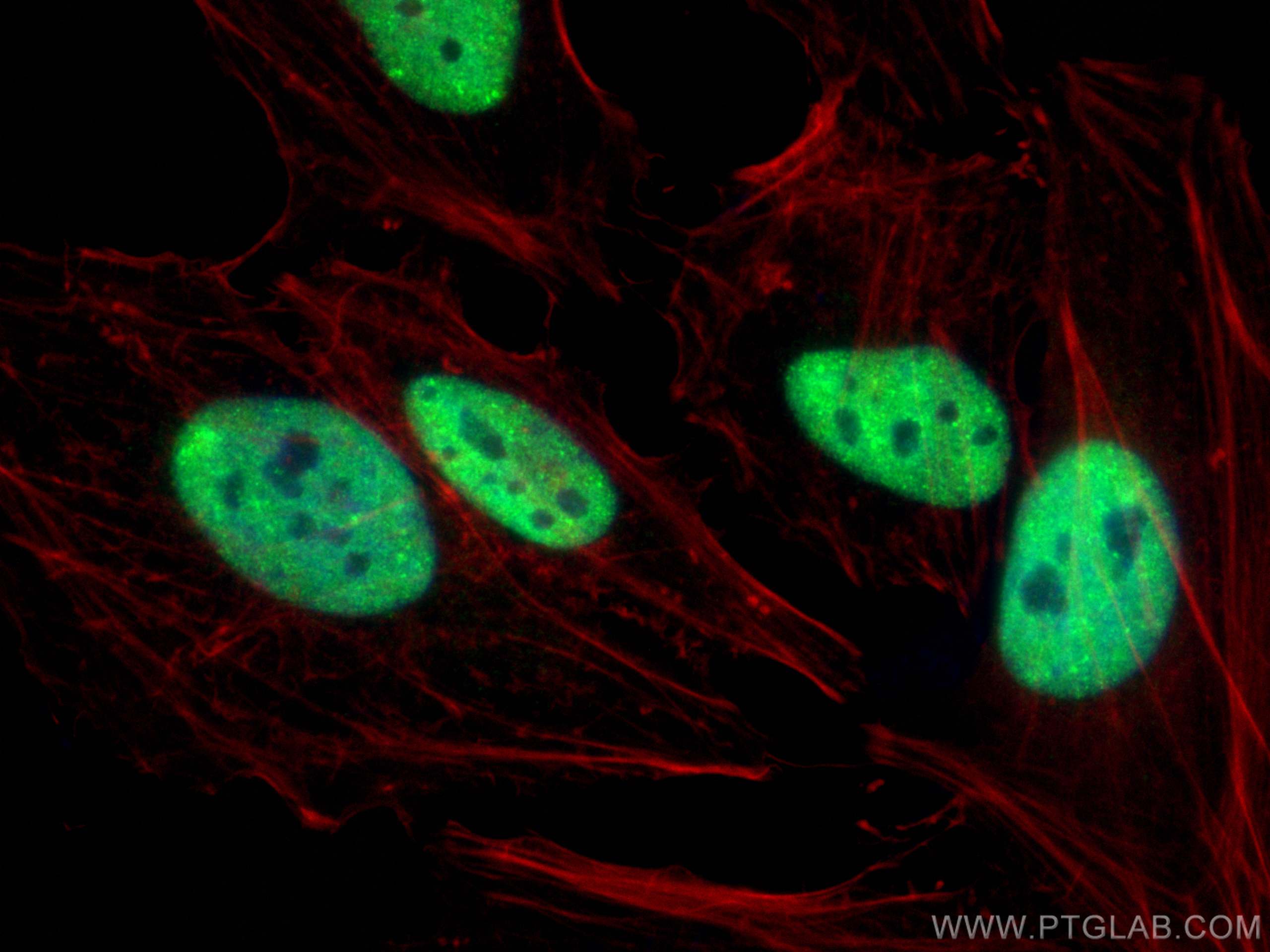
 fixed HeLa cells using TDP-43 antibody (10782-2-AP) at dilution of 1:6000 and CoraLite®488-Conjugated AffiniPure Goat Anti-Rabbit IgG(H+L), CL594-phalloidin (red).")
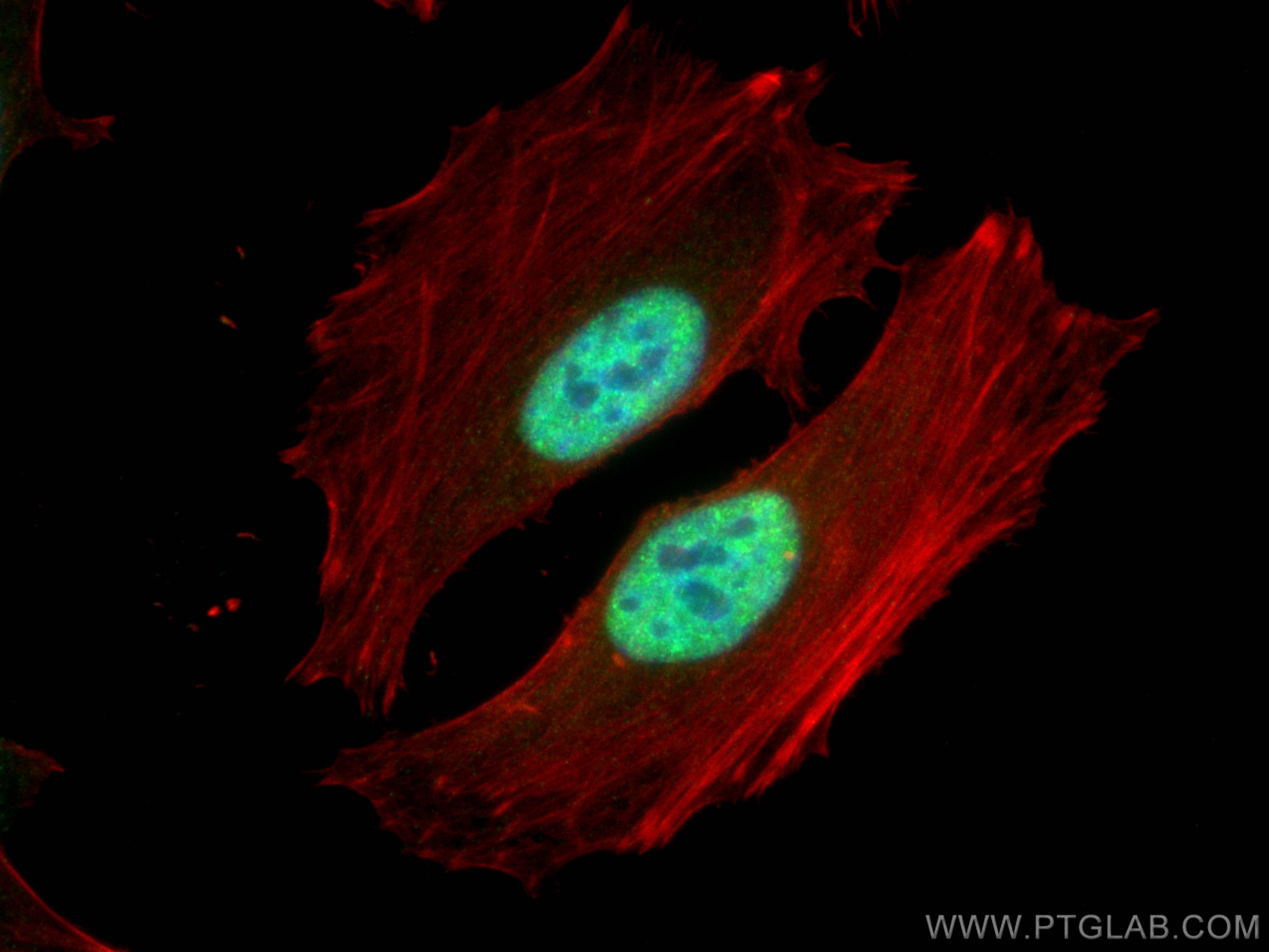
 fixed HeLa cells using TDP-43 antibody (10782-2-AP) at dilution of 1:584 and CoraLite®488-Conjugated AffiniPure Goat Anti-Rabbit IgG(H+L), CL594-Phalloidin (red).")
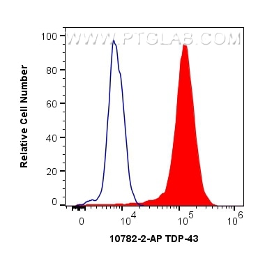
 and CoraLite®488-Conjugated AffiniPure Goat Anti-Rabbit IgG(H+L) at dilution 1:1000 (red), or 0.4 ug Isotype Control. Cells were fixed and permeabilized with Transcription Factor Staining Buffer Kit (PF00011).")
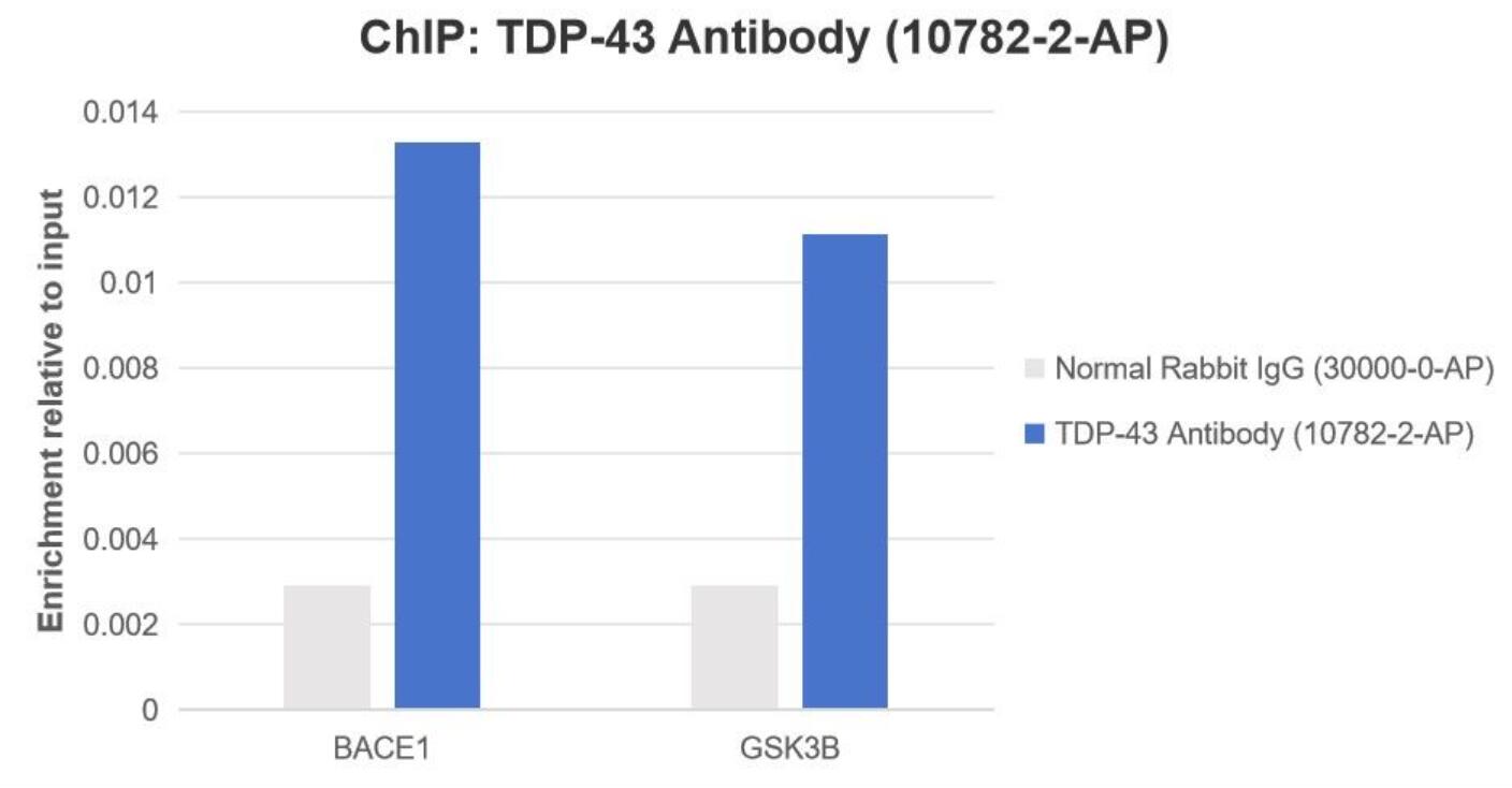
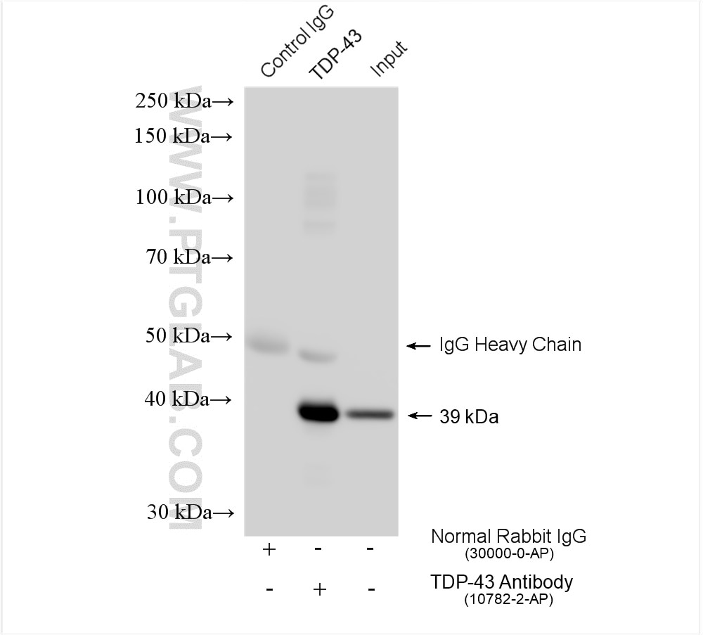
 or 5 ug of Normal Rabbit IgG (30000-0-AP), and 30 µl of Protein A Magarose Beads.
The immunoprecipitated DNA was quantified by real time PCR. Primers are located in the first kb of the transcribed region.")
Tested Applications
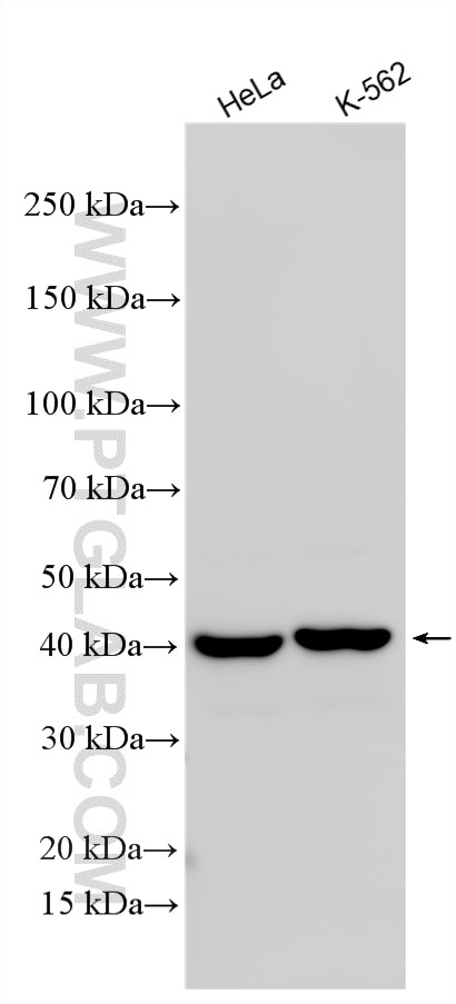
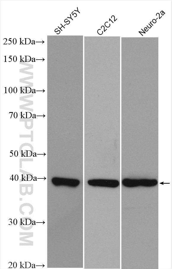
| Positive WB detected in | HeLa cells, SH-SY5Y cells, C2C12 cells, Neuro-2a cells |
| Positive IP detected in | HeLa cells |
| Positive IHC detected in | human gliomas tissue, human brain tissue, human brain (FTLD-U) tissue, mouse brain tissue Note: suggested antigen retrieval with TE buffer pH 9.0; (*) Alternatively, antigen retrieval may be performed with citrate buffer pH 6.0 |
| Positive IF-Fro detected in | rat brain tissue, mouse brain tissue |
| Positive IF/ICC detected in | HeLa cells |
| Positive FC (Intra) detected in | HeLa cells |
| Positive ChIP detected in | HeLa cells |
Recommended dilution
| Application | Dilution |
|---|---|
| Western Blot (WB) | WB : 1:20000-1:100000 |
| Immunoprecipitation (IP) | IP : 0.5-4.0 ug for 1.0-3.0 mg of total protein lysate |
| Immunohistochemistry (IHC) | IHC : 1:2000-1:8000 |
| Immunofluorescence (IF)-FRO | IF-FRO : 1:1000-1:4000 |
| Immunofluorescence (IF)/ICC | IF/ICC : 1:3000-1:12000 |
| Flow Cytometry (FC) (INTRA) | FC (INTRA) : 0.40 ug per 10^6 cells in a 100 µl suspension |
| Chromatin immunoprecipitation (ChIP) | CHIP : 1:10-1:100 |
| It is recommended that this reagent should be titrated in each testing system to obtain optimal results. | |
| Sample-dependent, Check data in validation data gallery. | |
Product Information
10782-2-AP targets TDP-43 in WB, IHC, IF/ICC, IF-Fro, FC (Intra), IP, CoIP, ChIP, RIP, ELISA, IEM applications and shows reactivity with human, mouse, rat, zebrafish samples.
| Tested Reactivity | human, mouse, rat, zebrafish |
| Cited Reactivity | human, mouse, canine, monkey, chicken, hamster, horse, drosophila, caenorhabditis elegans, macaca fascicularis (crab-eating macaque) |
| Host / Isotype | Rabbit / IgG |
| Class | Polyclonal |
| Type | Antibody |
| Immunogen |
Recombinant protein Predict reactive species |
| Full Name | TAR DNA binding protein |
| Calculated Molecular Weight | 43 kDa |
| Observed Molecular Weight | 44 kDa |
| GenBank Accession Number | BC001487 |
| Gene Symbol | TDP-43 |
| Gene ID (NCBI) | 23435 |
| RRID | AB_615042 |
| Conjugate | Unconjugated |
| Form | Liquid |
| Purification Method | Antigen affinity purification |
| UNIPROT ID | Q13148 |
| Storage Buffer | PBS with 0.02% sodium azide and 50% glycerol, pH 7.3. |
| Storage Conditions | Store at -20°C. Stable for one year after shipment. Aliquoting is unnecessary for -20oC storage. 20ul sizes contain 0.1% BSA. |
Background Information
The TARDBP gene encodes the TDP-43 protein, initially found to repress HIV-1 transcription by binding TAR DNA. TDP-43 has since been shown to bind RNA as well as DNA, and have multiple functions in transcriptional repression, translational regulation and pre-mRNA splicing. For instance, it is reported to regulate alternate splicing of the CTFR gene. In 2006 Neumann et al. found that hyperphosphorylated, ubiquitinated and/or cleaved forms of TDP-43, collectively known as pathological TDP-43, play a major role in the disease mechanisms of ubiquitin-positive, tau- and alpha-synuclein-negative frontotemporal dementia (FTLD-U) and in amyotrophic lateral sclerosis (ALS). Proteintech's 10782-2-AP antibody is a rabbit polyclonal antibody recognizing N-terminal TDP-43. It recognizes the intact 43 kDa protein as well as all posttranslationally modified and truncated forms in multiple applications. Various forms of TDP-43 exist, including 18-35 kDa of cleaved C-terminal fragments, 45-50 kDa phospho-protein, 55 kDa glycosylated form, 75 kDa hyperphosphorylated form, and 90-300 kDa cross-linked form. (17023659, 19823856, 21666678, 22193176) Recently TDP-43 has been reported to be overexpressed in triple negative breast cancer (TNBC) and it may be a potential target for TNBC diagnosis and drug design. (29581274)
Protocols
| Product Specific Protocols | |
|---|---|
| FC protocol for TDP-43 antibody 10782-2-AP | Download protocol |
| IF protocol for TDP-43 antibody 10782-2-AP | Download protocol |
| IHC protocol for TDP-43 antibody 10782-2-AP | Download protocol |
| IP protocol for TDP-43 antibody 10782-2-AP | Download protocol |
| WB protocol for TDP-43 antibody 10782-2-AP | Download protocol |
| Standard Protocols | |
|---|---|
| Click here to view our Standard Protocols |
Publications
| Species | Application | Title |
|---|---|---|
Lancet Neurol A C9orf72 promoter repeat expansion in a Flanders-Belgian cohort with disorders of the frontotemporal lobar degeneration-amyotrophic lateral sclerosis spectrum: a gene identification study. | ||
Cell Res Disruption of ER ion homeostasis maintained by an ER anion channel CLCC1 contributes to ALS-like pathologies | ||
Reviews
The reviews below have been submitted by verified Proteintech customers who received an incentive for providing their feedback.
FH Nicole (Verified Customer) (02-20-2026) | Worked great for our application!
|
FH Yuanmin (Verified Customer) (10-06-2025) | It works really nice for the IF, nice picture with less background
|
FH Emilie (Verified Customer) (09-24-2025) | I used it at 1:1000 for WB and at 1:200 for IF, and it worked as expected.
|
FH Manon (Verified Customer) (09-23-2025) | The antibody shows very nice staining by IF and also works well by WB
|
FH Nikita (Verified Customer) (08-14-2025) | Used this for checking our in house TDP production quality on WB. We did a lot of tests with other Ab and we trust this one here.
|
FH Cynthia (Verified Customer) (07-28-2025) | This antibody worked well for our lab.
|
FH Paloma (Verified Customer) (03-13-2025) | We love the results of this antibody
|
FH Jose (Verified Customer) (02-24-2025) | N/A
|
FH Kevin (Verified Customer) (01-31-2025) | Worked well for Opal staining of TDP-43 in human brain tissue sections and visualization on confocal
|
FH Scott (Verified Customer) (10-22-2024) | 10µg of protein was loaded and antibody was incubated overnight at 4oC following a total protein stain. The band appeared at the expected size but with two bands, blue bars, with Alpha-tubulin internal control (red band - 66031-1-Ig). Precision plus protein standard ladder #1610373.
 |
FH Makenna (Verified Customer) (07-16-2024) | Didn't work well for western blot, had high background
|
FH Paloma (Verified Customer) (03-26-2024) | We love this item since we start to use it.
|
FH MANOHAR (Verified Customer) (03-06-2024) |
|
FH David (Verified Customer) (01-02-2024) | Top notch antibody, single band at predicted molecular weight. Gold standard.
|
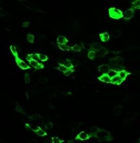
FH Parijat (Verified Customer) (09-09-2023) | Good antibody for IF
 |
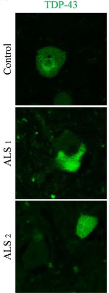
FH Florencia (Verified Customer) (07-17-2023) | This antibody is one of the most important ones we use in the lab for studying ALS! It works very well and we will continue to use it.
|
FH Pauline (Verified Customer) (02-23-2022) | everything ok
|
FH Xin (Verified Customer) (01-23-2022) | Very good in WB with a band of around 45 kD.
|
FH Jessica (Verified Customer) (06-10-2021) | good for WB and IF
|
FH Ana (Verified Customer) (03-14-2021) | TDP43 in human primary fibroblasts. 10 ug of total protein. Primary ab incubation 16h 4ºC 1:1000 in BSA 3% in PBST. Secondary ab Goat anti-rabbit HRP 1:5000 1h RT incubation.
 |
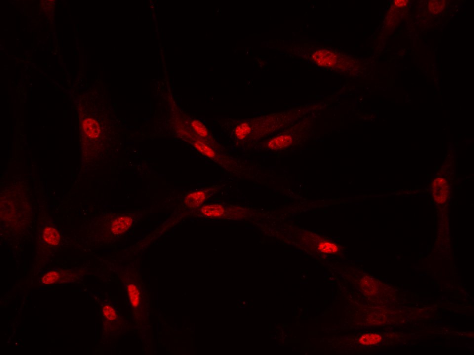
FH Yasuyo (Verified Customer) (01-20-2021) | Works good for ICC in fibroblasts. High nuclear signal. Cells fixed with 4% PFA 15 min. TDP43 ab dilution 1:200 in PBST O/N 4ºC. Donkey anti-rabbit Alexafluor 568 1:500 for 1h RT.
|
FH MEIMEI (Verified Customer) (11-12-2020) | the antibody work prefect for my staining.
|
FH Stephane (Verified Customer) (10-31-2020) | Good product for staining.
 |
FH Marina (Verified Customer) (10-25-2020) | TDP43 in human primary fibroblasts. Loading: 10 ug in LB with DTT. TGX 4-15% gel. Blocking: 1h 3% BSA in PBST (0.1% tween). Primary ab incubation 16h 4ºC 1:1000 in BSA 3% in PBST (0.1% Tween) Detection: Goat anti-rabbit HRP + ECL+.
 |
FH Uxoa (Verified Customer) (02-18-2020) | Good ICC in human primary fibroblats. Small amount of signal in cytoplasm in UT and control cells. High nuclear signal. Fibroblats fixed with 4% PFA 15 min. TDP43 ab dilution 1:100 in PBST (0.1% triton) + 10% DS O/N 4ºC. Donkey anti-rabbit alexa fluor 555 1:500 in PBST+10%DS 1h RT.
 |
FH Paul (Verified Customer) (01-15-2020) | Produces good IF images with minimal background.
|
FH Laura (Verified Customer) (01-10-2020) | Works very well
|
FH Benjamin (Verified Customer) (01-07-2020) | Works well in both western blotting and Immunofluorescence. Nice clear images obtained with IF.
|
FH Apoorva (Verified Customer) (12-18-2019) | It has great specificity over wide range of species.
|
FH Tilly (Verified Customer) (11-27-2019) | Great N-terminal TDP-43 antibody, works really well for immunohistochemistry, immunofluorescence and western blot from mouse brain tissue. Unfortunately, I have not been able to get it to work for precipitation in mouse brain.
|
FH Azita (Verified Customer) (10-04-2019) | I used it for ICC and it had low signal and high background with following (1/500, 1/300, 1/100,1/50) dilutions. Cell were fixed by 4% PFA.
|
FH Francisco (Verified Customer) (09-28-2019) | Antibody worked well. My lab has used the antibody in various experiments for quantifications, many of which were included in published works.
|
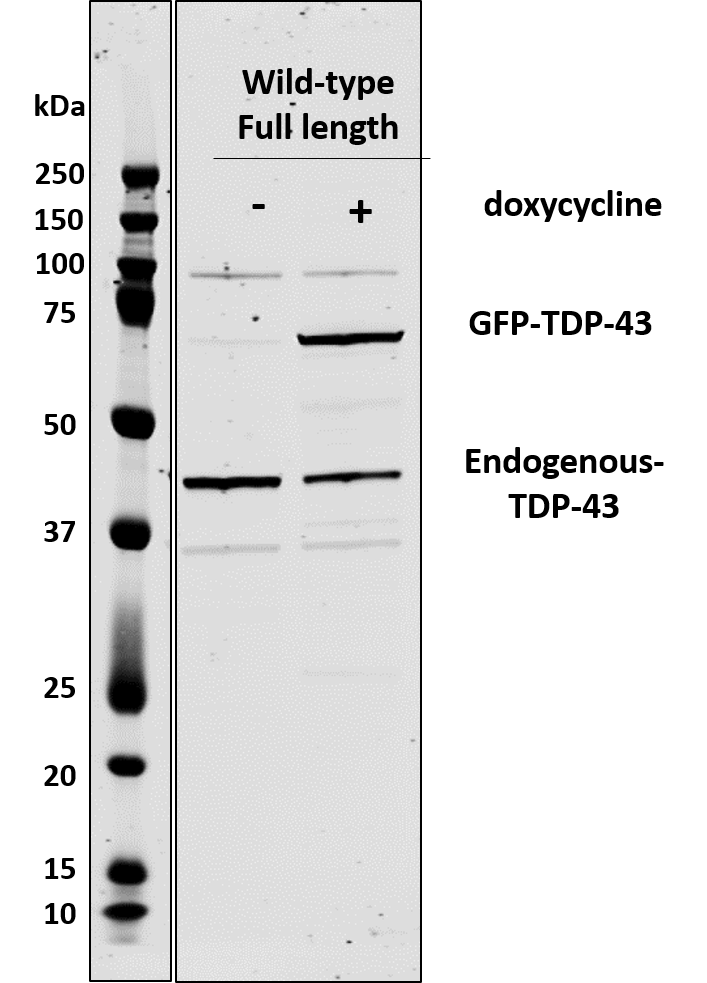
FH Alinda (Verified Customer) (09-11-2019) | Doxycycline-inducible SHSY5Y show GFP-TDP-43 expression upon dox addition.
 |
FH David (Verified Customer) (07-18-2019) | Excellent for both immunoblot and immunocytochemistry. Strong signal and low background in both cases. Single band in control cells for immunoblot.
|
FH George (Verified Customer) (07-07-2019) | Good antibody to show mislocalisation in transgenic ALS disease mouse model tissue culture. Needs a high concentration to show up for microscopy but very strong and clear signal.
|
FH Elena (Verified Customer) (08-17-2018) |
|
FH Petra (Verified Customer) (03-06-2018) |
|