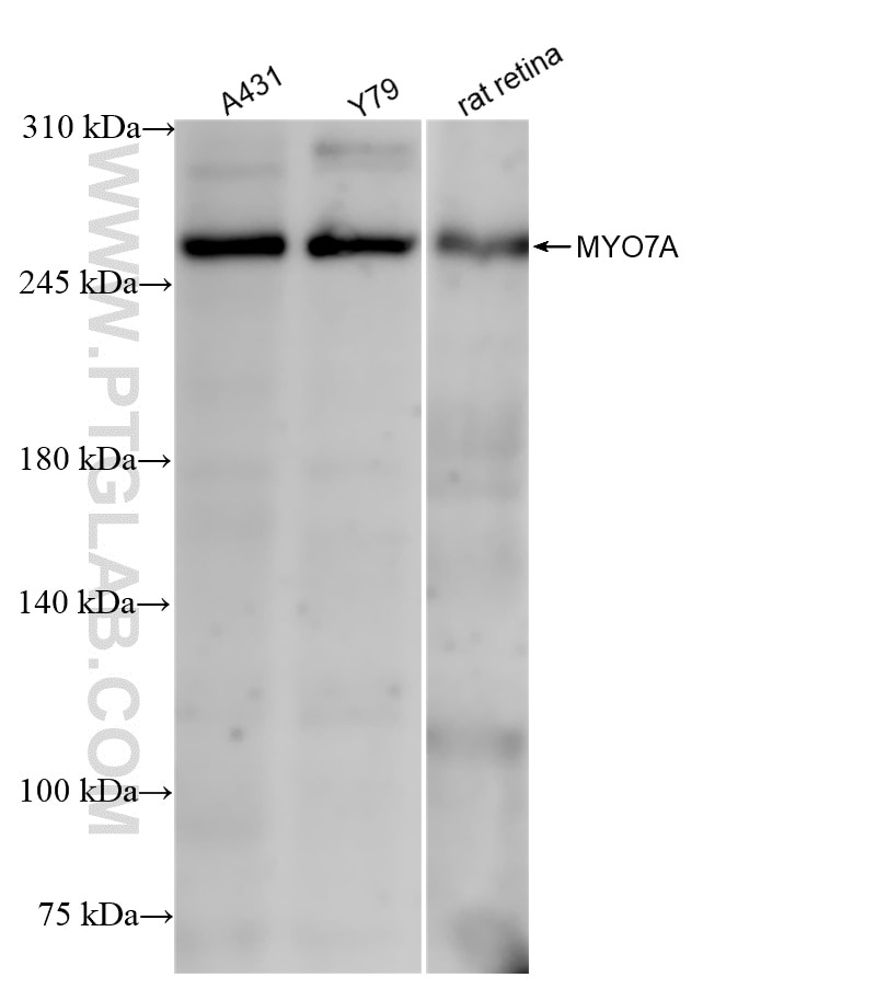
Various lysates were subjected to SDS PAGE followed by western blot with 83807-1-RR (MYO7A antibody) at dilution of 1:1000 incubated at room temperature for 1.5 hours.
Various lysates were subjected to SDS PAGE followed by western blot with 83807-1-RR (MYO7A antibody) at dilution of 1:1000 incubated at room temperature for 1.5 hours.
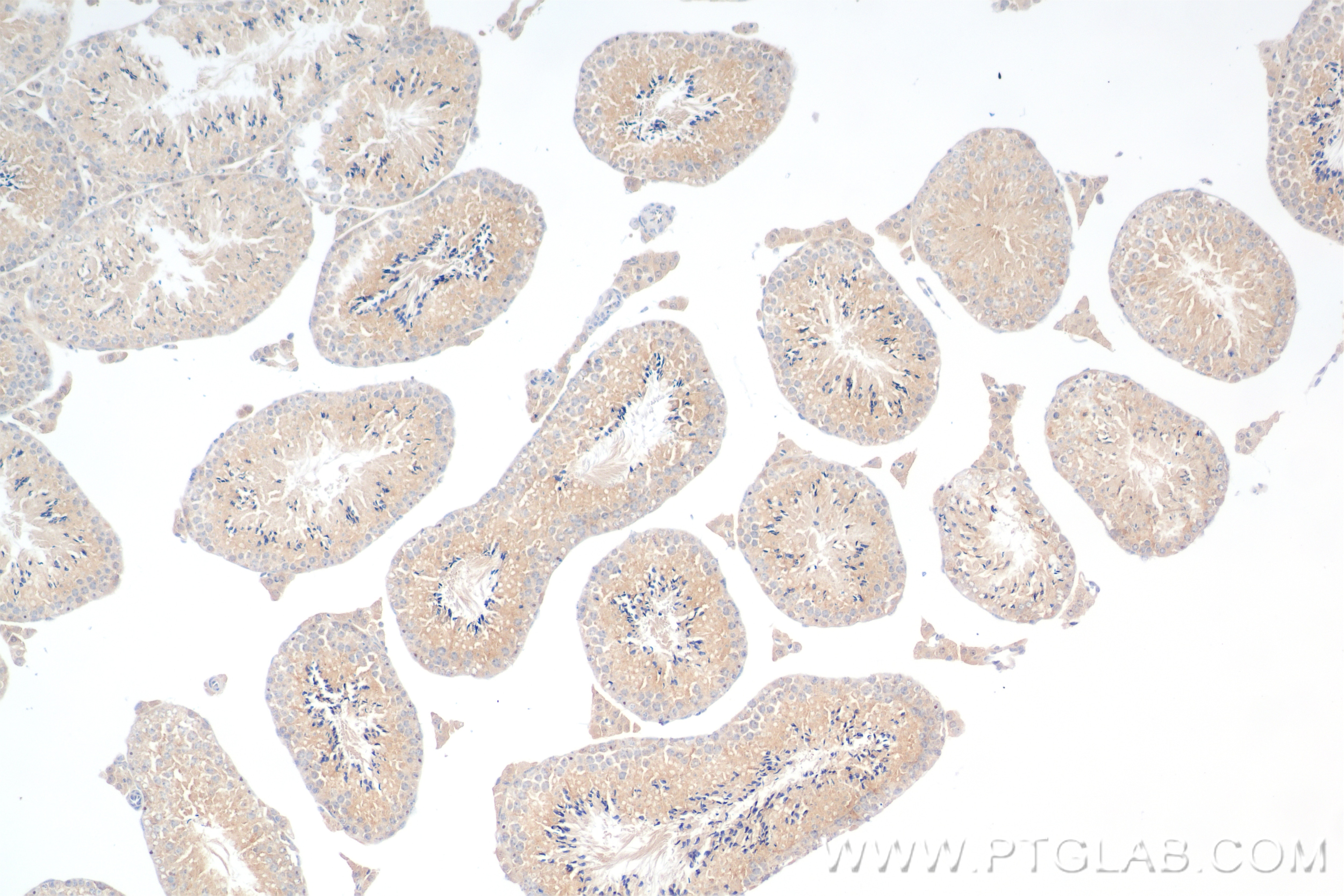
IHC staining of mouse testis using 83807-1-RR
Immunohistochemical analysis of paraffin-embedded mouse testis tissue slide using 83807-1-RR (MYO7A antibody) at dilution of 1:400 (under 10x lens). Heat mediated antigen retrieval with Tris-EDTA buffer (pH 9.0).
Immunohistochemical analysis of paraffin-embedded mouse testis tissue slide using 83807-1-RR (MYO7A antibody) at dilution of 1:400 (under 10x lens). Heat mediated antigen retrieval with Tris-EDTA buffer (pH 9.0).
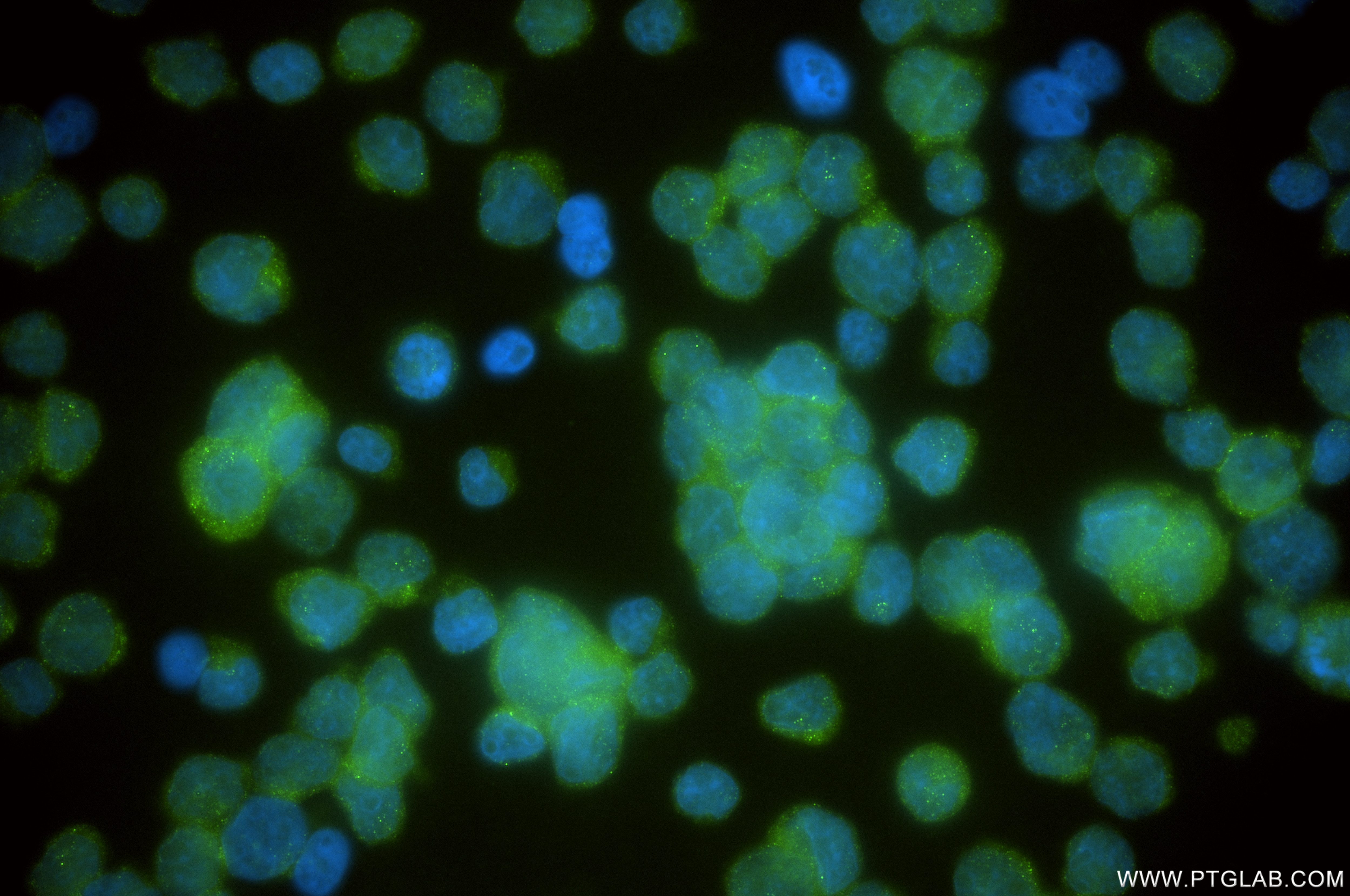
IF Staining of Y79 using 83807-1-RR
Immunofluorescent analysis of (4% PFA) fixed Y79 cells using MYO7A antibody (83807-1-RR, Clone: 240612C11 ) at dilution of 1:250 and CoraLite®488-Conjugated Goat Anti-Rabbit IgG(H+L) (SA00013-2).
Immunofluorescent analysis of (4% PFA) fixed Y79 cells using MYO7A antibody (83807-1-RR, Clone: 240612C11 ) at dilution of 1:250 and CoraLite®488-Conjugated Goat Anti-Rabbit IgG(H+L) (SA00013-2).
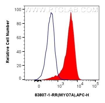
FC experiment of HepG2 using 83807-1-RR
1x10^6 HepG2 cells were intracellularly stained with 0.25 ug MYO7A Recombinant antibody (83807-1-RR, Clone:240612C11) and APC-Conjugated AffiniPure Goat Anti-Rabbit IgG(H+L)(red), or 0.25 ug Isotype Control (blue). Cells were fixed with 4% PFA and permeabilized with Flow Cytometry Perm Buffer.
1x10^6 HepG2 cells were intracellularly stained with 0.25 ug MYO7A Recombinant antibody (83807-1-RR, Clone:240612C11) and APC-Conjugated AffiniPure Goat Anti-Rabbit IgG(H+L)(red), or 0.25 ug Isotype Control (blue). Cells were fixed with 4% PFA and permeabilized with Flow Cytometry Perm Buffer.
The Proteintech guarantee covers Proteintech antibodies in any species and any application, including those not listed on the datasheet. If the antibody doesn’t perform, you can receive a hassle-free refund or credit note.
mouse testis tissue Note: suggested antigen retrieval with TE buffer pH 9.0; (*) Alternatively, antigen retrieval may be performed with citrate buffer pH 6.0
Positive IF/ICC detected in
Y79 cells
Positive FC (Intra) detected in
HepG2 cells
Recommended dilution
Application
Dilution
Western Blot (WB)
WB : 1:500-1:2000
Immunohistochemistry (IHC)
IHC : 1:200-1:800
Immunofluorescence (IF)/ICC
IF/ICC : 1:125-1:500
Flow Cytometry (FC) (INTRA)
FC (INTRA) : 0.25 ug per 10^6 cells in a 100 µl suspension
It is recommended that this reagent should be titrated in each testing system to obtain optimal results.
Sample-dependent, Check data in validation data gallery.
Product Information
83807-1-RR targets MYO7A in WB, IHC, IF/ICC, FC (Intra), ELISA applications and shows reactivity with human, mouse, rat samples.
PBS with 0.02% sodium azide and 50% glycerol, pH 7.3.
Storage Conditions
Store at -20°C. Stable for one year after shipment. Aliquoting is unnecessary for -20oC storage. 20ul sizes contain 0.1% BSA.
Background Information
MYO7A, also named a USH1B, is one of myosins protein which are actin-based motor molecules with ATPase activity. Unconventional myosins serve in intracellular movements. Their highly divergent tails are presumed to bind to membranous compartments, which would be moved relative to actin filaments. In retina, MYO7A might play a role in trafficking of ribbon-synaptic vesicle complexes and renewal of the outer photoreceptors disks. In inner ear, it might maintain the rigidity of stereocilia during the dynamic movements of the bundle. It is involved in hair-cell vesicle trafficking of aminoglycosides, which are known to induce ototoxicity. Defects in MYO7A are the cause of Usher syndrome type 1B (USH1B). Defects in MYO7A are the cause of deafness autosomal recessive type 2 (DFNB2). Defects in MYO7A are the cause of deafness autosomal dominant type 11 (DFNA11). The antibody is specific to MYO7A.
Various lysates were subjected to SDS PAGE followed by western blot with 83807-1-RR (MYO7A antibody) at dilution of 1:1000 incubated at room temperature for 1.5 hours.
IHC Figures
IHC staining of mouse testis using 83807-1-RR
Immunohistochemical analysis of paraffin-embedded mouse testis tissue slide using 83807-1-RR (MYO7A antibody) at dilution of 1:400 (under 10x lens). Heat mediated antigen retrieval with Tris-EDTA buffer (pH 9.0).
IF/ICC Figures
IF Staining of Y79 using 83807-1-RR
Immunofluorescent analysis of (4% PFA) fixed Y79 cells using MYO7A antibody (83807-1-RR, Clone: 240612C11 ) at dilution of 1:250 and CoraLite®488-Conjugated Goat Anti-Rabbit IgG(H+L) (SA00013-2).
FC (INTRA) Figures
FC experiment of HepG2 using 83807-1-RR
1x10^6 HepG2 cells were intracellularly stained with 0.25 ug MYO7A Recombinant antibody (83807-1-RR, Clone:240612C11) and APC-Conjugated AffiniPure Goat Anti-Rabbit IgG(H+L)(red), or 0.25 ug Isotype Control (blue). Cells were fixed with 4% PFA and permeabilized with Flow Cytometry Perm Buffer.
The species listed in Tested Reactivity are in-house verified and applicable species. For unlisted species, please refer to the homology analysis of the immunogen sequence and related species. For rabbit polyclonal antibodies, homology >70% is recommended. For mouse monoclonal antibodies and rabbit recombinant antibodies, homology >90% is recommended. Generally, the higher the homology, the greater the applicability. However, there will be certain differences in protein expression in different species, tissues or cells. Therefore, the homology analysis results are for reference only and do not serve as a guarantee.
At Proteintech, we pride ourselves on our antibody quality, customer service and transparency. As such, we are comparing our antibodies with other vendors, enabling easy identification and comparisons of key data to help you choose the suitable antibody for your needs.
We have selected the top cited antibodies from these vendors for you to compare.
Proteintech
MYO7A Recombinant antibody
Catalog Number
83807-1-RR
Citations
-
Dilutions
WB : 1:500-1:2000 IHC : 1:200-1:800 IF/ICC : 1:125-1:500 FC (INTRA) : 0.25 ug per 10^6 cells in a 100 µl suspension
Applications
WB, IHC, IF/ICC, FC (Intra), ELISA
Reactivity
human, mouse, rat
Product Guarantee
Covers any species including not listed on datasheet
Covers any applications including not listed on datasheet

 at dilution of 1:1000 incubated at room temperature for 1.5 hours.")
 at dilution of 1:400 (under 10x lens). Heat mediated antigen retrieval with Tris-EDTA buffer (pH 9.0).")
 fixed Y79 cells using MYO7A antibody (83807-1-RR, Clone: 240612C11 ) at dilution of 1:250 and CoraLite®488-Conjugated Goat Anti-Rabbit IgG(H+L) (SA00013-2).")
 and APC-Conjugated AffiniPure Goat Anti-Rabbit IgG(H+L)(red), or 0.25 ug Isotype Control (blue). Cells were fixed with 4% PFA and permeabilized with Flow Cytometry Perm Buffer.")