at dilution of 1:5000 incubated at room temperature for 1.5 hours.")
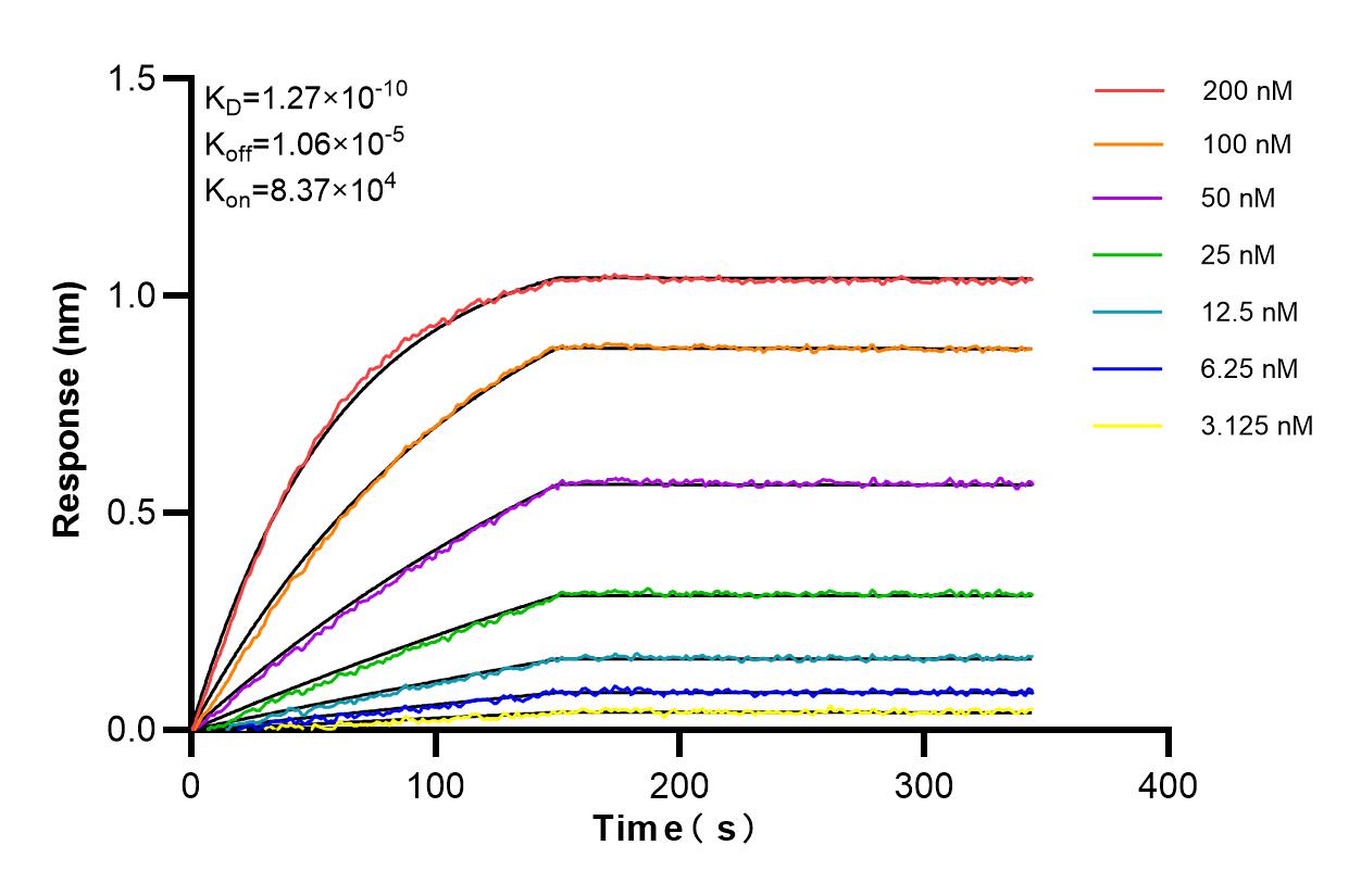
 kinetic assays of 86020-2-RR against Human FA2H were performed. The affinity constant is 0.127 nM.")
Tested Applications
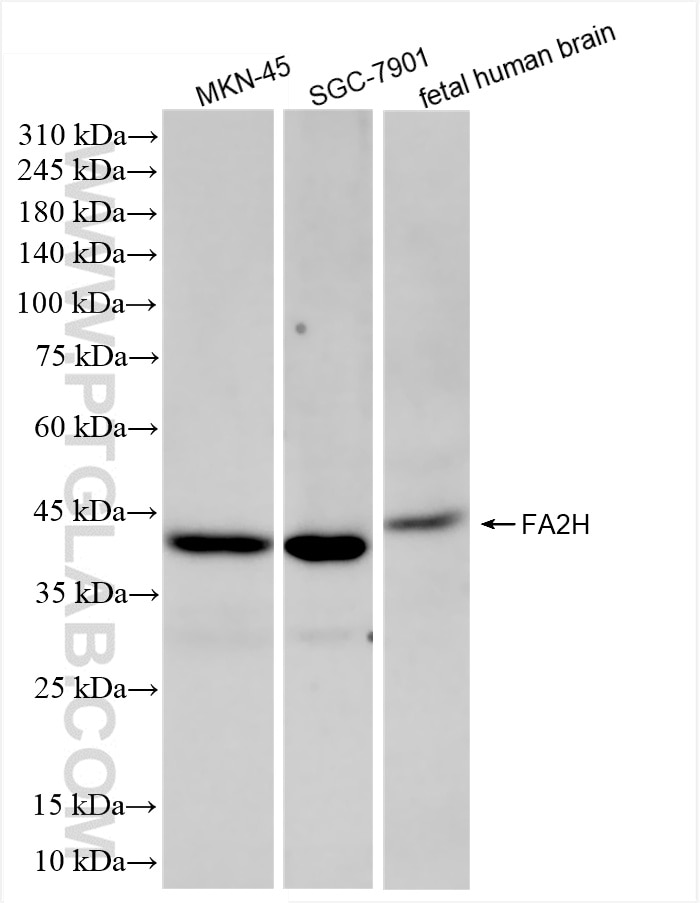
| Positive WB detected in | MKN-45 cells, SGC-7901 cells, fetal human brain tissue |
Recommended dilution
| Application | Dilution |
|---|---|
| Western Blot (WB) | WB : 1:2000-1:10000 |
| It is recommended that this reagent should be titrated in each testing system to obtain optimal results. | |
| Sample-dependent, Check data in validation data gallery. | |
Product Information
86020-2-RR targets FA2H in WB, ELISA applications and shows reactivity with human samples.
| Tested Reactivity | human |
| Host / Isotype | Rabbit / IgG |
| Class | Recombinant |
| Type | Antibody |
| Immunogen |
CatNo: Ag7717 Product name: Recombinant human FA2H protein Source: e coli.-derived, PGEX-4T Tag: GST Domain: 1-164 aa of BC002679 Sequence: MAPAPPPAASFSPSEVQRRLAAGACWVRRGARLYDLSSFVRHHPGGEQLLRARAGQDISADLDGPPHRHSANARRWLEQYYVGELRGEQQGSMENEPVALEETQKTDPAMEPRFKVVDWDKDLVDWRKPLLWQVGHLGEKYDEWVHQPVTRPIRLFHSDLIEGL Predict reactive species |
| Full Name | fatty acid 2-hydroxylase |
| Calculated Molecular Weight | 43 kDa |
| Observed Molecular Weight | 43 kDa |
| GenBank Accession Number | BC002679 |
| Gene Symbol | FA2H |
| Gene ID (NCBI) | 79152 |
| Conjugate | Unconjugated |
| Form | Liquid |
| Purification Method | Protein A purification |
| UNIPROT ID | Q7L5A8 |
| Storage Buffer | PBS with 0.02% sodium azide and 50% glycerol, pH 7.3. |
| Storage Conditions | Store at -20°C. Stable for one year after shipment. Aliquoting is unnecessary for -20oC storage. 20ul sizes contain 0.1% BSA. |
Background Information
FA2H(Fatty acid 2-hydroxylase) required for alpha-hydroxylation of free fatty acids and the formation of alpha-hydroxylated sphingolipids. The deduced 372-amino acid protein has a calculated molecular mass of 42.8 kD .Both mouse and human FA2H have a C-terminal endoplasmic reticulum (ER) retention motif(PMID: 15658937).
Protocols
| Product Specific Protocols | |
|---|---|
| WB protocol for FA2H antibody 86020-2-RR | Download protocol |
| Standard Protocols | |
|---|---|
| Click here to view our Standard Protocols |