Tested Applications
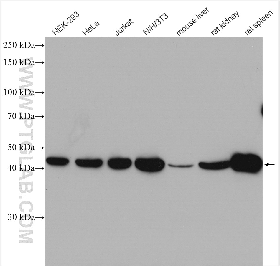
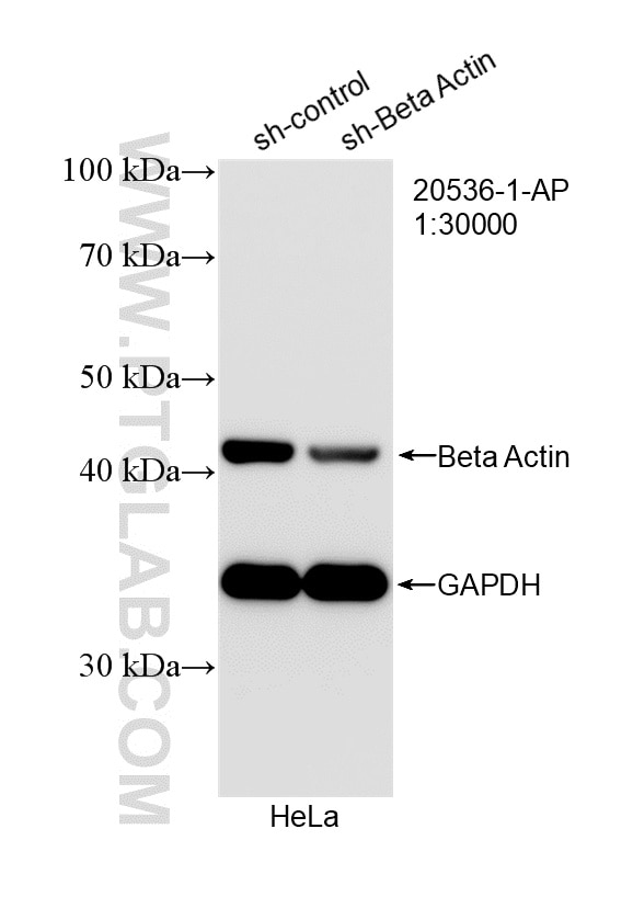
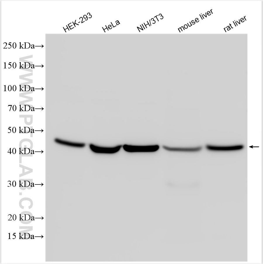
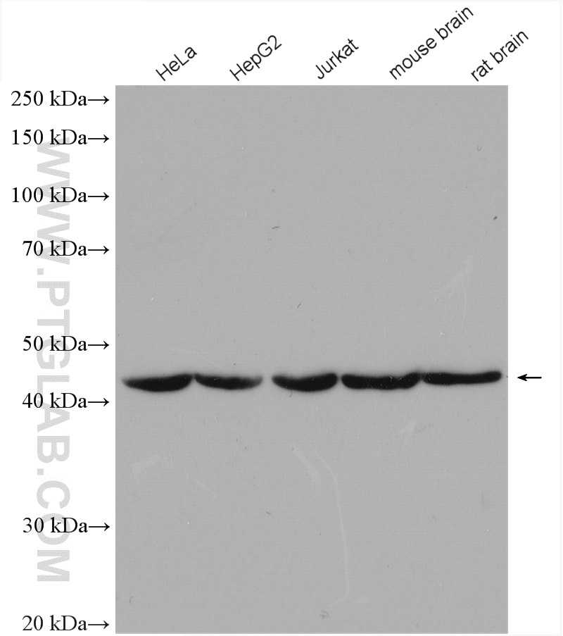
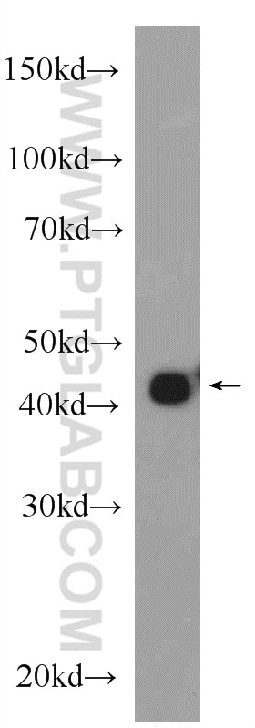
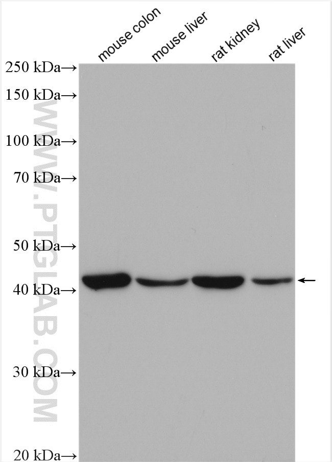
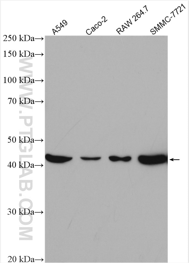
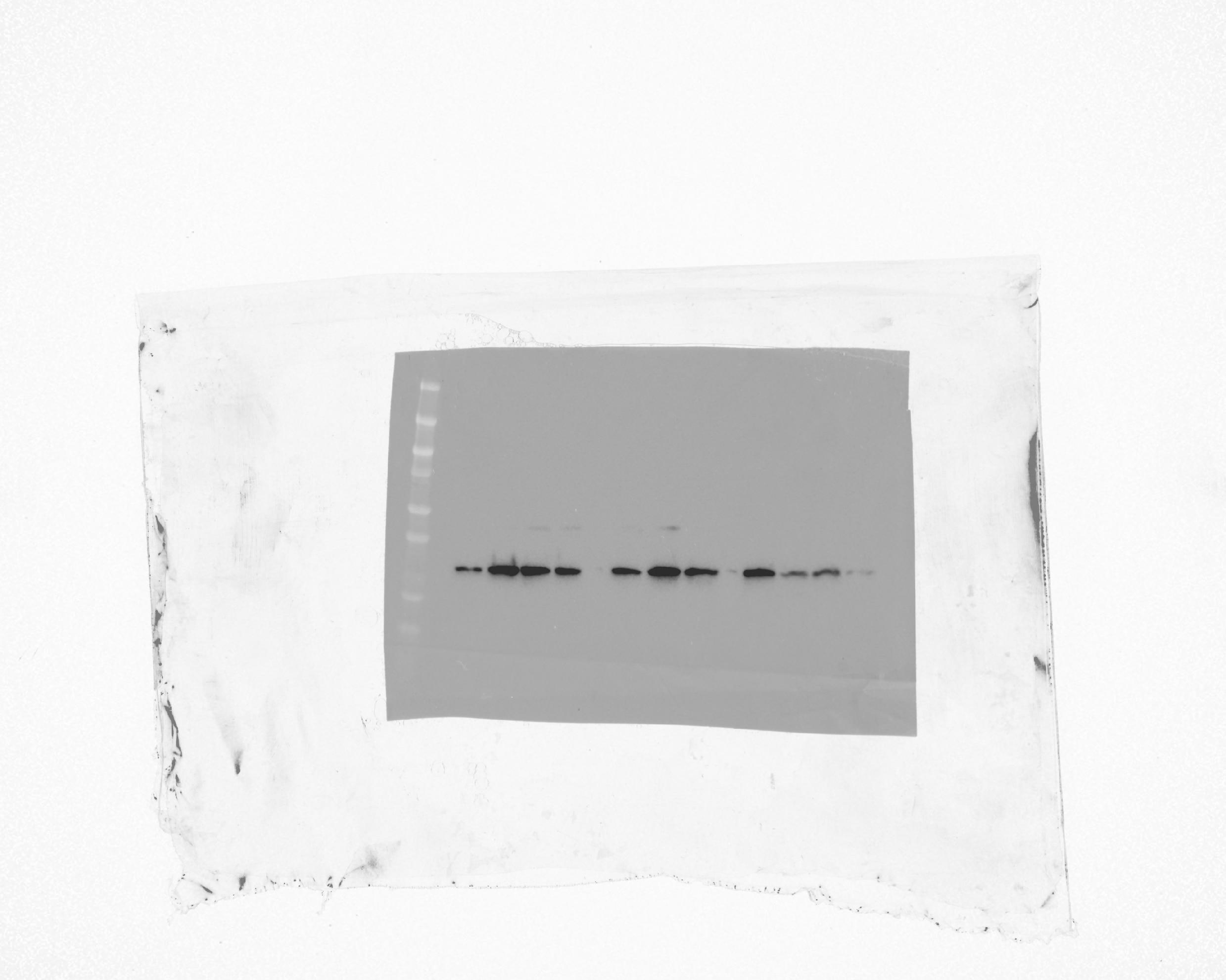
| Positive WB detected in | HEK-293 cells, A549 cells, C6 cells, HeLa cells, mouse colon tissue, Caco-2 cells, RAW 264.7 cells, SMMC-7721 cells, HepG2 cells, Jurkat cells, mouse brain tissue, rat brain tissue, NIH/3T3 cells, mouse liver tissue, rat kidney tissue, rat spleen tissue, rat liver tissue |
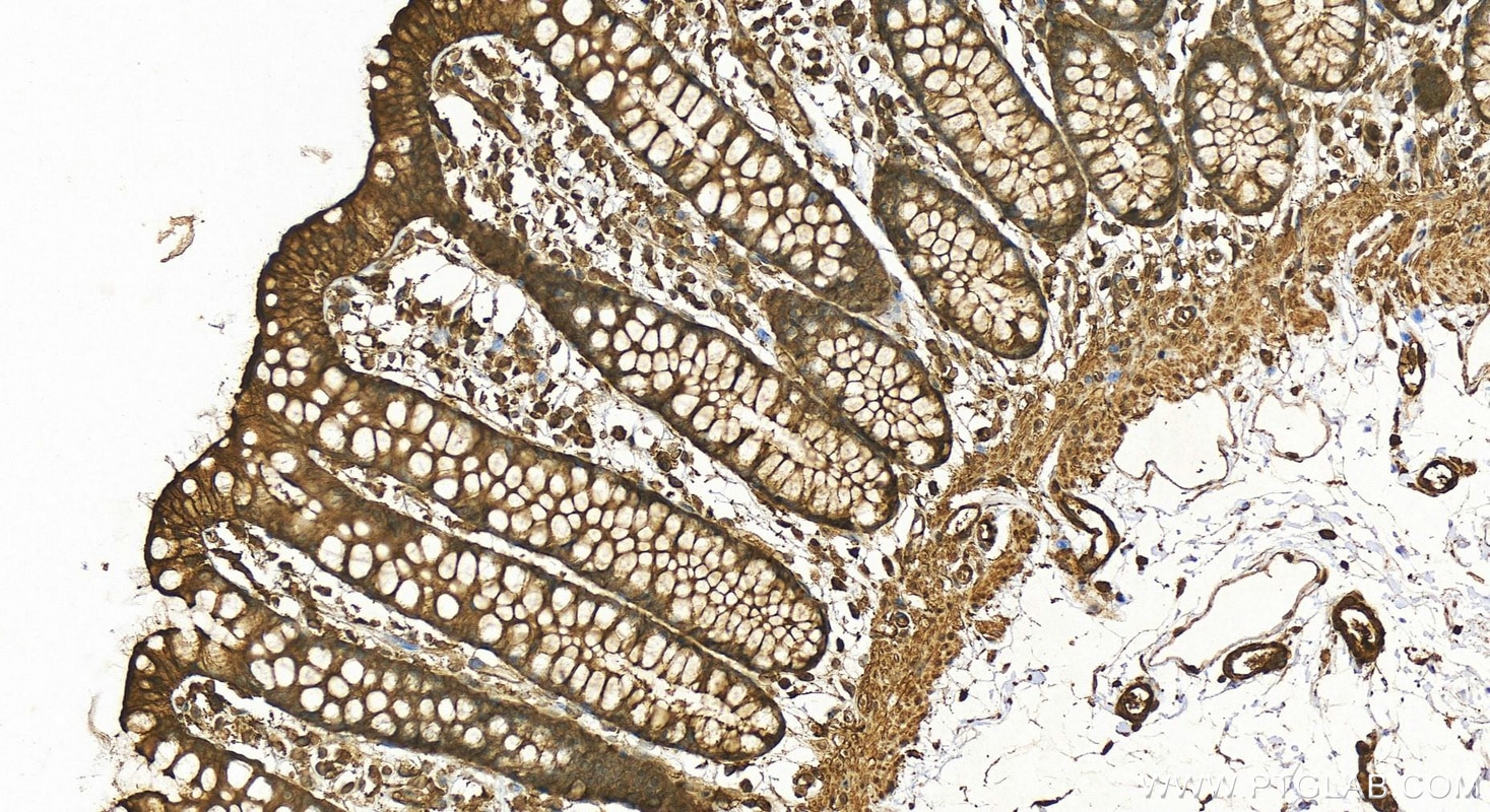
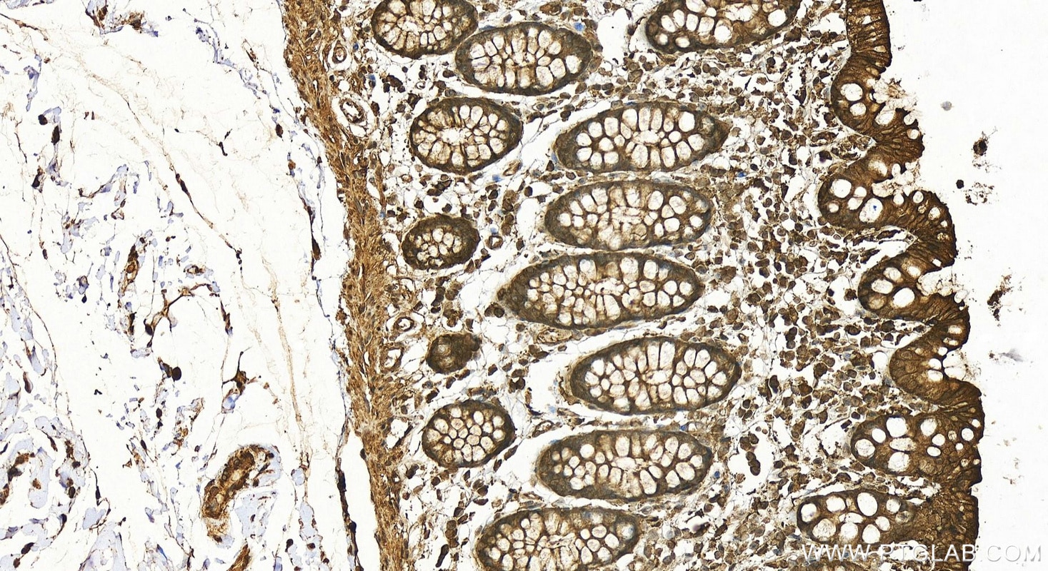
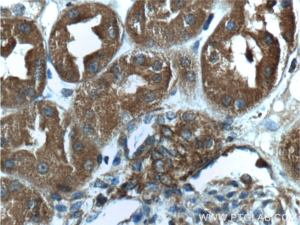
| Positive IHC detected in | human colon tissue, human kidney tissue Note: suggested antigen retrieval with TE buffer pH 9.0; (*) Alternatively, antigen retrieval may be performed with citrate buffer pH 6.0 |
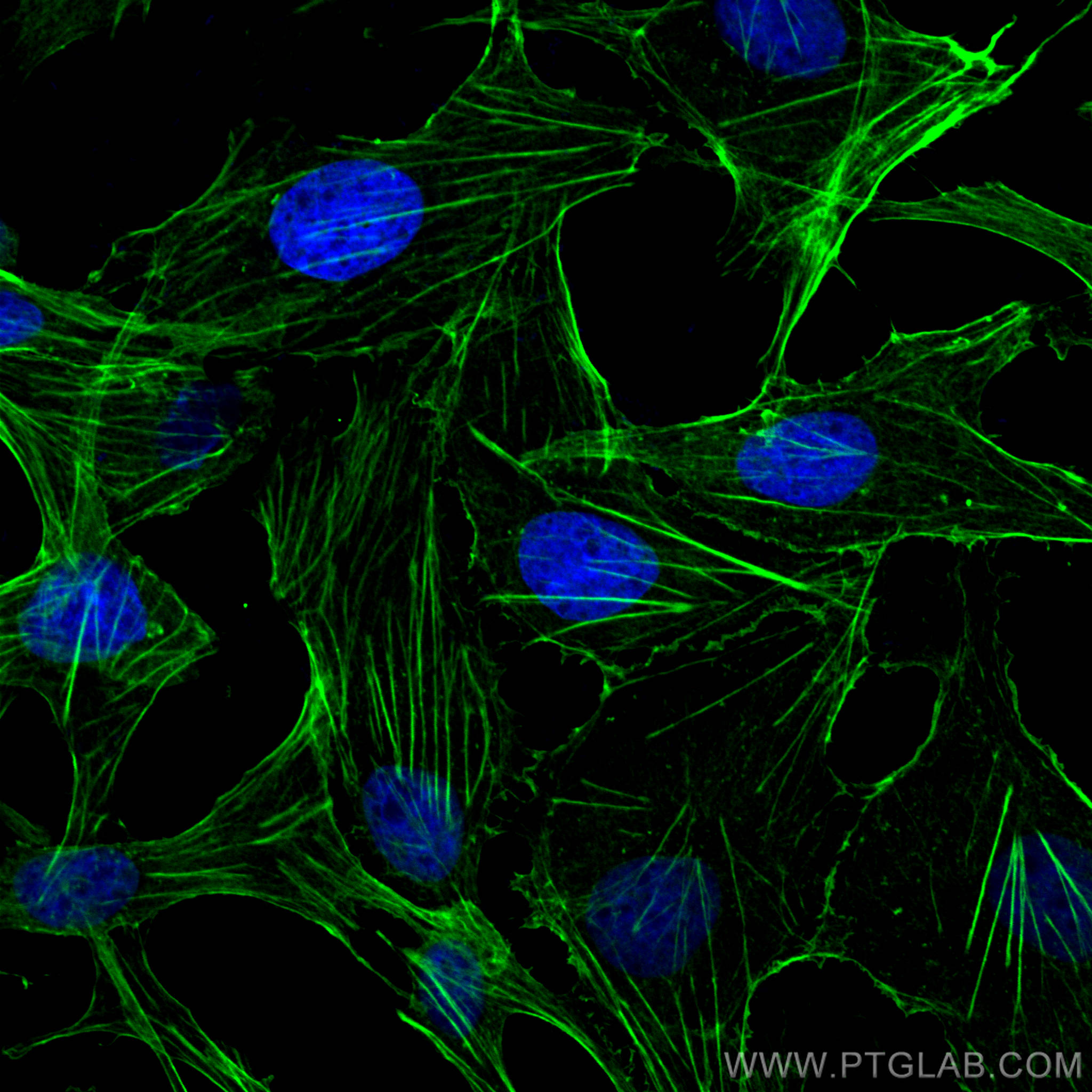
| Positive IF/ICC detected in | MDCK cells |
Recommended dilution
| Application | Dilution |
|---|---|
| Western Blot (WB) | WB : 1:4000-1:10000 |
| Immunohistochemistry (IHC) | IHC : 1:50-1:500 |
| Immunofluorescence (IF)/ICC | IF/ICC : 1:200-1:800 |
| It is recommended that this reagent should be titrated in each testing system to obtain optimal results. | |
| Sample-dependent, Check data in validation data gallery. | |
Published Applications
| KD/KO | See 7 publications below |
| WB | See 5675 publications below |
| IHC | See 11 publications below |
| IF | See 31 publications below |
| IP | See 11 publications below |
| ELISA | See 2 publications below |
| CoIP | See 4 publications below |
Product Information
20536-1-AP targets Beta Actin in WB, IHC, IF/ICC, IP, CoIP, ELISA applications and shows reactivity with human, mouse, rat, canine, monkey samples.
| Tested Reactivity | human, mouse, rat, canine, monkey |
| Cited Reactivity | human, mouse, rat, chicken, goat, sheep, yeast, ochotona curzoniae, camelus bactrianus, carp |
| Host / Isotype | Rabbit / IgG |
| Class | Polyclonal |
| Type | Antibody |
| Immunogen |
CatNo: Ag14521 Product name: Recombinant human beta actin protein Source: e coli.-derived, PGEX-4T Tag: GST Domain: 1-50 aa of BC002409 Sequence: MDDDIAALVVDNGSGMCKAGFAGDDAPRAVFPSIVGRPRHQGVMVGMGQK Predict reactive species |
| Full Name | actin, beta |
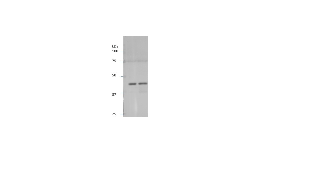
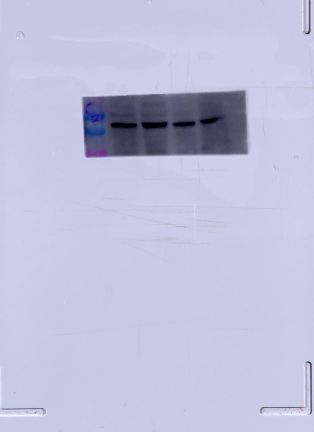
| Calculated Molecular Weight | 375 aa, 42 kDa |
| Observed Molecular Weight | 42 kDa |
| GenBank Accession Number | BC002409 |
| Gene Symbol | Beta Actin |
| Gene ID (NCBI) | 60 |
| RRID | AB_10700003 |
| Conjugate | Unconjugated |
| Form | Liquid |
| Purification Method | Antigen affinity purification |
| UNIPROT ID | P60709 |
| Storage Buffer | PBS with 0.02% sodium azide and 50% glycerol, pH 7.3. |
| Storage Conditions | Store at -20°C. Stable for one year after shipment. Aliquoting is unnecessary for -20oC storage. 20ul sizes contain 0.1% BSA. |
Background Information
Beta Actin, also named as ACTB and F-Actin, belongs to the actin family. Actins are highly conserved globular proteins that are involved in various types of cell motility and are ubiquitously expressed in all eukaryotic cells. At least six isoforms of actins are known in mammals and other vertebrates: alpha (ACTC1, cardiac muscle 1), alpha 1 (ACTA1, skeletal muscle) and 2 (ACTA2, aortic smooth muscle), beta (ACTB), gamma 1 (ACTG1) and 2 (ACTG2, enteric smooth muscle). Beta and gamma 1 are two non-muscle actin proteins. Most actins consist of 376aa, while ACTG2 (rich in muscles) has 375aa and ACTG1(found in non-muscle cells) has only 374aa. Beta actin has been widely used as the internal control in RT-PCR and Western Blotting as a 42-kDa protein. However, the 37-40, 31, 15 kDa cleaved fragment of beta actin can be generated during apoptosis process. This antibody was generated against N-terminal region of human beta actin protein and can cross-react with other actins. (9173887, 11217076, 10229193 )
Protocols
| Product Specific Protocols | |
|---|---|
| IF protocol for Beta Actin antibody 20536-1-AP | Download protocol |
| IHC protocol for Beta Actin antibody 20536-1-AP | Download protocol |
| WB protocol for Beta Actin antibody 20536-1-AP | Download protocol |
| Standard Protocols | |
|---|---|
| Click here to view our Standard Protocols |
Publications
| Species | Application | Title |
|---|---|---|
Cancer Cell Targeting the immune privilege of tumor-initiating cells to enhance cancer immunotherapy | ||
Nature Aspm knockout ferret reveals an evolutionary mechanism governing cerebral cortical size. | ||
Signal Transduct Target Ther Identifying genetic targets in clinical subtypes of Parkinson's disease for optimizing pharmacological treatment strategies | ||
Cell Microglia jointly degrade fibrillar alpha-synuclein cargo by distribution through tunneling nanotubes. |
Reviews
The reviews below have been submitted by verified Proteintech customers who received an incentive for providing their feedback.
FH Mounika (Verified Customer) (03-24-2026) | Great Saviour for our research projects to validate all the cell lines!
|
FH Sai Sindhura (Verified Customer) (01-08-2026) |
|
FH Shivam (Verified Customer) (01-02-2026) | prominent bands. Very few unspecific binding.
|
FH Matthieu (Verified Customer) (09-24-2025) | Produces a clear band in WB at the expected molecular weight
|
FH Manon (Verified Customer) (09-17-2025) | Very good housekeeping antibody
|
FH YINGJIAN (Verified Customer) (05-27-2025) | This primary antibody is effective, even at a low concentration (1:10000).
 |
FH Julia (Verified Customer) (06-20-2023) | Our lab has been using Proteintech's beta actin polyclonal antibody for years. We use this antibody in all our western blot experiments for normalization. This antibody works consistently at a 1:1000 dilution for human and mouse retina and for HRECs. We have always gotten a beautiful result, with nice thick and specific bands. The only downside is that this antibody is hard to strip. Even after stripping, the membrane tends to have residual beta actin bands. Even so, we would highly recommend this product for routine western blots.
 |
FH Chun (Verified Customer) (06-19-2022) | This antibody worked very well.
|
FH Aaron (Verified Customer) (12-08-2020) | nice bands achieved
|
FH Wei (Verified Customer) (01-30-2020) | Strong, clear band for basal heart tissues.Good for internal control for heart diseases.
|
FH SCOTT (Verified Customer) (10-11-2019) | Used 1/1000 overnight 4 C. overnightHuman Grey matter cortex post mortem material
 |
FH Dan (Verified Customer) (08-19-2019) | The antibody works
|
FH Kishor (Verified Customer) (01-30-2019) | Working well for Western Blotting (1:5000)
 |