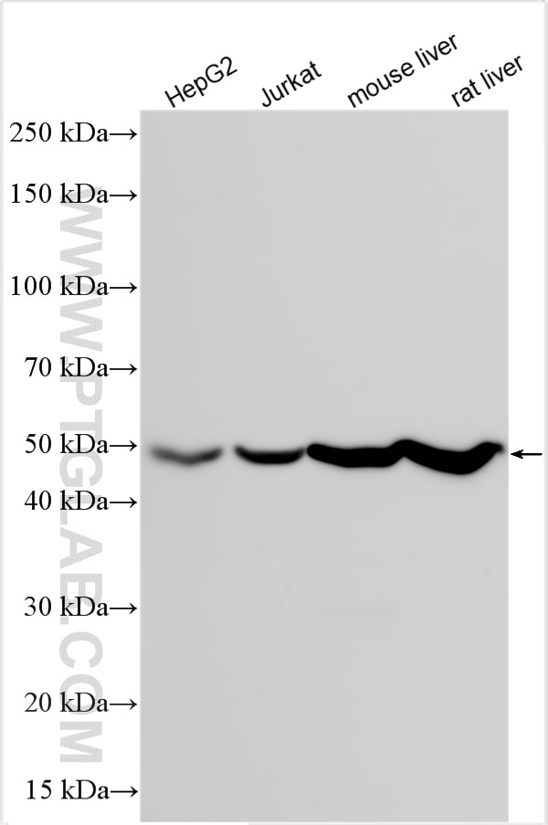
Various lysates were subjected to SDS PAGE followed by western blot with RMX00023 (ATP5A1 antibody) at dilution of 1:50000 incubated at room temperature for 1.5 hours.
Various lysates were subjected to SDS PAGE followed by western blot with RMX00023 (ATP5A1 antibody) at dilution of 1:50000 incubated at room temperature for 1.5 hours.
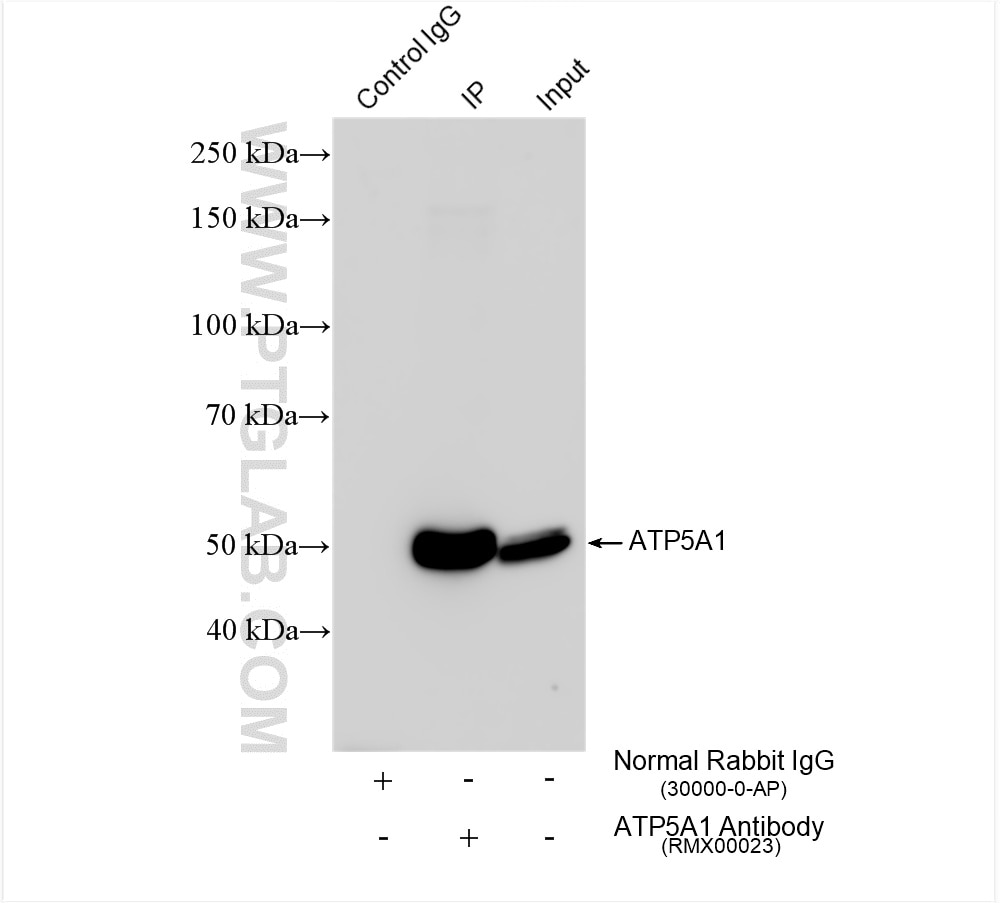
IP experiment of HepG2 using RMX00023
IP result of anti-ATP5A1 (IP:RMX00023, 4ug; Detection:RMX00023 1:30000) with HepG2 cells lysate 1480 ug.
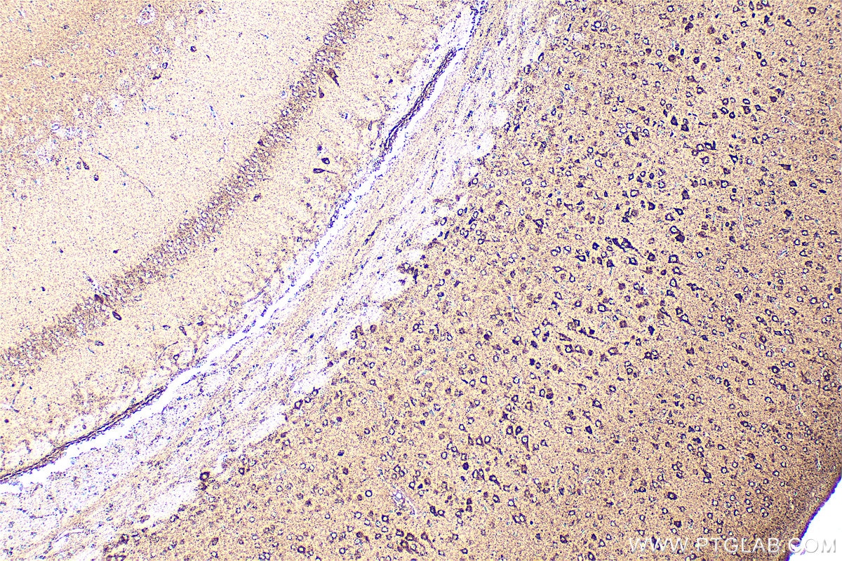
Immunohistochemical analysis of paraffin-embedded mouse brain tissue slide using RMX00023 (ATP5A1 antibody) at dilution of 1:1000 (under 10x lens). Heat mediated antigen retrieval with Tris-EDTA buffer (pH 9.0).
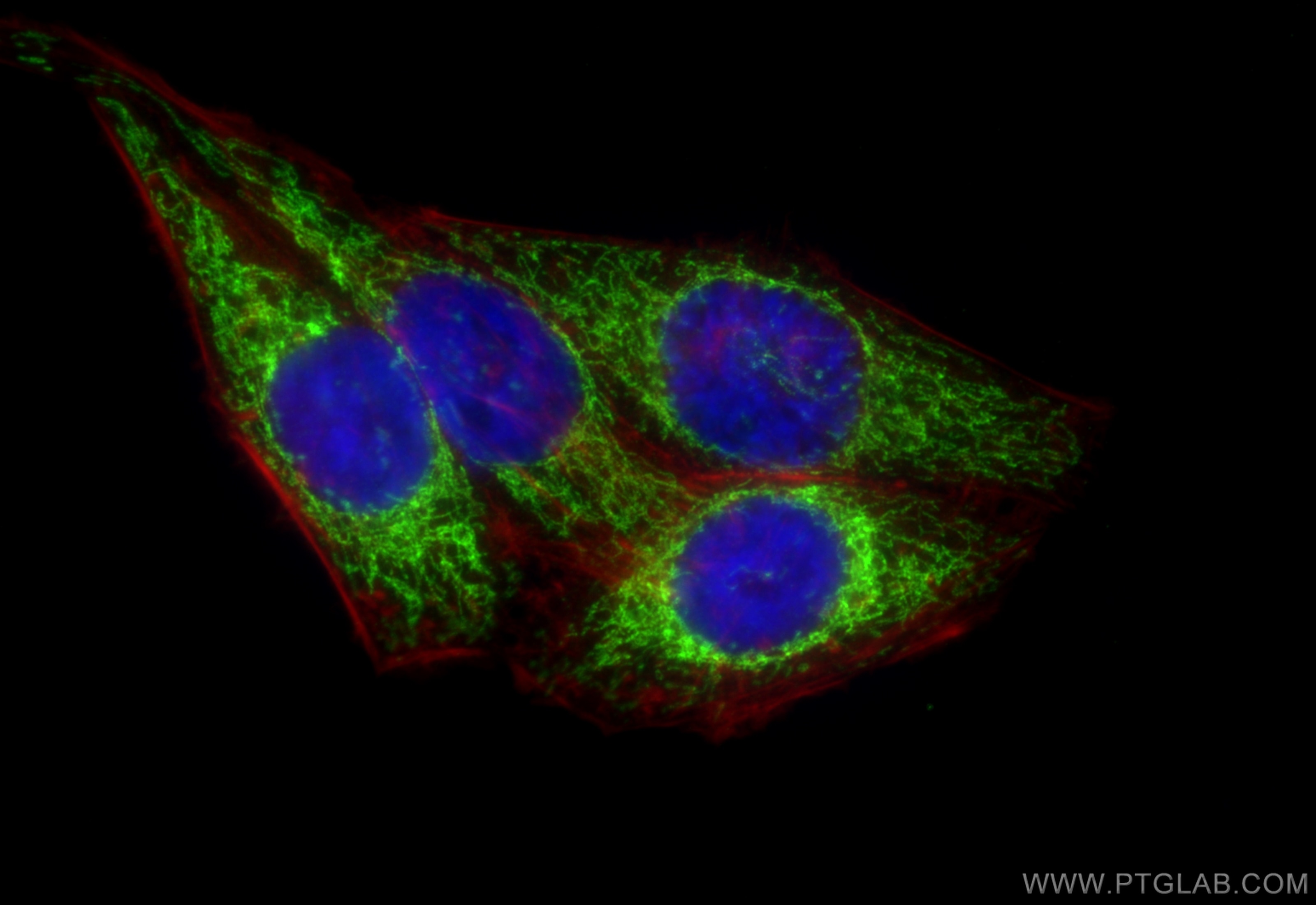
IF Staining of HepG2 using RMX00023
Immunofluorescent analysis of (4% PFA) fixed HepG2 cells using ATP5A1 antibody (RMX00023) at dilution of 1:200 and CoraLite®488-Conjugated Goat Anti-Rabbit IgG(H+L) (SA00013-2).
Immunofluorescent analysis of (4% PFA) fixed HepG2 cells using ATP5A1 antibody (RMX00023) at dilution of 1:200 and CoraLite®488-Conjugated Goat Anti-Rabbit IgG(H+L) (SA00013-2).
The Proteintech guarantee covers Proteintech antibodies in any species and any application, including those not listed on the datasheet. If the antibody doesn’t perform, you can receive a hassle-free refund or credit note.
HepG2 cells, Jurkat cells, mouse liver tissue, rat liver tissue
Positive IP detected in
HepG2 cells
Positive IHC detected in
mouse brain tissue Note: suggested antigen retrieval with TE buffer pH 9.0; (*) Alternatively, antigen retrieval may be performed with citrate buffer pH 6.0
Positive IF/ICC detected in
HepG2 cells
Recommended dilution
Application
Dilution
Western Blot (WB)
WB : 1:20000-1:100000
Immunoprecipitation (IP)
IP : 0.5-4.0 ug for 1.0-3.0 mg of total protein lysate
Immunohistochemistry (IHC)
IHC : 1:500-1:2000
Immunofluorescence (IF)/ICC
IF/ICC : 1:50-1:500
It is recommended that this reagent should be titrated in each testing system to obtain optimal results.
Sample-dependent, Check data in validation data gallery.
Product Information
RMX00023 targets ATP5A1 in WB, IHC, IF/ICC, IP, ELISA applications and shows reactivity with human, mouse, rat samples.
PBS with 0.02% sodium azide and 50% glycerol, pH 7.3.
Storage Conditions
Store at -20°C. Stable for one year after shipment. Aliquoting is unnecessary for -20oC storage. 20ul sizes contain 0.1% BSA.
Background Information
The ATP5A1 gene encodes the α subunit of mitochondrial ATP synthase which produces ATP from ADP in the presence of a proton gradient across the membrane. The mitochondrial ATP synthase, also known as Complex V or F1F0 ATP synthase, is a multi-subunit enzyme complex consisting of two functional domains, the F1-containing the catalytic core and the Fo-containing the membrane proton channel. F0 domain has 10 subunits: a,b, c, d, e, f, g, OSCP, A6L, and F6. F1 is composed of subunits α, β, γ, δ, ε, and a loosely attached inhibitor protein IF1. Recently defect in ATP5A1 has been linked to the fatal neonatal mitochondrial encephalopathy. ATP5A1 is localized in the mitochondria and anti-ATP5A1 can be used as the loading control for mitochondrial or Complex V proteins. This antibody recognizes the endogenous ATP5A1 protein in lysates from various cell lines and tissues. The predicted MW of ATP5A1 is 60 kDa, while it undergoes the transit peptide cleavage to become a mature form around 50-55 kDa. Several isoforms of ATP5A1 exist due to the alternative splicing.
Various lysates were subjected to SDS PAGE followed by western blot with RMX00023 (ATP5A1 antibody) at dilution of 1:50000 incubated at room temperature for 1.5 hours.
IHC Figures
IHC staining of mouse brain using RMX00023
Immunohistochemical analysis of paraffin-embedded mouse brain tissue slide using RMX00023 (ATP5A1 antibody) at dilution of 1:1000 (under 10x lens). Heat mediated antigen retrieval with Tris-EDTA buffer (pH 9.0).
IP Figures
IP experiment of HepG2 using RMX00023
IP result of anti-ATP5A1 (IP:RMX00023, 4ug; Detection:RMX00023 1:30000) with HepG2 cells lysate 1480 ug.
IF/ICC Figures
IF Staining of HepG2 using RMX00023
Immunofluorescent analysis of (4% PFA) fixed HepG2 cells using ATP5A1 antibody (RMX00023) at dilution of 1:200 and CoraLite®488-Conjugated Goat Anti-Rabbit IgG(H+L) (SA00013-2).
The species listed in Tested Reactivity are in-house verified and applicable species. For unlisted species, please refer to the homology analysis of the immunogen sequence and related species. For rabbit polyclonal antibodies, homology >70% is recommended. For mouse monoclonal antibodies and rabbit recombinant antibodies, homology >90% is recommended. Generally, the higher the homology, the greater the applicability. However, there will be certain differences in protein expression in different species, tissues or cells. Therefore, the homology analysis results are for reference only and do not serve as a guarantee.
At Proteintech, we pride ourselves on our antibody quality, customer service and transparency. As such, we are comparing our antibodies with other vendors, enabling easy identification and comparisons of key data to help you choose the suitable antibody for your needs.
We have selected the top cited antibodies from these vendors for you to compare.
Proteintech
ATP5A1 Multi-Recombinant antibody
Catalog Number
RMX00023
Citations
-
Dilutions
WB : 1:20000-1:100000 IP : 0.5-4.0 ug for IP and 0.5-4.0 ug for 1.0-3.0 mg of total protein lysate for WB IHC : 1:500-1:2000 IF/ICC : 1:50-1:500
Applications
WB, IHC, IF/ICC, IP, ELISA
Reactivity
human, mouse, rat
Product Guarantee
Covers any species including not listed on datasheet
Covers any applications including not listed on datasheet