Filter:


Tested Applications
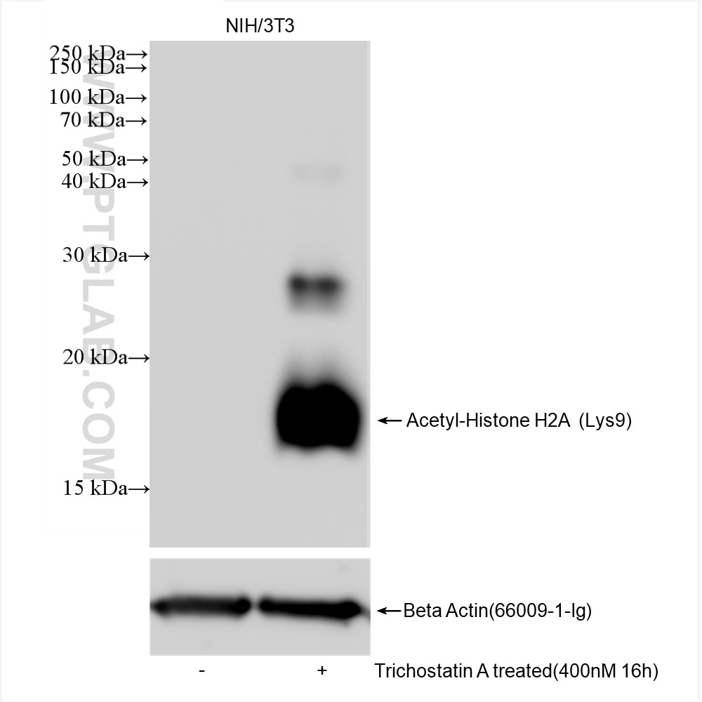
| Positive WB detected in | Trichostatin A treated NIH/3T3 cells |
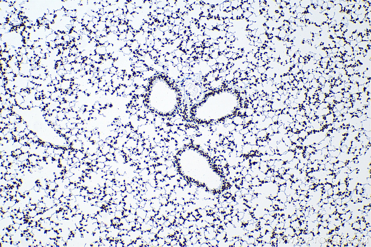
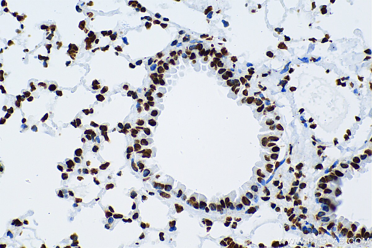
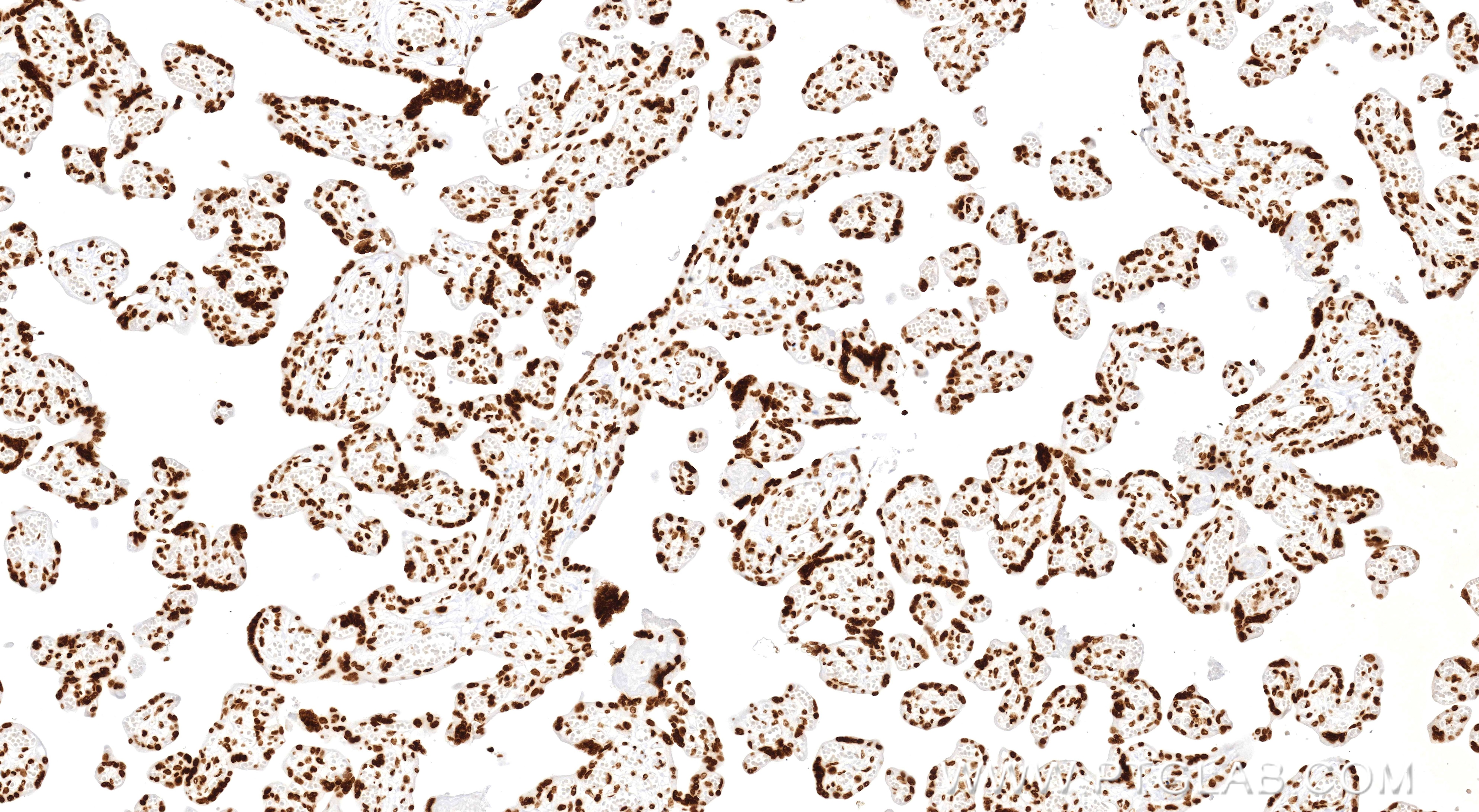
| Positive IHC detected in | mouse lung tissue, human placenta tissue Note: suggested antigen retrieval with TE buffer pH 9.0; (*) Alternatively, antigen retrieval may be performed with citrate buffer pH 6.0 |
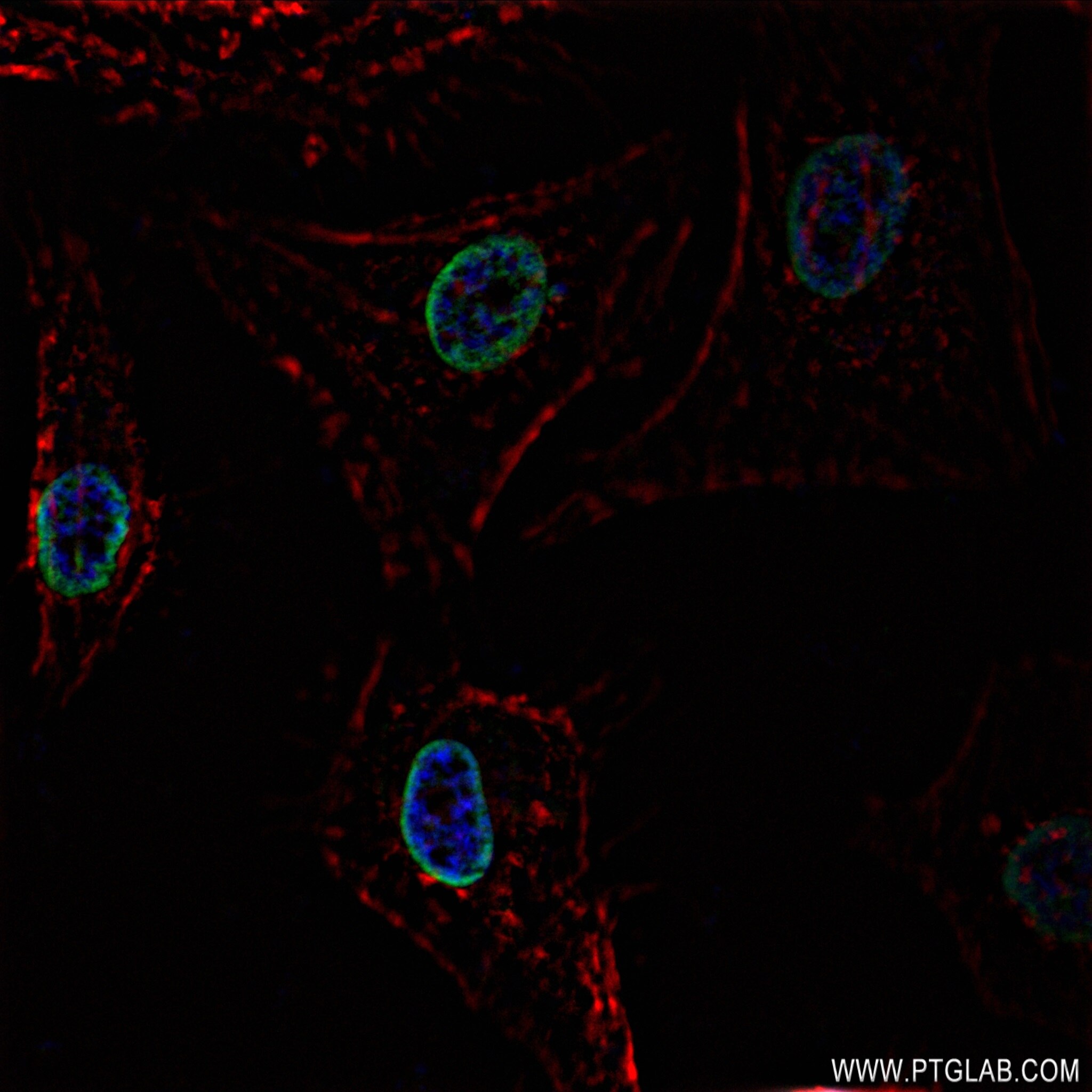
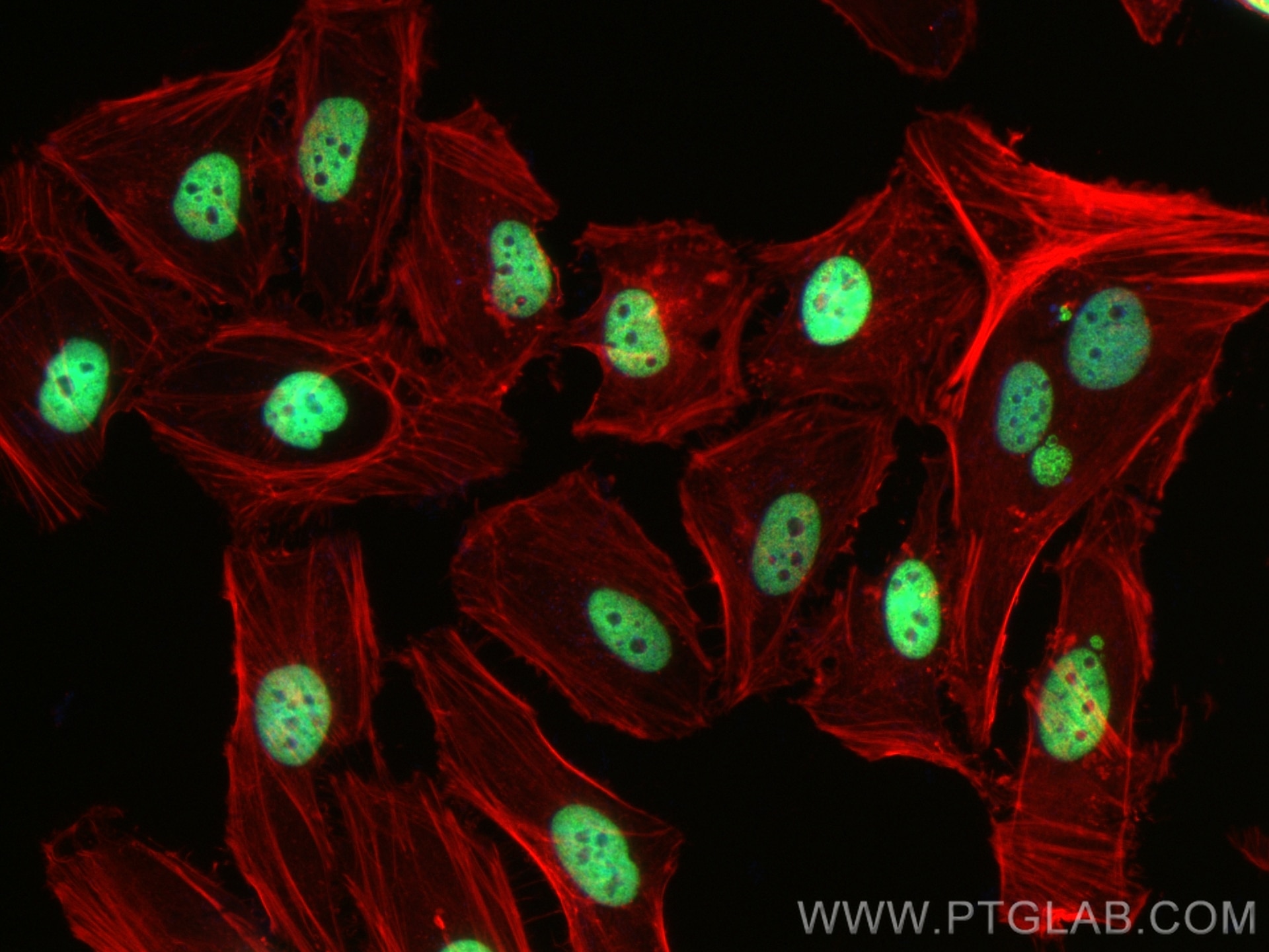
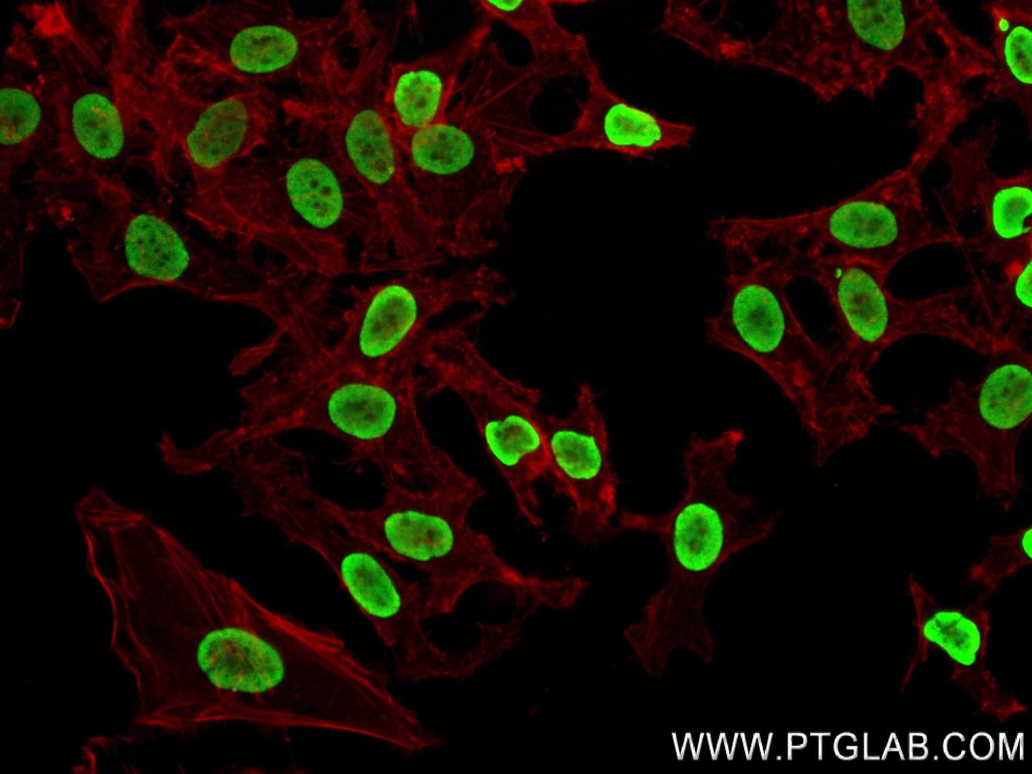
| Positive IF/ICC detected in | HeLa cells |
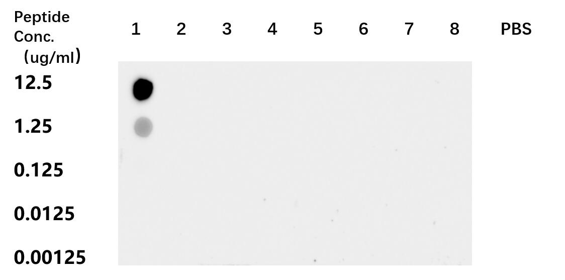
| Positive Dot Blot detected in | peptide |
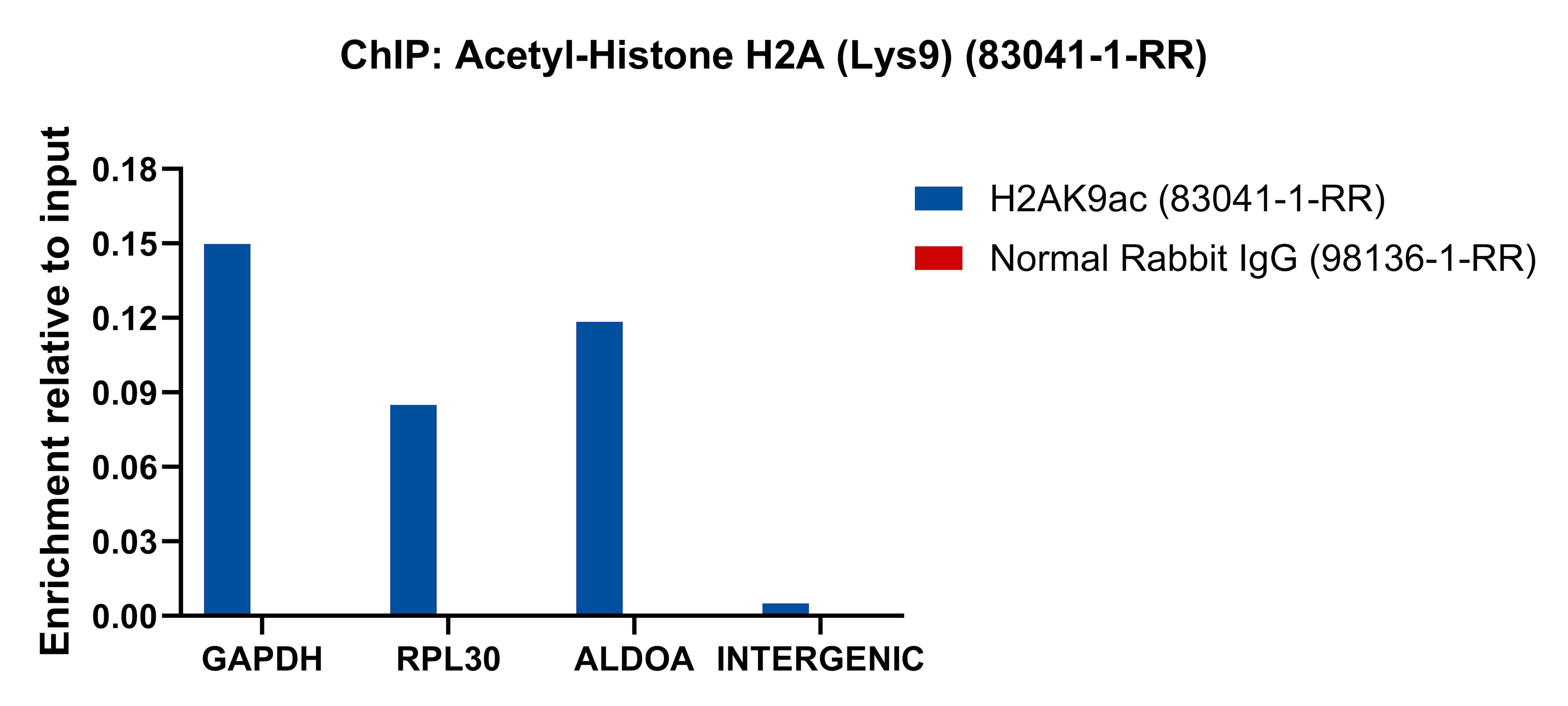
| Positive ChIP-qPCR detected in | HeLa cells |
Recommended dilution
| Application | Dilution |
|---|---|
| Western Blot (WB) | WB : 1:2000-1:10000 |
| Immunohistochemistry (IHC) | IHC : 1:250-1:1000 |
| Immunofluorescence (IF)/ICC | IF/ICC : 1:200-1:800 |
| DOT BLOT | DOT BLOT : 1:10-1:100 |
| CHIP-QPCR | CHIP-QPCR : 1:10-1:100 |
| It is recommended that this reagent should be titrated in each testing system to obtain optimal results. | |
| Sample-dependent, Check data in validation data gallery. | |
Product Information
83041-1-RR targets Acetyl-Histone H2A (Lys9) in WB, IHC, IF/ICC, Dot Blot, ELISA, ChIP-qPCR applications and shows reactivity with human, mouse samples.
| Tested Reactivity | human, mouse |
| Host / Isotype | Rabbit / IgG |
| Class | Recombinant |
| Type | Antibody |
| Immunogen |
Peptide Predict reactive species |
| Full Name | histone cluster 1, H2ae |
| Calculated Molecular Weight | 14 kDa |
| Observed Molecular Weight | 14 kDa |
| GenBank Accession Number | BC093836 |
| Gene Symbol | HIST1H2AE |
| Gene ID (NCBI) | 3012 |
| RRID | AB_3670771 |
| Conjugate | Unconjugated |
| Form | Liquid |
| Purification Method | Protein A purification |
| UNIPROT ID | P04908 |
| Storage Buffer | PBS with 0.02% sodium azide and 50% glycerol, pH 7.3. |
| Storage Conditions | Store at -20°C. Stable for one year after shipment. Aliquoting is unnecessary for -20oC storage. 20ul sizes contain 0.1% BSA. |
Background Information
Histone H2A is a core component of nucleosome. Histone variants contribute to chromatin complexity by creating specialized nucleosomes. Within nucleosomes, either one canonical H2A or both of them can be exchanged with a particular variant (heterotypic and homotypic nucleosomes, respectively), and such changes can have profound influences on nucleosome stability and biological outcome.
Protocols
| Product Specific Protocols | |
|---|---|
| IF protocol for Acetyl-Histone H2A (Lys9) antibody 83041-1-RR | Download protocol |
| IHC protocol for Acetyl-Histone H2A (Lys9) antibody 83041-1-RR | Download protocol |
| WB protocol for Acetyl-Histone H2A (Lys9) antibody 83041-1-RR | Download protocol |
| Standard Protocols | |
|---|---|
| Click here to view our Standard Protocols |