Tested Applications
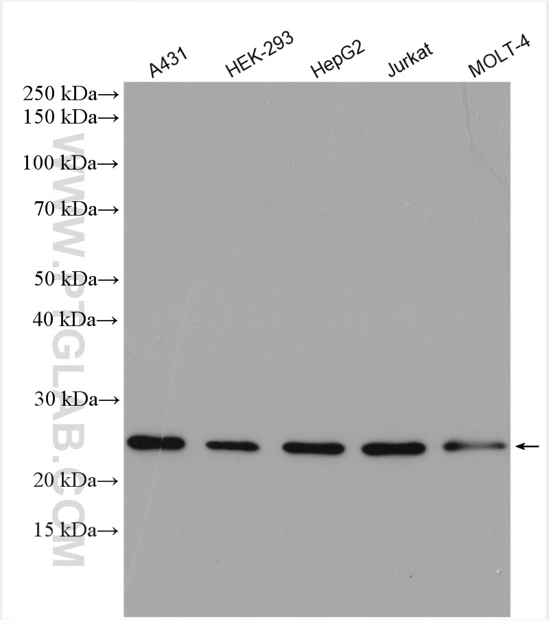
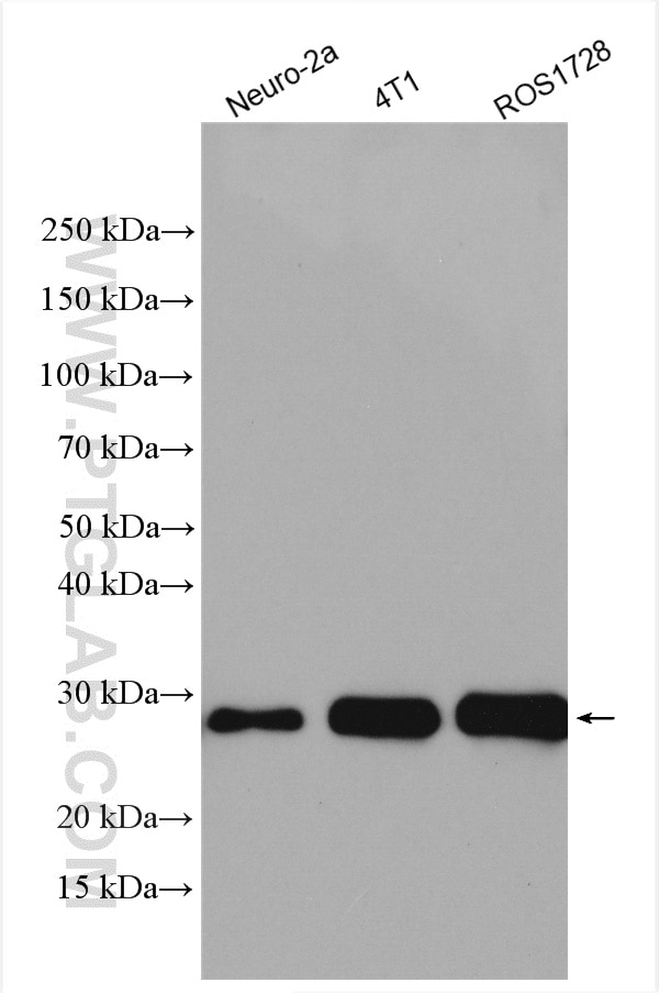
| Positive WB detected in | A431 cells, Neuro-2a cells, HEK-293 cells, HepG2 cells, Jurkat cells, MOLT-4 cells, 4T1 cells, ROS1728 cells |
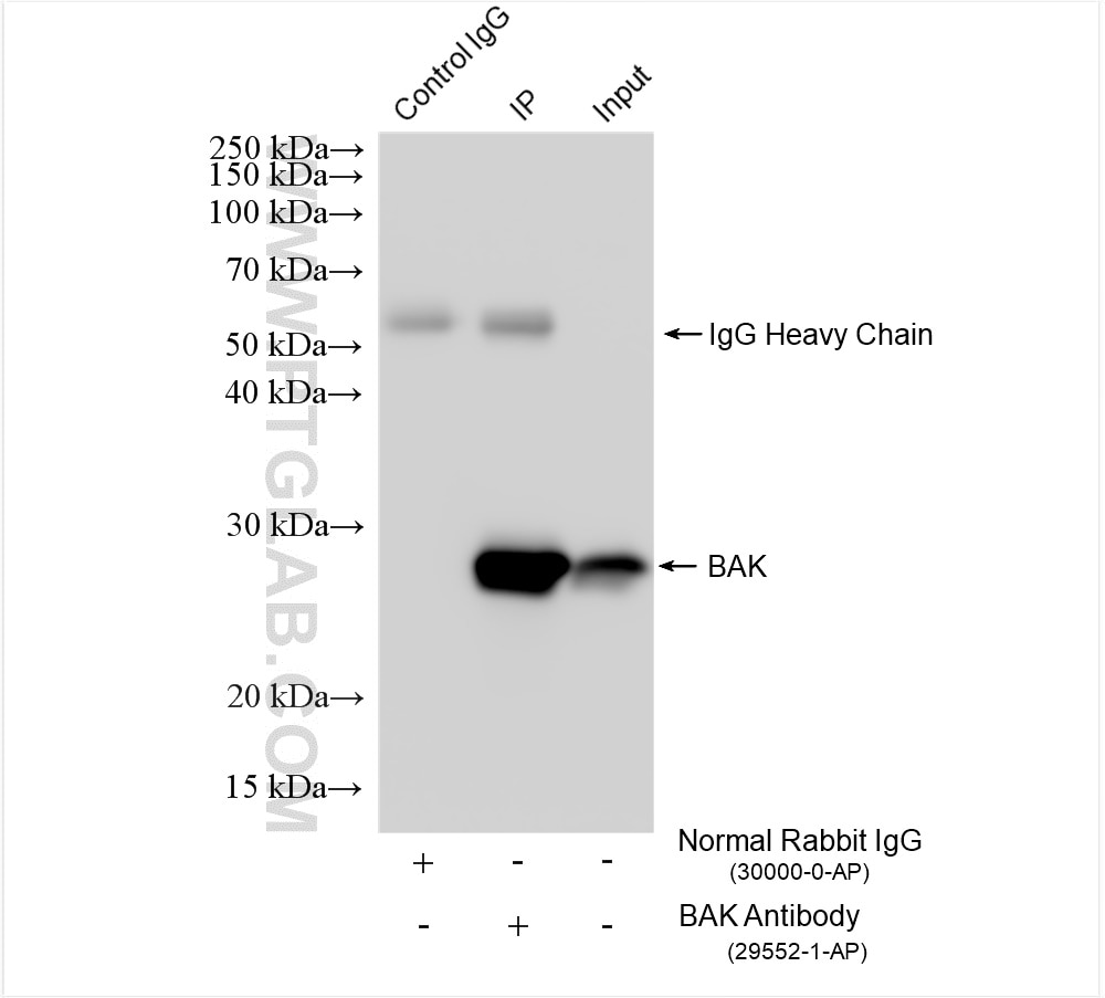
| Positive IP detected in | A431 cells |
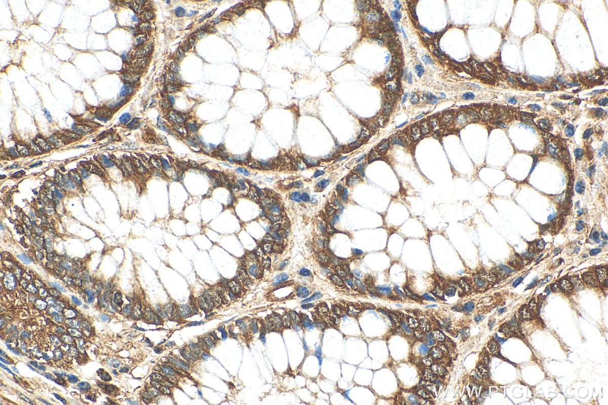
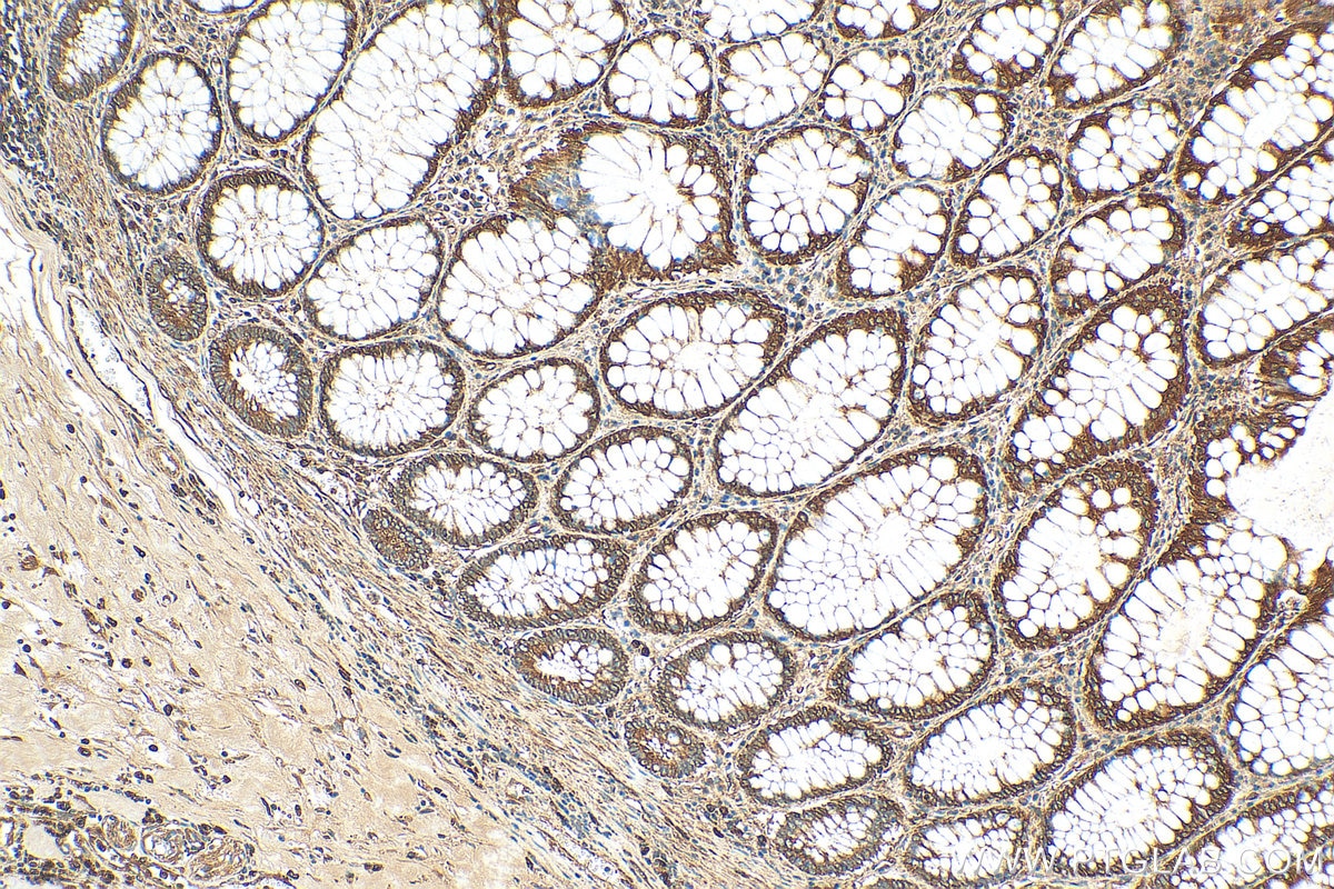
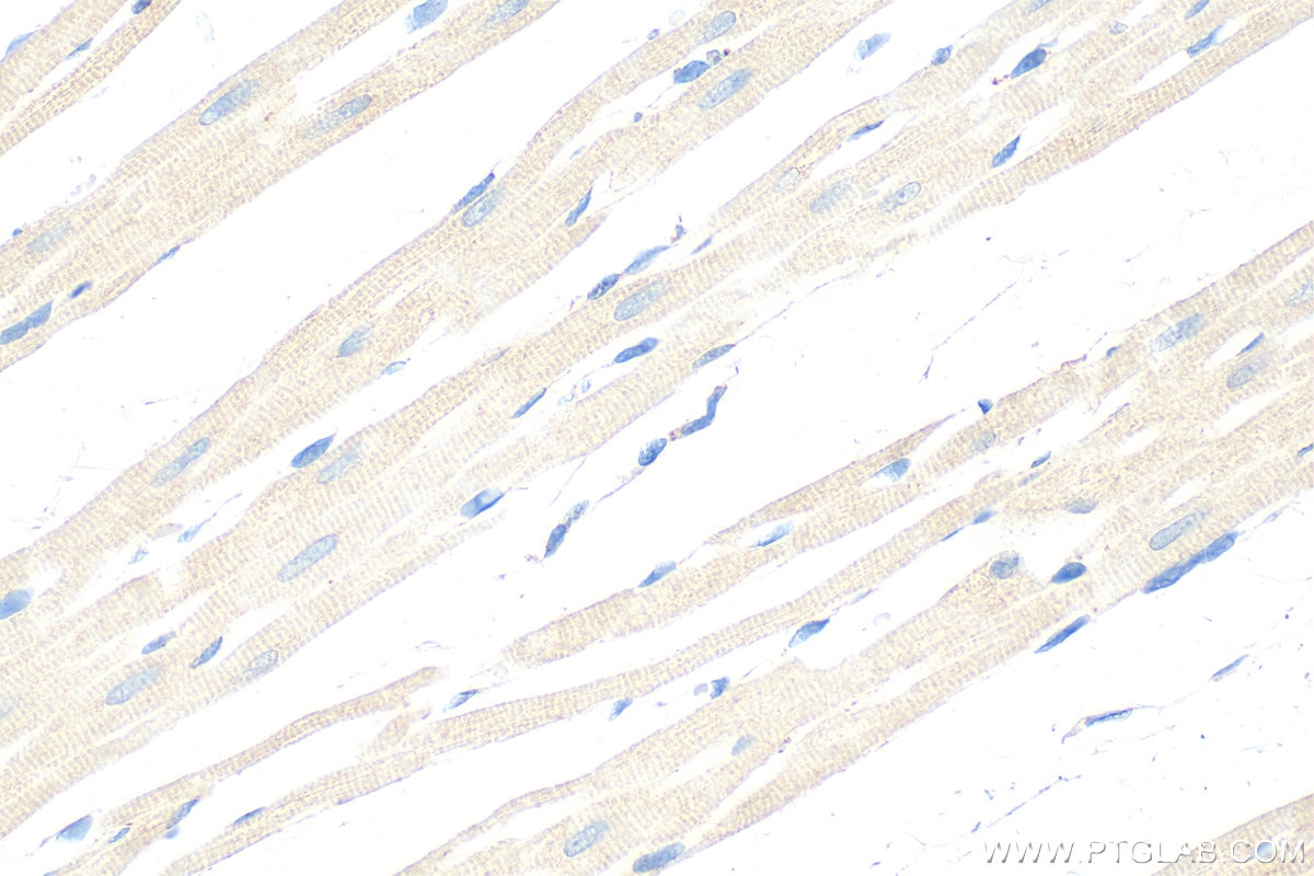
| Positive IHC detected in | human colon cancer tissue, rat heart tissue Note: suggested antigen retrieval with TE buffer pH 9.0; (*) Alternatively, antigen retrieval may be performed with citrate buffer pH 6.0 |
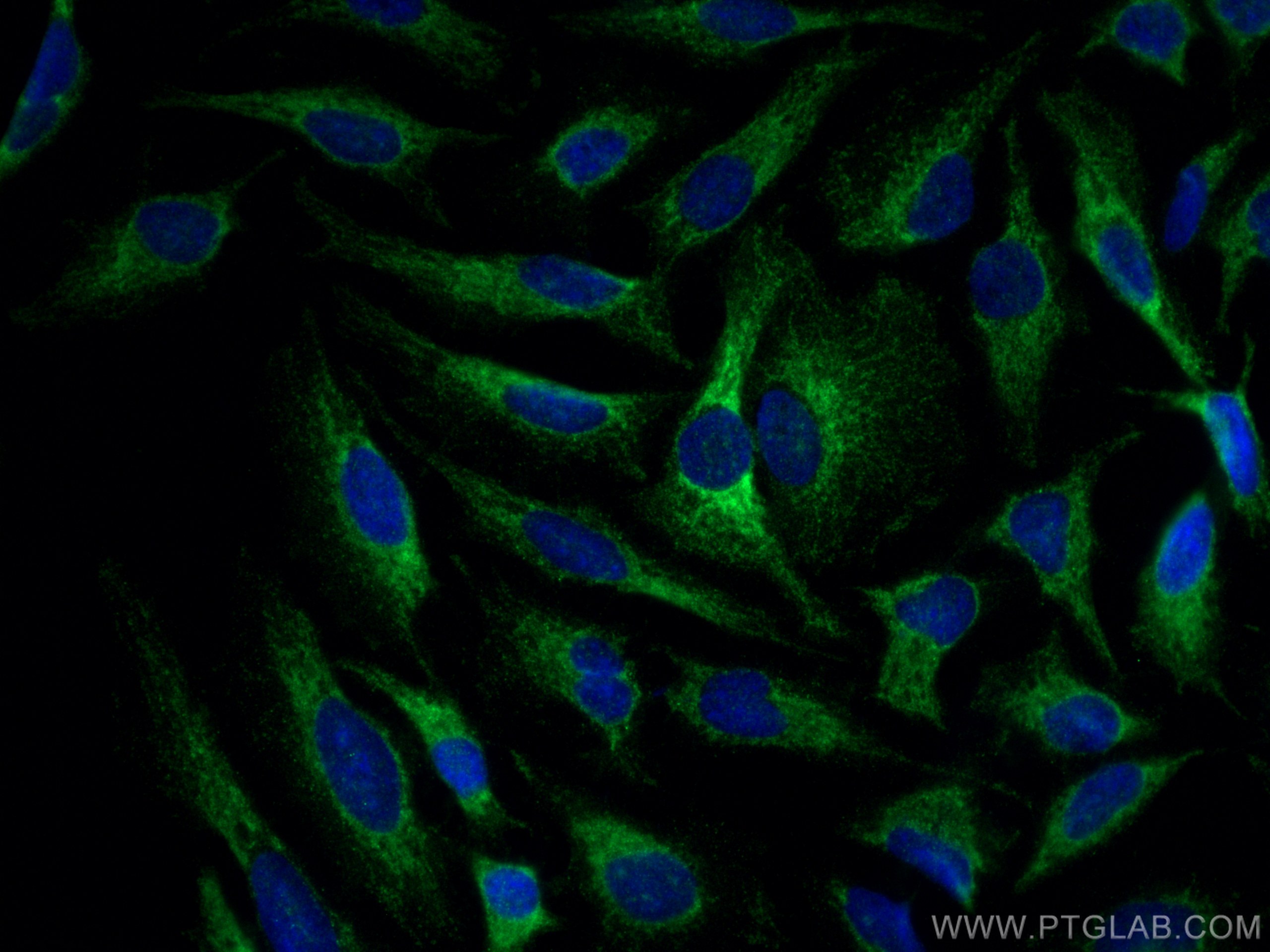
| Positive IF/ICC detected in | HeLa cells |
Recommended dilution
| Application | Dilution |
|---|---|
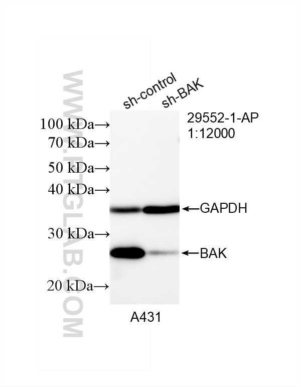
| Western Blot (WB) | WB : 1:2000-1:12000 |
| Immunoprecipitation (IP) | IP : 0.5-4.0 ug for 1.0-3.0 mg of total protein lysate |
| Immunohistochemistry (IHC) | IHC : 1:50-1:500 |
| Immunofluorescence (IF)/ICC | IF/ICC : 1:50-1:500 |
| It is recommended that this reagent should be titrated in each testing system to obtain optimal results. | |
| Sample-dependent, Check data in validation data gallery. | |
Published Applications
| KD/KO | See 2 publications below |
| WB | See 31 publications below |
| IHC | See 4 publications below |
Product Information
29552-1-AP targets BAK in WB, IHC, IF/ICC, IP, ELISA applications and shows reactivity with human, mouse, rat samples.
| Tested Reactivity | human, mouse, rat |
| Cited Reactivity | human, mouse, rat, pig, chicken, hamster |
| Host / Isotype | Rabbit / IgG |
| Class | Polyclonal |
| Type | Antibody |
| Immunogen |
CatNo: Ag31042 Product name: Recombinant human BAK protein Source: e coli.-derived, PGEX-4T Tag: GST Domain: 1-100 aa of NM_001188 Sequence: MASGQGPGPPRQECGEPALPSASEEQVAQDTEEVFRSYVFYRHQQEQEAEGVAAPADPEMVTLPLQPSSTMGQVGRQLAIIGDDINRRYDSEFQTMLQHL Predict reactive species |
| Full Name | BCL2-antagonist/killer 1 |
| Calculated Molecular Weight | 23 kDa |
| Observed Molecular Weight | 23-25 kDa |
| GenBank Accession Number | NM_001188 |
| Gene Symbol | BAK1 |
| Gene ID (NCBI) | 578 |
| RRID | AB_2923596 |
| Conjugate | Unconjugated |
| Form | Liquid |
| Purification Method | Antigen affinity purification |
| UNIPROT ID | Q16611 |
| Storage Buffer | PBS with 0.02% sodium azide and 50% glycerol, pH 7.3. |
| Storage Conditions | Store at -20°C. Stable for one year after shipment. Aliquoting is unnecessary for -20oC storage. 20ul sizes contain 0.1% BSA. |
Background Information
The protein encoded by this gene belongs to the BCL2 protein family. BCL2 family members form oligomers or heterodimers and act as anti- or pro-apoptotic regulators that are involved in a wide variety of cellular activities. This protein localizes to mitochondria, and functions to induce apoptosis. It interacts with and accelerates the opening of the mitochondrial voltage-dependent anion channel, which leads to a loss in membrane potential and the release of cytochrome c. This protein also interacts with the tumor suppressor P53 after exposure to cell stress.
Protocols
| Product Specific Protocols | |
|---|---|
| IF protocol for BAK antibody 29552-1-AP | Download protocol |
| IHC protocol for BAK antibody 29552-1-AP | Download protocol |
| IP protocol for BAK antibody 29552-1-AP | Download protocol |
| WB protocol for BAK antibody 29552-1-AP | Download protocol |
| Standard Protocols | |
|---|---|
| Click here to view our Standard Protocols |
Publications
| Species | Application | Title |
|---|---|---|
ACS Nano Mitochondria-Targeted Microneedles Reverse Doxorubicin Resistance via Apoptosis-Ferroptosis Synergy | ||
Cell Rep Med High-dose ascorbic acid selectively induces pyroptosis in LKB1-deficient lung cancer and sensitizes immunotherapy | ||
Apoptosis Investigation of GPR143 as a promising novel marker for the progression of skin cutaneous melanoma through bioinformatic analyses and cell experiments | ||
Life Sci A novel antimycin analogue antimycin A2c, derived from marine Streptomyces sp., suppresses HeLa cells via disrupting mitochondrial function and depleting HPV oncoproteins E6/E7 | ||
Mar Drugs Astaxanthin Inhibits H2O2-Induced Excessive Mitophagy and Apoptosis in SH-SY5Y Cells by Regulation of Akt/mTOR Activation | ||