Tested Applications
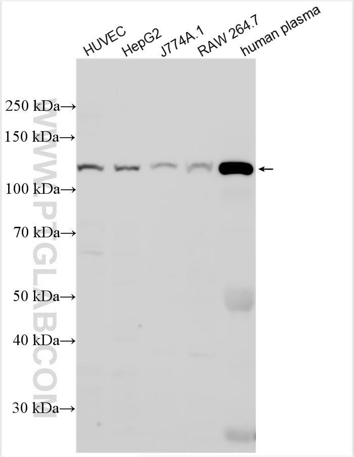
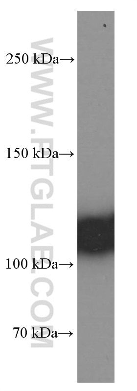
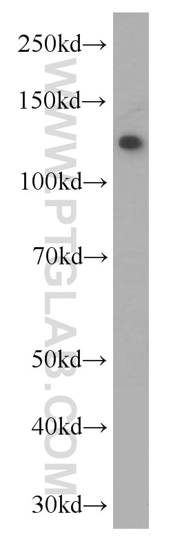
| Positive WB detected in | HUVEC cells, human blood, human plasma, HepG2 cells, J774A.1 cells, RAW 264.7 cells |
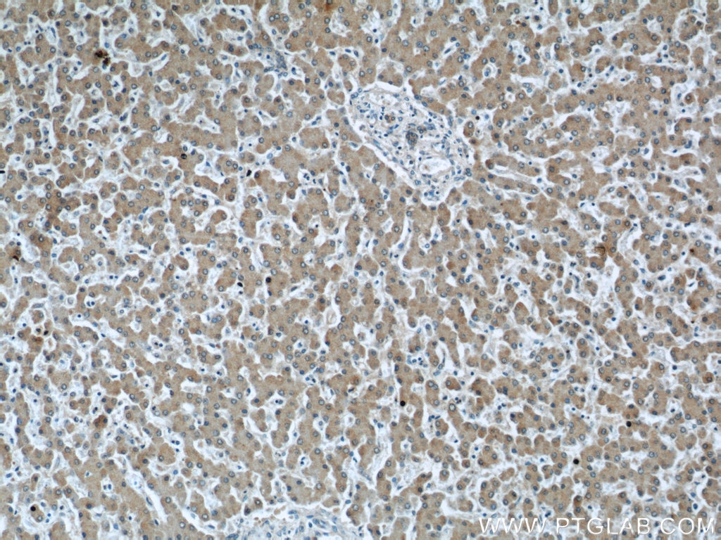
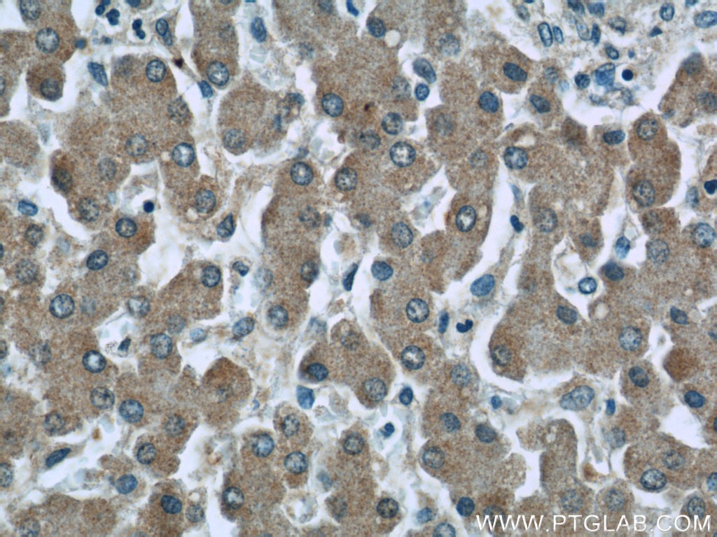
| Positive IHC detected in | human liver tissue Note: suggested antigen retrieval with TE buffer pH 9.0; (*) Alternatively, antigen retrieval may be performed with citrate buffer pH 6.0 |
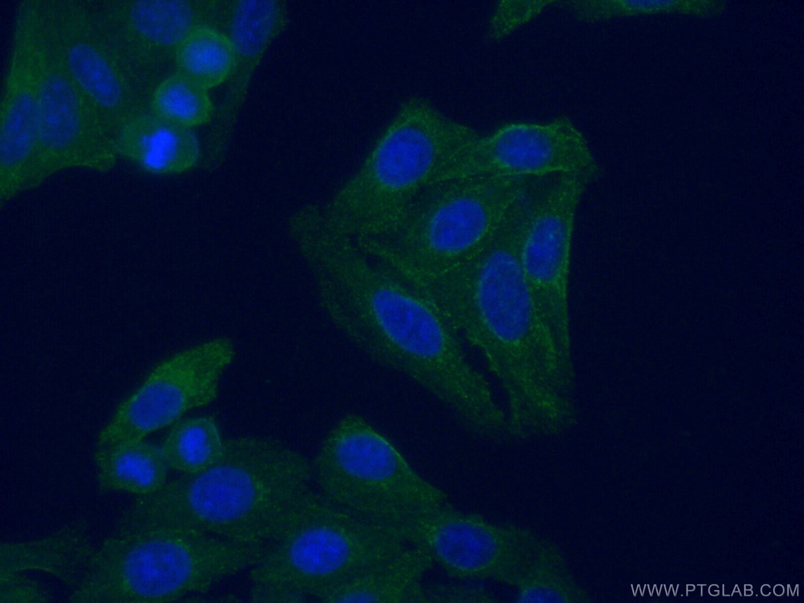
| Positive IF/ICC detected in | HepG2 cells |
Recommended dilution
| Application | Dilution |
|---|---|
| Western Blot (WB) | WB : 1:1000-1:8000 |
| Immunohistochemistry (IHC) | IHC : 1:20-1:200 |
| Immunofluorescence (IF)/ICC | IF/ICC : 1:50-1:500 |
| It is recommended that this reagent should be titrated in each testing system to obtain optimal results. | |
| Sample-dependent, Check data in validation data gallery. | |
Published Applications
| WB | See 3 publications below |
| IF | See 4 publications below |
| ELISA | See 1 publications below |
Product Information
66157-1-Ig targets C3/C3b/C3c in WB, IHC, IF/ICC, ELISA applications and shows reactivity with human, mouse samples.
| Tested Reactivity | human, mouse |
| Cited Reactivity | mouse, rat |
| Host / Isotype | Mouse / IgG1 |
| Class | Monoclonal |
| Type | Antibody |
| Immunogen |
CatNo: Ag15955 Product name: Recombinant human C3 protein Source: e coli.-derived, PET28a Tag: 6*His Domain: 1314-1663 aa of BC150179 Sequence: ESASLLRSEETKENEGFTVTAEGKGQGTLSVVTMYHAKAKDQLTCNKFDLKVTIKPAPETEKRPQDAKNTMILEICTRYRGDQDATMSILDISMMTGFAPDTDDLKQLANGVDRYISKYELDKAFSDRNTLIIYLDKVSHSEDDCLAFKVHQYFNVELIQPGAVKVYAYYNLEESCTRFYHPEKEDGKLNKLCRDELCRCAEENCFIQKSDDKVTLEERLDKACEPGVDYVYKTRLVKVQLSNDFDEYIMAIEQTIKSGSDEVQVGQQRTFISPIKCREALKLEEKKHYLMWGLSSDFWGEKPNLSYIIGKDTWVEHWPEEDECQDEENQKQCQDLGAFTESMVVFGCPN Predict reactive species |
| Full Name | complement component 3 |
| Calculated Molecular Weight | 1663 aa, 187 kDa |
| Observed Molecular Weight | 115 kDa |
| GenBank Accession Number | BC150179 |
| Gene Symbol | C3 |
| Gene ID (NCBI) | 718 |
| RRID | AB_2881553 |
| Conjugate | Unconjugated |
| Form | Liquid |
| Purification Method | Protein G purification |
| UNIPROT ID | P01024 |
| Storage Buffer | PBS with 0.02% sodium azide and 50% glycerol, pH 7.3. |
| Storage Conditions | Store at -20°C. Stable for one year after shipment. Aliquoting is unnecessary for -20oC storage. 20ul sizes contain 0.1% BSA. |
Background Information
The complement system is an important effector that bridges the innate and adaptive immune systems (PMID: 20010915). The third component of complement, C3, plays a central role in the activation of the complement system. Its processing by C3 convertase is the central reaction in both classical and alternative complement pathways (PMID: 11414361). Human C3, composed of α and β chains (115 and 75 kDa, respectively), is cleaved into C3a and C3b by C3 convertase. C3b is further cleaved into iC3b, C3c, C3dg and C3f. This antibody raised against 1314-1663 aa of human C3 protein recognizes the C3 alpha chain, C3b alpha' chain, and C3c alpha' chain fragment 2.
Protocols
| Product Specific Protocols | |
|---|---|
| IF protocol for C3/C3b/C3c antibody 66157-1-Ig | Download protocol |
| IHC protocol for C3/C3b/C3c antibody 66157-1-Ig | Download protocol |
| WB protocol for C3/C3b/C3c antibody 66157-1-Ig | Download protocol |
| Standard Protocols | |
|---|---|
| Click here to view our Standard Protocols |
Publications
| Species | Application | Title |
|---|---|---|
J Neuroinflammation Apelin alleviated neuroinflammation and promoted endogenous neural stem cell proliferation and differentiation after spinal cord injury in rats. | ||
Int J Oral Sci Author Correction: Fibroblast derived C3 promotes the progression of experimental periodontitis through macrophage M1 polarization and osteoclast differentiation | ||
Clin Kidney J Complement abnormality predisposes to the development of malignant hypertension-associated thrombotic microangiopathy disease | ||
Virulence HylS', a fragment of truncated hyaluronidase of Streptococcus suis, contributes to immune evasion by interaction with host complement factor C3b | ||
Int J Oral Sci Fibroblast derived C3 promotes the progression of experimental periodontitis through macrophage M1 polarization and osteoclast differentiation | ||
Neurochem Int Quantitative iTRAQ-based proteomic analysis of piperine protected cerebral ischemia/reperfusion injury in rat brain. |
Reviews
The reviews below have been submitted by verified Proteintech customers who received an incentive for providing their feedback.
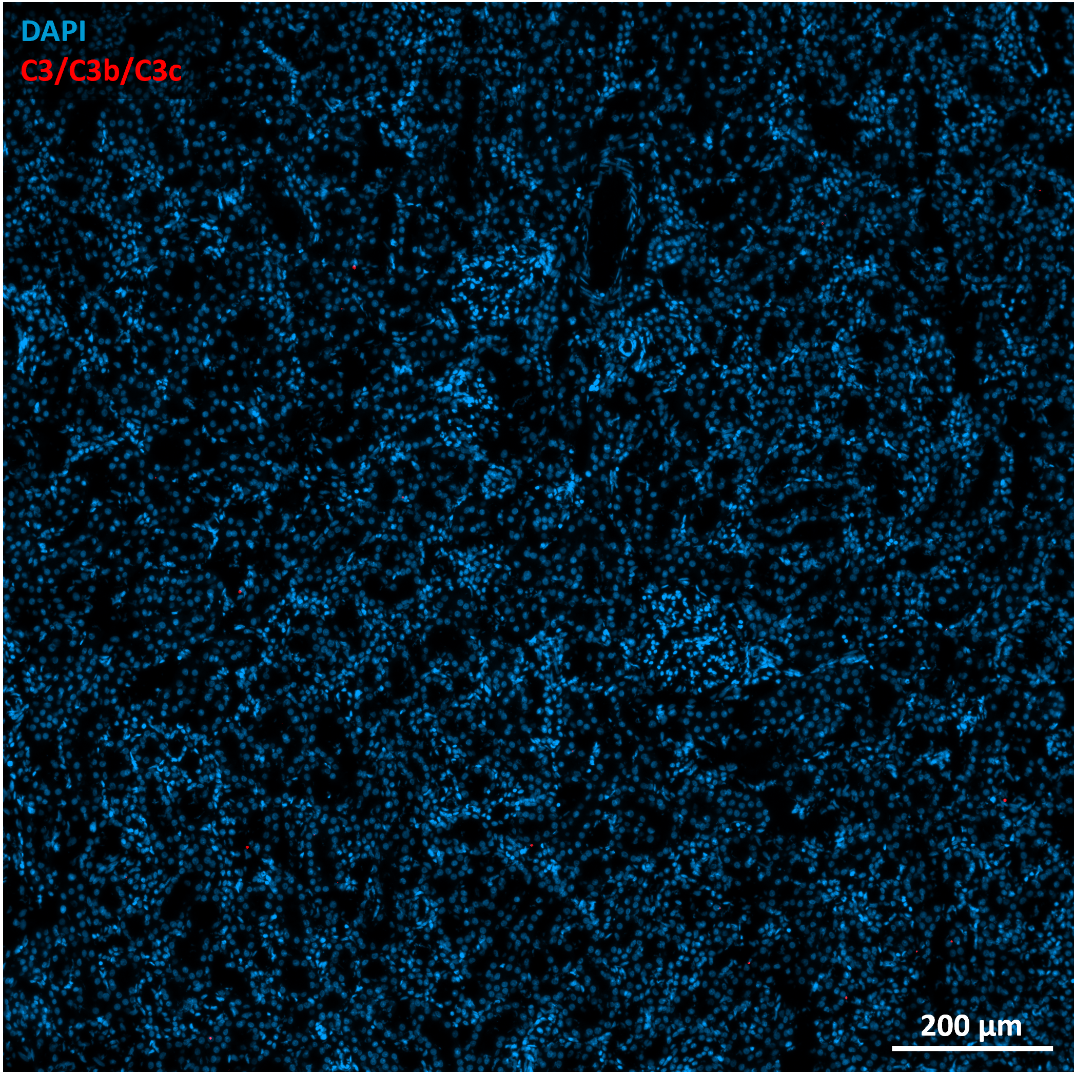
FH Marion (Verified Customer) (08-01-2024) | LPS induction model Fixative: Paraformaldehyde 4% Permeabilization: 10 min; RT; 0.2% Triton X-100 Blocking: 2 h; RT; 10% Normal donkey serum; 1% BSA Primary antibody: 1 night; 4°C Secondary antibody: ab150112 (Abcam); 1h30; RT Antibodies diluted in blocking solution
 |
FH Neil (Verified Customer) (05-10-2019) | The Western Blot came out spectacularly! I saw the bands I expected to see, and everything was clear and distinct. I would recommend this to anyone looking to identify C3.
|

