Tested Applications
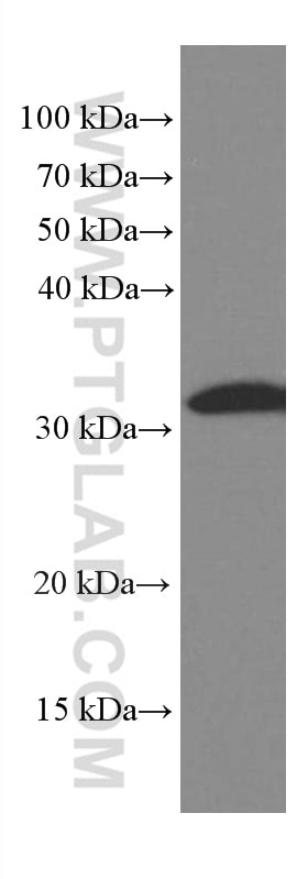
| Positive WB detected in | Jurkat cells, HEK-293 cells |
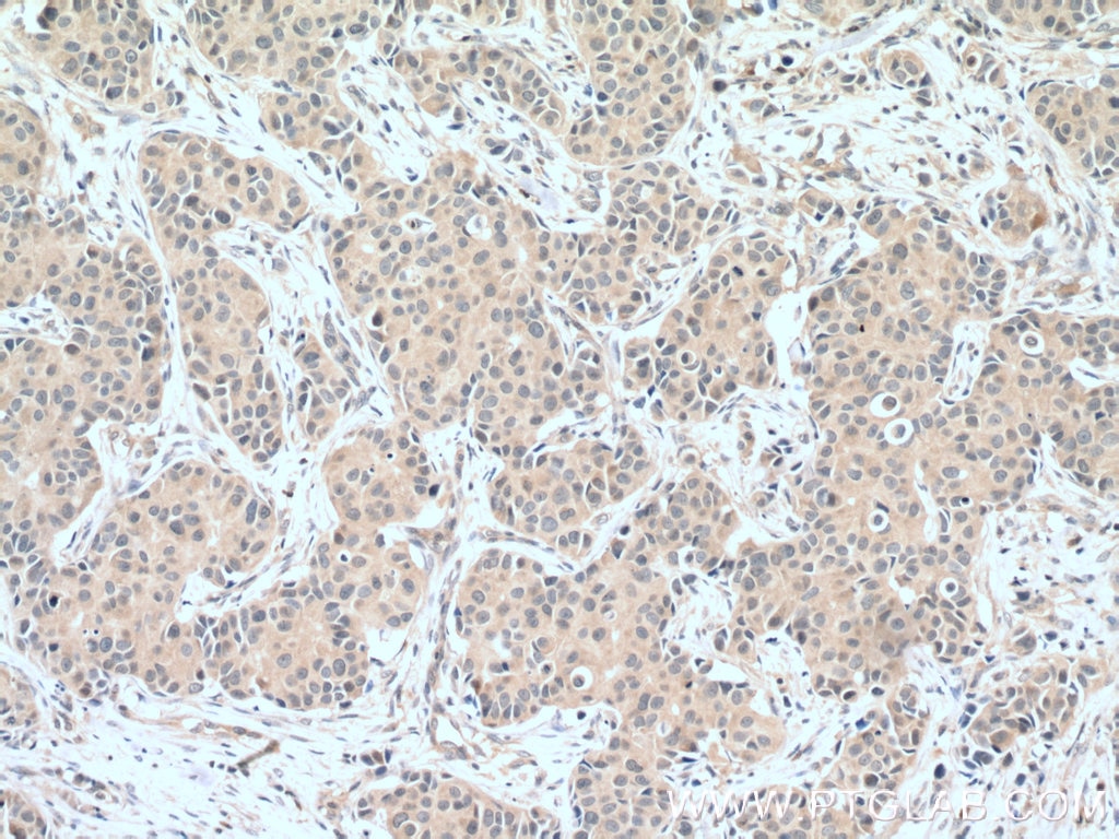
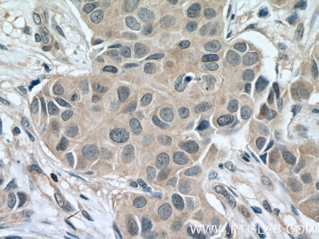
| Positive IHC detected in | human breast cancer tissue Note: suggested antigen retrieval with TE buffer pH 9.0; (*) Alternatively, antigen retrieval may be performed with citrate buffer pH 6.0 |
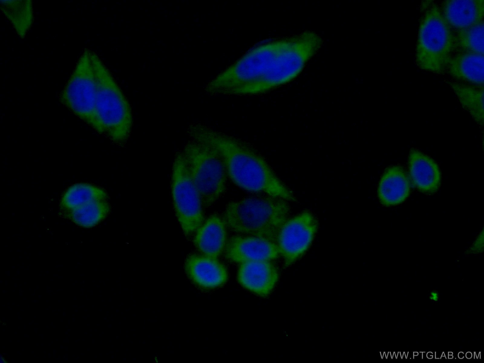
| Positive IF/ICC detected in | HeLa cells |
Recommended dilution
| Application | Dilution |
|---|---|
| Western Blot (WB) | WB : 1:500-1:2000 |
| Immunohistochemistry (IHC) | IHC : 1:100-1:400 |
| Immunofluorescence (IF)/ICC | IF/ICC : 1:50-1:500 |
| It is recommended that this reagent should be titrated in each testing system to obtain optimal results. | |
| Sample-dependent, Check data in validation data gallery. | |
Published Applications
| WB | See 72 publications below |
| IHC | See 2 publications below |
| IF | See 5 publications below |
Product Information
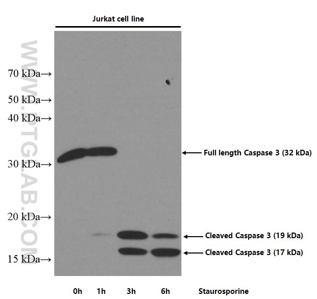
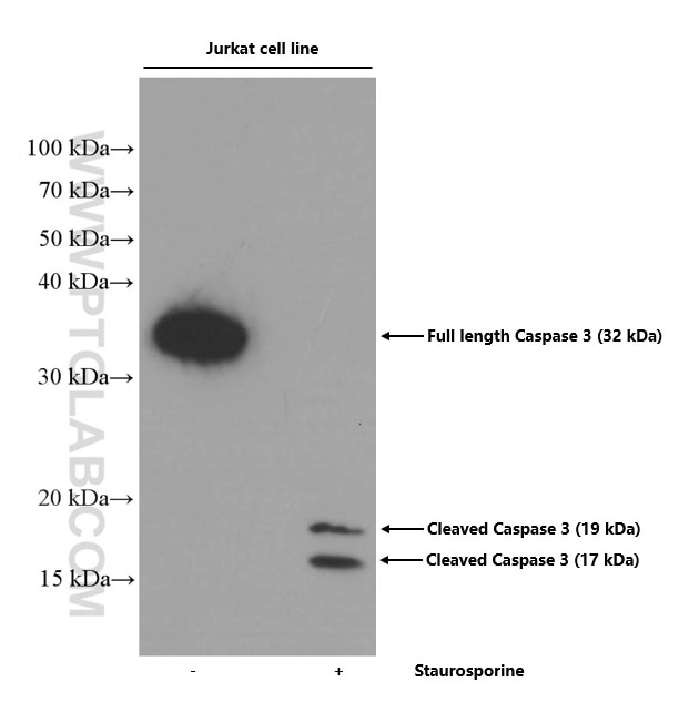
66470-1-Ig targets Caspase 3(human specific) in WB, IHC, IF/ICC, ELISA applications and shows reactivity with human samples.
| Tested Reactivity | human |
| Cited Reactivity | human, mouse, rat, pig, zebrafish, sheep, cow |
| Host / Isotype | Mouse / IgG2b |
| Class | Monoclonal |
| Type | Antibody |
| Immunogen |
CatNo: Ag25029 Product name: Recombinant human CASP3 protein Source: e coli.-derived, PET30a Tag: 6*His Domain: 31-176 aa of BC016926 Sequence: ISLDNSYKMDYPEMGLCIIINNKNFHKSTGMTSRSGTDVDAANLRETFRNLKYEVRNKNDLTREEIVELMRDVSKEDHSKRSSFVCVLLSHGEEGIIFGTNGPVDLKKITNFFRGDRCRSLTGKPKLFIIQACRGTELDCGIETDS Predict reactive species |
| Full Name | caspase 3, apoptosis-related cysteine peptidase |
| Calculated Molecular Weight | 277 aa, 32 kDa |
| Observed Molecular Weight | 32-35 kDa, 19 kDa, 17 kDa |
| GenBank Accession Number | BC016926 |
| Gene Symbol | Caspase 3 |
| Gene ID (NCBI) | 836 |
| Conjugate | Unconjugated |
| Form | Liquid |
| Purification Method | Protein A purification |
| UNIPROT ID | P42574 |
| Storage Buffer | PBS with 0.02% sodium azide and 50% glycerol, pH 7.3. |
| Storage Conditions | Store at -20°C. Stable for one year after shipment. Aliquoting is unnecessary for -20oC storage. 20ul sizes contain 0.1% BSA. |
Background Information
Casp3 (caspase 3), also named as CPP32, SCA-1 and Apopain, belongs to the peptidase C14A family. Caspase 3 is involved in the activation cascade of caspases responsible for apoptosis execution. At the onset of apoptosis it proteolytically cleaves poly(ADP-ribose) polymerase (PARP) at a '216-Asp-|-Gly-217' bond. Caspase 3 cleaves and activates sterol regulatory element binding proteins (SREBPs) between the basic helix-loop-helix leucine zipper domain and the membrane attachment domain. Cleaves and activates caspase-6, -7 and -9. CASP3 is involved in the cleavage of huntingtin. This antibody is specific for human caspase 3, it can recognize the 32 kDa pro-caspase 3 as well as 17 and 19 kDa cleaved-caspase 3.
Publications
| Species | Application | Title |
|---|---|---|
Sci Adv The deacetylation-phosphorylation regulation of SIRT2-SMC1A axis as a mechanism of antimitotic catastrophe in early tumorigenesis. | ||
Redox Biol Restoration of L-OPA1 alleviates acute ischemic stroke injury in rats via inhibiting neuronal apoptosis and preserving mitochondrial function. | ||
Environ Int Chemical conjugation of FITC to track silica nanoparticles in vivo and in vitro: An emerging method to assess the reproductive toxicity of industrial nanomaterials. | ||
Cancer Lett UM-6 induces autophagy and apoptosis via the Hippo-YAP signaling pathway in cervical cancer. | ||
Oxid Med Cell Longev Gastrodin Promotes the Survival of Random-Pattern Skin Flaps via Autophagy Flux Stimulation. | ||
Oxid Med Cell Longev Farrerol Enhances Nrf2-Mediated Defense Mechanisms against Hydrogen Peroxide-Induced Oxidative Damage in Human Retinal Pigment Epithelial Cells by Activating Akt and MAPK. |