Tested Applications
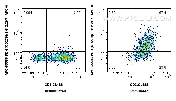
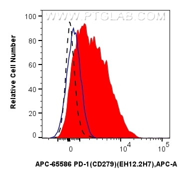
| Positive FC detected in | PHA treated human PBMCs |
Recommended dilution
| Application | Dilution |
|---|---|
| Flow Cytometry (FC) | FC : 5 ul per 10^6 cells in 100 μl suspension |
| This reagent has been pre-titrated and tested for flow cytometric analysis. The suggested use of this reagent is 5 ul per 10^6 cells in a 100 µl suspension or 5 ul per 100 µl of whole blood. | |
| Sample-dependent, Check data in validation data gallery. | |
Product Information
APC-65586 targets PD-1/CD279 in FC applications and shows reactivity with human samples.
| Tested Reactivity | human |
| Host / Isotype | Mouse / IgG2a |
| Class | Recombinant |
| Type | Antibody |
| Immunogen |
N/A Predict reactive species |
| Full Name | programmed cell death 1 |
| Calculated Molecular Weight | 288 aa, 32 kDa |
| GenBank Accession Number | BC074740 |
| Gene Symbol | PD-1 |
| Gene ID (NCBI) | 5133 |
| RRID | AB_3672352 |
| Conjugate | APC Fluorescent Dye |
| Excitation/Emission Maxima Wavelengths | 650 nm / 660 nm |
| Excitation Laser | Red Laser (633 nm) |
| Form | Liquid |
| Purification Method | Protein A purification |
| UNIPROT ID | Q15116 |
| Storage Buffer | PBS with 0.09% sodium azide and 0.5% BSA, pH 7.3. |
| Storage Conditions | Store at 2-8°C. Avoid exposure to light. Stable for one year after shipment. |
Background Information
Programmed cell death 1 (PD-1, also known as CD279) is an immunoinhibitory receptor that belongs to the CD28/CTLA-4 subfamily of the Ig superfamily. It is a 288 amino acid (aa) type I transmembrane protein composed of one Ig superfamily domain, a stalk, a transmembrane domain, and an intracellular domain containing an immunoreceptor tyrosine-based inhibitory motif (ITIM) as well as an immunoreceptor tyrosine-based switch motif (ITSM) (PMID: 18173375). PD-1 is expressed during thymic development and is induced in a variety of hematopoietic cells in the periphery by antigen receptor signaling and cytokines (PMID: 20636820). Engagement of PD-1 by its ligands PD-L1 or PD-L2 transduces a signal that inhibits T-cell proliferation, cytokine production, and cytolytic function (PMID: 19426218). It is critical for the regulation of T cell function during immunity and tolerance. Blockade of PD-1 can overcome immune resistance and also has been shown to have antitumor activity (PMID: 22658127; 23169436).
Protocols
| Product Specific Protocols | |
|---|---|
| FC protocol for APC PD-1/CD279 antibody APC-65586 | Download protocol |
| Standard Protocols | |
|---|---|
| Click here to view our Standard Protocols |