Tested Applications
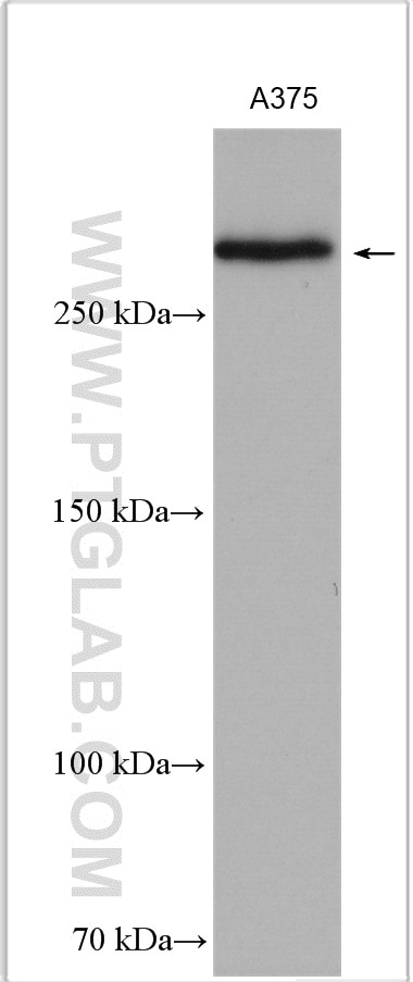
| Positive WB detected in | A375 cells |
Recommended dilution
| Application | Dilution |
|---|---|
| Western Blot (WB) | WB : 1:1000-1:4000 |
| It is recommended that this reagent should be titrated in each testing system to obtain optimal results. | |
| Sample-dependent, Check data in validation data gallery. | |
Published Applications
| KD/KO | See 1 publications below |
| WB | See 15 publications below |
| IHC | See 8 publications below |
Product Information
55027-1-AP targets CSPG4 in WB, IHC, ELISA applications and shows reactivity with human, mouse, rat samples.
| Tested Reactivity | human, mouse, rat |
| Cited Reactivity | human, mouse, rat |
| Host / Isotype | Rabbit / IgG |
| Class | Polyclonal |
| Type | Antibody |
| Immunogen |
Peptide Predict reactive species |
| Full Name | chondroitin sulfate proteoglycan 4 |
| Calculated Molecular Weight | 251 kDa |
| Observed Molecular Weight | 240-250 kDa, 450kDa |
| GenBank Accession Number | NM_001897 |
| Gene Symbol | CSPG4 |
| Gene ID (NCBI) | 1464 |
| RRID | AB_10791888 |
| Conjugate | Unconjugated |
| Form | Liquid |
| Purification Method | Antigen affinity purification |
| UNIPROT ID | Q6UVK1 |
| Storage Buffer | PBS with 0.02% sodium azide and 50% glycerol, pH 7.3. |
| Storage Conditions | Store at -20°C. Stable for one year after shipment. Aliquoting is unnecessary for -20oC storage. 20ul sizes contain 0.1% BSA. |
Background Information
CSPG4, also named as HMW-MAA, MCSP, MCSPG, MEL-CSPG, MSK16 and NG2, is a proteoglycan playing a role in cell proliferation and migration which stimulates endothelial cells motility during microvascular morphogenesis. CSPG4 may inhibit neurite outgrowth and growth cone collapse during axon regeneration. It is cell surface receptor for collagen alpha 2(VI) which may confer cells ability to migrate on that substrate. CSPG4 may regulate MPP16-dependent degradation and invasion of type I collagen participating in melanoma cells invasion properties. It modulates the plasminogen system by enhancing plasminogen activation and inhibiting angiostatin. CSPG4 functions as a signal transducing protein by binding through its cytoplasmic C-terminus scaffolding and signaling proteins. It promotes retraction fiber formation and cell polarization through Rho GTPase activation and stimulates alpha-4, beta-1 integrin-mediated adhesion and spreading by recruiting and activating a signaling cascade through CDC42, ACK1 and BCAR1. CSPG4 activates FAK and ERK1/ERK2 signaling cascades. The antibody is specific to CSPG4.
Protocols
| Product Specific Protocols | |
|---|---|
| WB protocol for CSPG4 antibody 55027-1-AP | Download protocol |
| Standard Protocols | |
|---|---|
| Click here to view our Standard Protocols |
Publications
| Species | Application | Title |
|---|---|---|
Nat Cell Biol A global analysis of SNX27-retromer assembly and cargo specificity reveals a function in glucose and metal ion transport. | ||
Nat Commun FNIP1 abrogation promotes functional revascularization of ischemic skeletal muscle by driving macrophage recruitment | ||
J Clin Invest Lysyl hydroxylase 2 glucosylates collagen VI to drive lung cancer progression | ||
J Pineal Res Melatonin pretreatment alleviates the long-term synaptic toxicity and dysmyelination induced by neonatal Sevoflurane exposure via MT1 receptor-mediated Wnt signaling modulation. | ||
Glia ST8SIA2 promotes oligodendrocyte differentiation and the integrity of myelin and axons. | ||
Commun Biol Complete remission of diabetes with a transient HDAC inhibitor and insulin in streptozotocin mice |
Reviews
The reviews below have been submitted by verified Proteintech customers who received an incentive for providing their feedback.
FH X (Verified Customer) (07-11-2022) | Although there is a weak band at the right MW, there are many other strong nonspecific bands.
|
FH Kinan (Verified Customer) (04-22-2022) | No specific staining on human lung and skin sections. Background signal was very high also. HIER pH6 in citrate buffer.
|
FH Nadine (Verified Customer) (01-31-2019) | We had high background and no specific staining of IHC-paraffin slides of human brain cortex. We tried both pH6 and pH9 antigen retrieval solutions. We have not tested other applications.
|