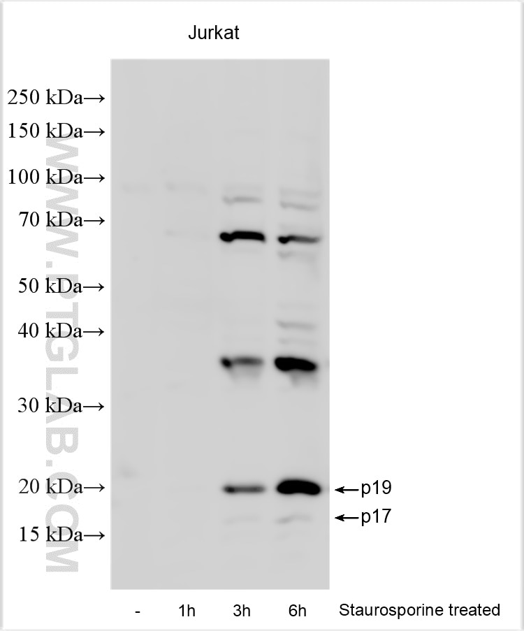
Staurosporine treated and untreated Jurkat cells were subjected to SDS PAGE followed by western blot with RMX00003 (Cleaved Caspase 3/P17/P19) at dilution of 1:3000 incubated at room temperature for 1.5 hours.
Staurosporine treated and untreated Jurkat cells were subjected to SDS PAGE followed by western blot with RMX00003 (Cleaved Caspase 3/P17/P19) at dilution of 1:3000 incubated at room temperature for 1.5 hours.
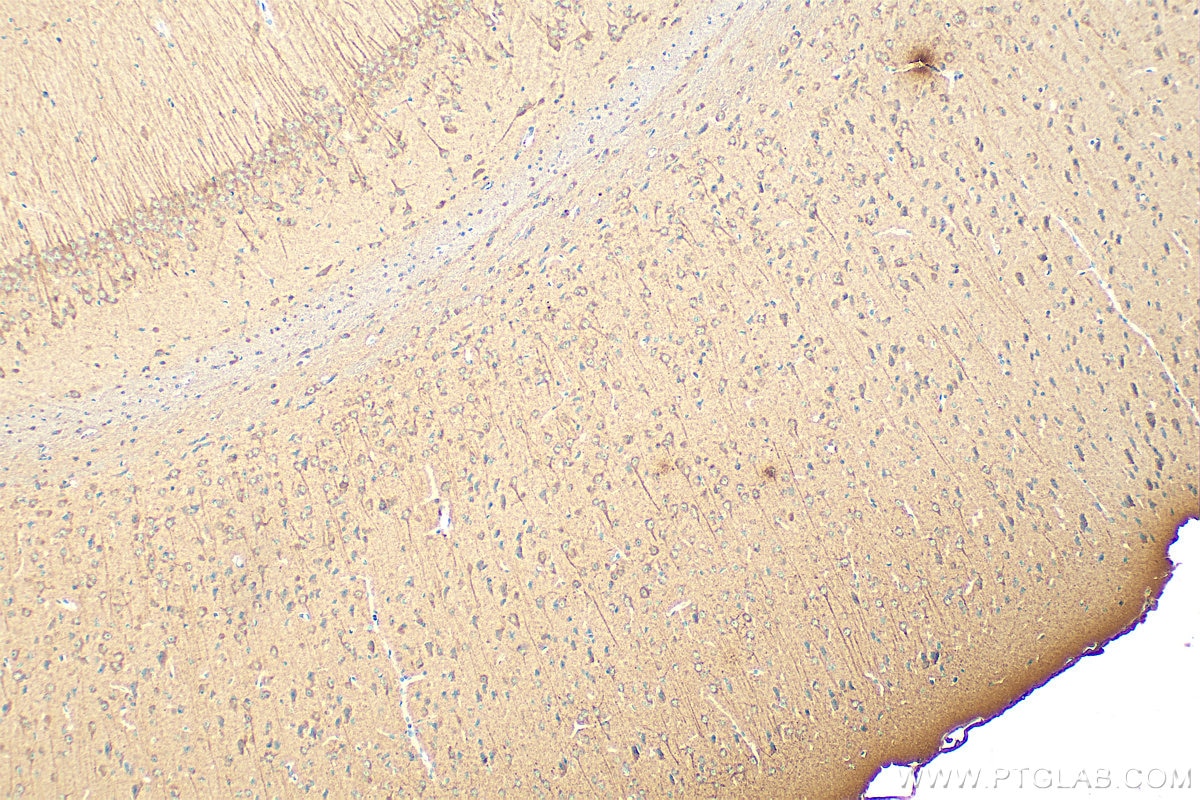
IHC staining of mouse brain using RMX00003
Immunohistochemical analysis of paraffin-embedded mouse brain tissue slide using RMX00003 (Cleaved Caspase 3/P17/P19 antibody) at dilution of 1:500 (under 10x lens). Heat mediated antigen retrieval with Tris-EDTA buffer (pH 9.0).
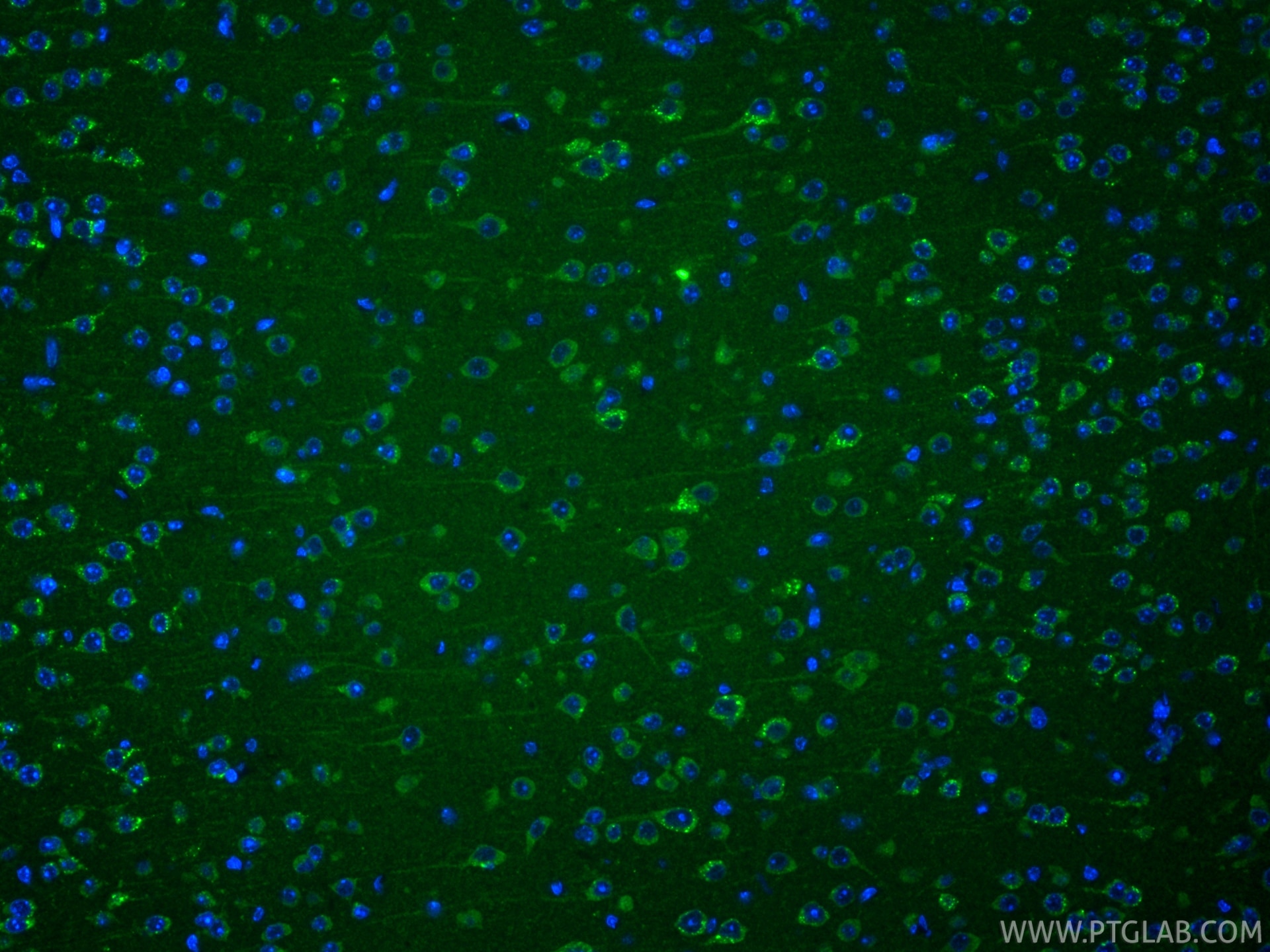
Immunofluorescent analysis of (4% PFA) fixed frozen OCT-embedded mouse brain tissue using Cleaved Caspase 3/P17/P19 antibody (RMX00003) at dilution of 1:400 and CoraLite®488-Conjugated Goat Anti-Rabbit IgG(H+L) (SA00013-2).
IF Staining of HeLa using RMX00003
Immunofluorescent analysis of (-20°C Ethanol) fixed HeLa cells using Cleaved Caspase 3/P17/P19 antibody (RMX00003) at dilution of 1:400 and Multi-rAb CoraLite ® Plus 488-Goat Anti-Rabbit Recombinant Secondary Antibody (H+L) (RGAR002).
Immunofluorescent analysis of (-20°C Ethanol) fixed HeLa cells using Cleaved Caspase 3/P17/P19 antibody (RMX00003) at dilution of 1:400 and Multi-rAb CoraLite ® Plus 488-Goat Anti-Rabbit Recombinant Secondary Antibody (H+L) (RGAR002).
The Proteintech guarantee covers Proteintech antibodies in any species and any application, including those not listed on the datasheet. If the antibody doesn’t perform, you can receive a hassle-free refund or credit note.
mouse brain tissue Note: suggested antigen retrieval with TE buffer pH 9.0; (*) Alternatively, antigen retrieval may be performed with citrate buffer pH 6.0
Positive IF-P detected in
mouse brain tissue, mouse embryo tissue
Positive IF-Fro detected in
mouse brain tissue
Positive IF/ICC detected in
HeLa cells
Recommended dilution
Application
Dilution
Western Blot (WB)
WB : 1:1000-1:6000
Immunohistochemistry (IHC)
IHC : 1:250-1:1000
Immunofluorescence (IF)-P
IF-P : 1:50-1:500
Immunofluorescence (IF)-FRO
IF-FRO : 1:200-1:800
Immunofluorescence (IF)/ICC
IF/ICC : 1:200-1:800
It is recommended that this reagent should be titrated in each testing system to obtain optimal results.
Sample-dependent, Check data in validation data gallery.
Product Information
RMX00003 targets Cleaved Caspase 3/P17/P19 in WB, IHC, IF/ICC, IF-P, IF-Fro, ELISA applications and shows reactivity with human, mouse samples.
PBS with 0.02% sodium azide and 50% glycerol, pH 7.3.
Storage Conditions
Store at -20°C. Stable for one year after shipment. Aliquoting is unnecessary for -20oC storage. 20ul sizes contain 0.1% BSA.
Background Information
Caspases, a family of endoproteases, are critical players in cell regulatory networks controlling inflammation and cell death. Initiator caspases (caspase-2, -8, -9, -10, -11, and -12) cleave and activate downstream effector caspases (caspase-3, -6, and -7), which in turn execute apoptosis by cleaving targeted cellular proteins. Caspase 3 plays a key role in the activation of sterol regulatory element binding proteins (SREBPs) between the basic helix-loop-helix leucine zipper domain and the membrane attachment domain. Caspase 3 can form heterocomplex with other proteins. This antibody can recognize p17 of Caspase 3. This antibody is specific for cleaved caspase 3, and does not recognize full length caspase-3, it can recognize the 17 kDa cleaved-caspase 3. The cleaved P17 and P19 fragment might form complex and shows at around 30-35 kDa by western blot (PMID: 25501826).
Protocols
Product Specific Protocols
IF protocol for Cleaved Caspase 3/P17/P19 antibody RMX00003
Staurosporine treated and untreated Jurkat cells were subjected to SDS PAGE followed by western blot with RMX00003 (Cleaved Caspase 3/P17/P19) at dilution of 1:3000 incubated at room temperature for 1.5 hours.
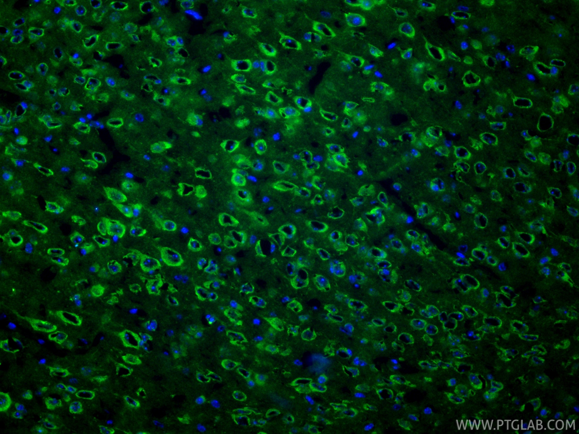
IHC Figures
IHC staining of mouse brain using RMX00003
Immunohistochemical analysis of paraffin-embedded mouse brain tissue slide using RMX00003 (Cleaved Caspase 3/P17/P19 antibody) at dilution of 1:500 (under 10x lens). Heat mediated antigen retrieval with Tris-EDTA buffer (pH 9.0).
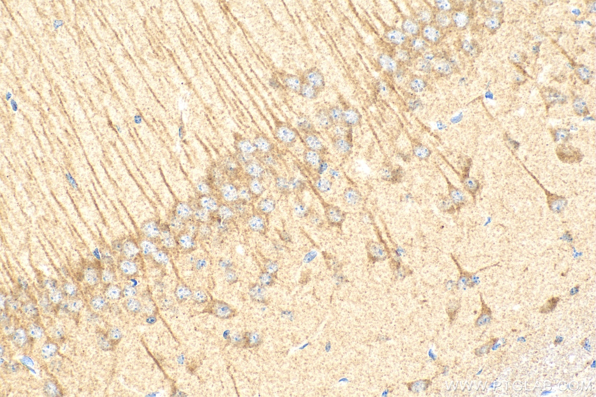
IHC staining of mouse brain using RMX00003
Immunohistochemical analysis of paraffin-embedded mouse brain tissue slide using RMX00003 (Cleaved Caspase 3/P17/P19 antibody) at dilution of 1:500 (under 40x lens). Heat mediated antigen retrieval with Tris-EDTA buffer (pH 9.0).
IF-P Figures
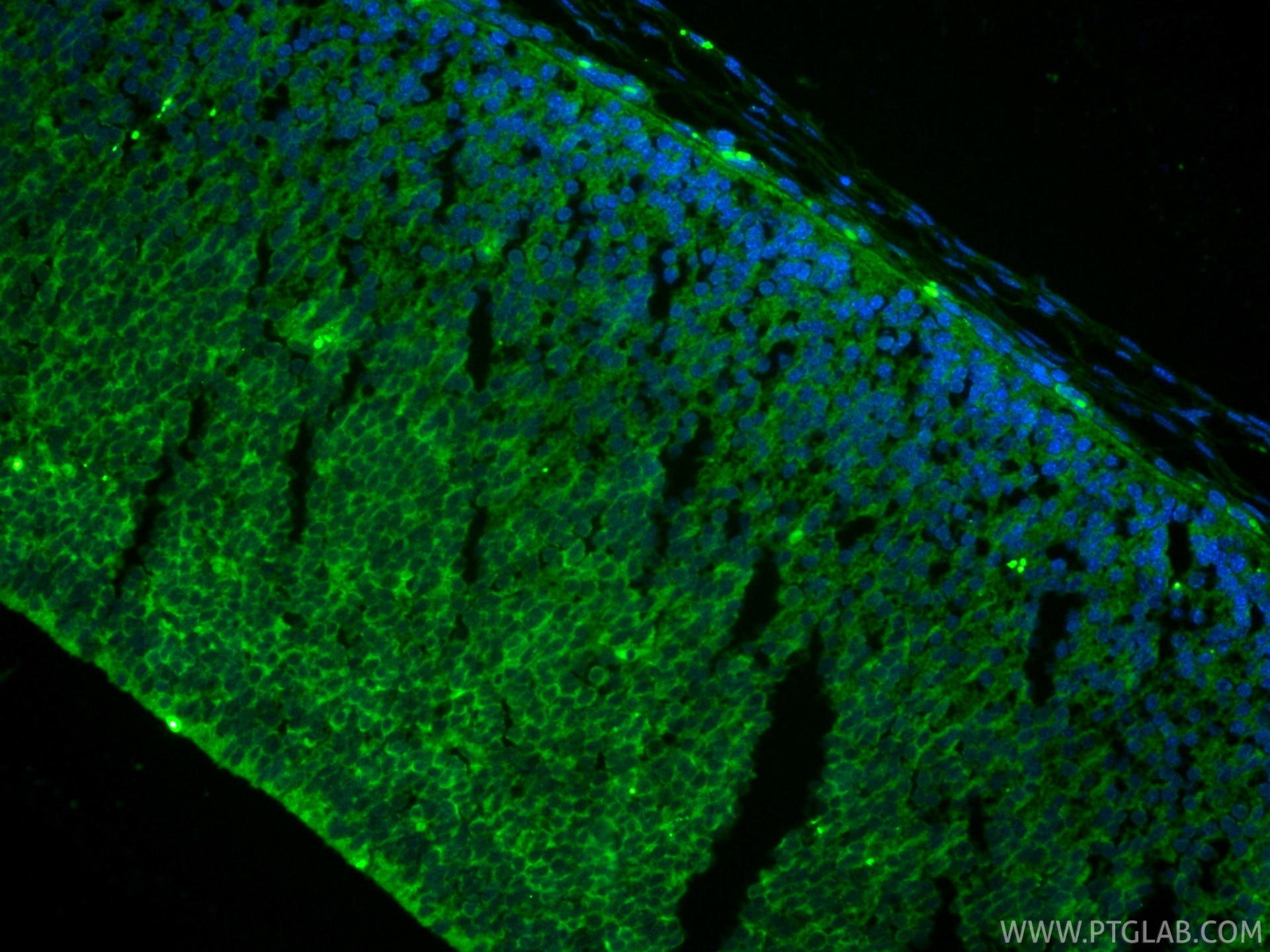
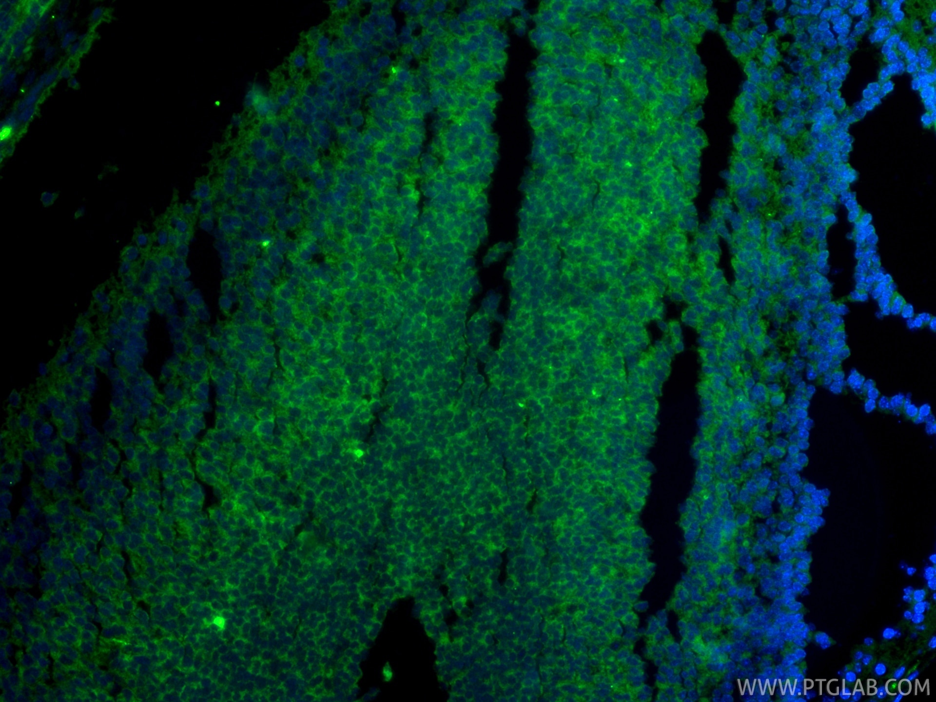
IF Staining of mouse brain using RMX00003
Immunofluorescent analysis of (4% PFA) fixed paraffin-embedded mouse brain tissue using Cleaved Caspase 3/P17/P19 antibody (RMX00003) at dilution of 1:200 and CoraLite®488-Conjugated Goat Anti-Rabbit IgG(H+L) (SA00013-2). Heat mediated antigen retrieval with Tris-EDTA buffer (pH 9.0).
IF Staining of mouse embryo using RMX00003
Immunofluorescent analysis of (4% PFA) fixed paraffin-embedded mouse embryo tissue using Cleaved Caspase 3/P17/P19 antibody (RMX00003) at dilution of 1:200 and CoraLite®488-Conjugated Goat Anti-Rabbit IgG(H+L) (SA00013-2). Heat mediated antigen retrieval with Tris-EDTA buffer (pH 9.0).
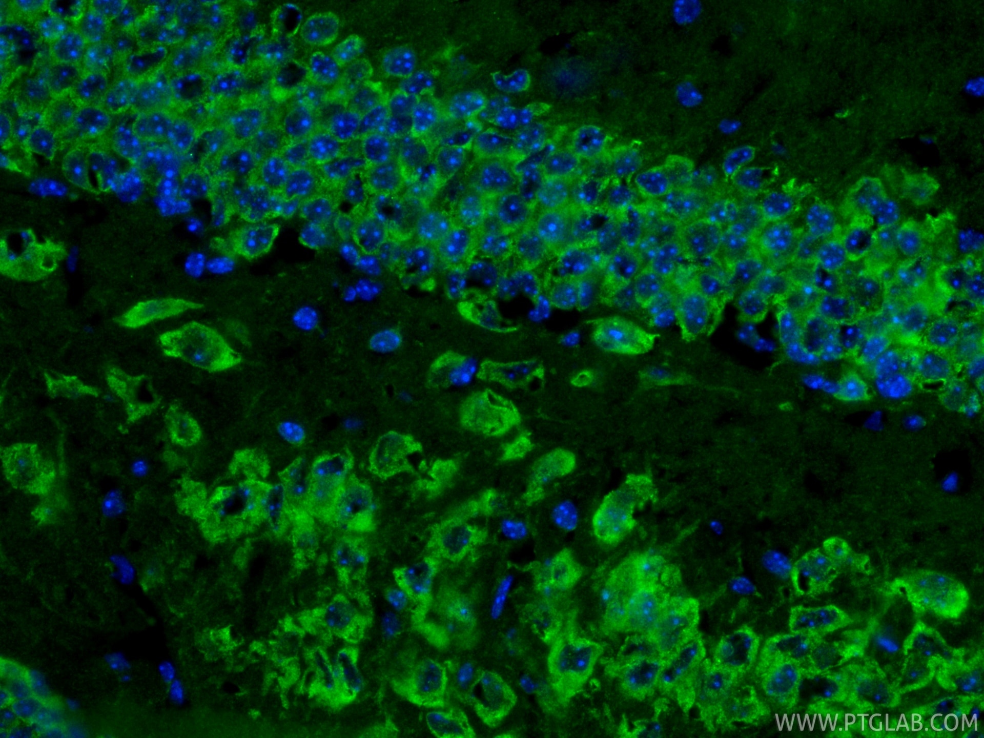
IF-FRO Figures
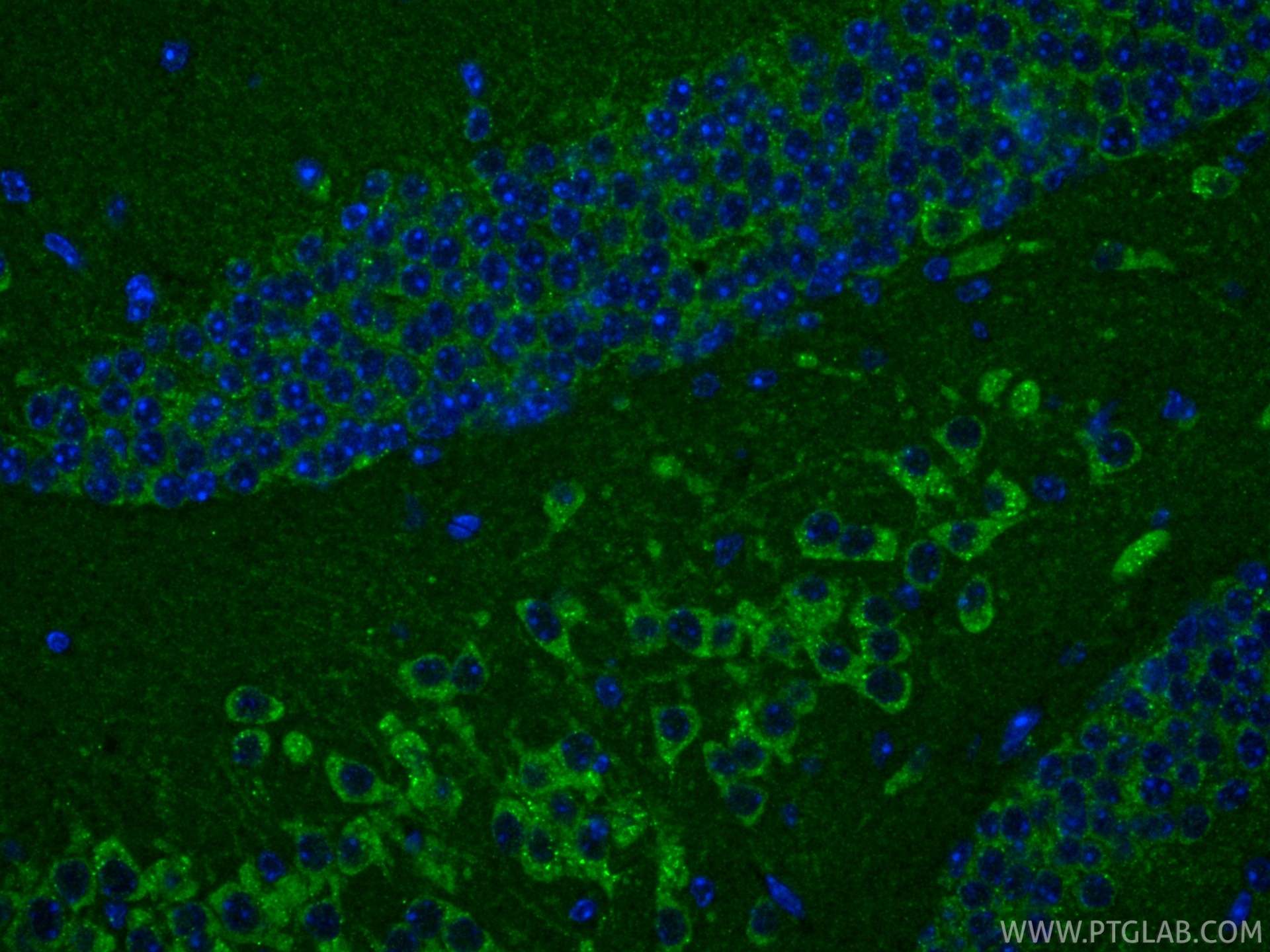
IF Staining of mouse brain using RMX00003
Immunofluorescent analysis of (4% PFA) fixed frozen OCT-embedded mouse brain tissue using Cleaved Caspase 3/P17/P19 antibody (RMX00003) at dilution of 1:400 and CoraLite®488-Conjugated Goat Anti-Rabbit IgG(H+L) (SA00013-2).
IF Staining of mouse brain using RMX00003
Immunofluorescent analysis of (4% PFA) fixed frozen OCT-embedded mouse brain tissue using Cleaved Caspase 3/P17/P19 antibody (RMX00003) at dilution of 1:400 and CoraLite®488-Conjugated Goat Anti-Rabbit IgG(H+L) (SA00013-2).
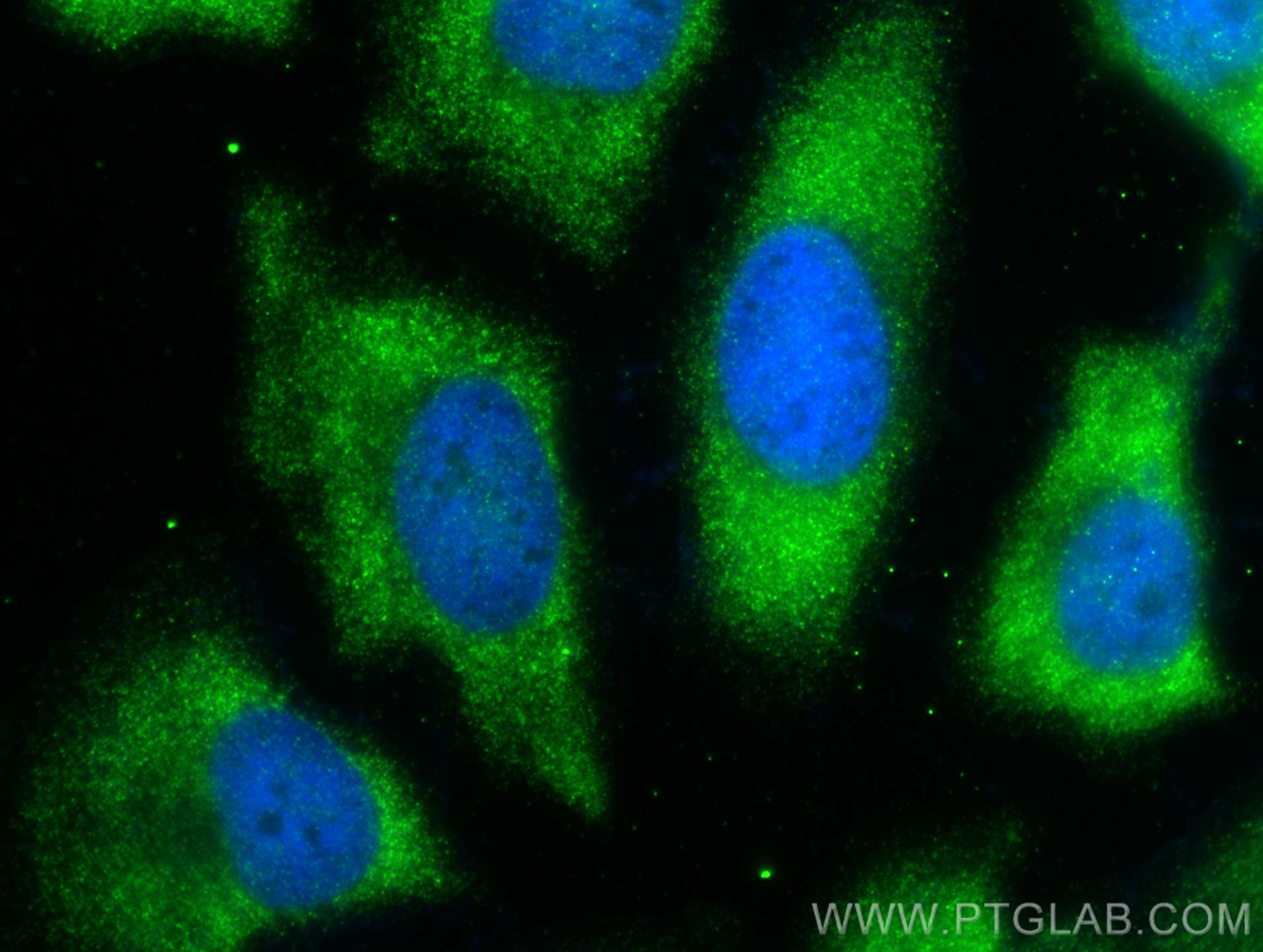
IF/ICC Figures
IF Staining of HeLa using RMX00003
Immunofluorescent analysis of (-20°C Ethanol) fixed HeLa cells using Cleaved Caspase 3/P17/P19 antibody (RMX00003) at dilution of 1:400 and Multi-rAb CoraLite ® Plus 488-Goat Anti-Rabbit Recombinant Secondary Antibody (H+L) (RGAR002).
The species listed in Tested Reactivity are in-house verified and applicable species. For unlisted species, please refer to the homology analysis of the immunogen sequence and related species. For rabbit polyclonal antibodies, homology >70% is recommended. For mouse monoclonal antibodies and rabbit recombinant antibodies, homology >90% is recommended. Generally, the higher the homology, the greater the applicability. However, there will be certain differences in protein expression in different species, tissues or cells. Therefore, the homology analysis results are for reference only and do not serve as a guarantee.
At Proteintech, we pride ourselves on our antibody quality, customer service and transparency. As such, we are comparing our antibodies with other vendors, enabling easy identification and comparisons of key data to help you choose the suitable antibody for your needs.
We have selected the top cited antibodies from these vendors for you to compare.