Tested Applications
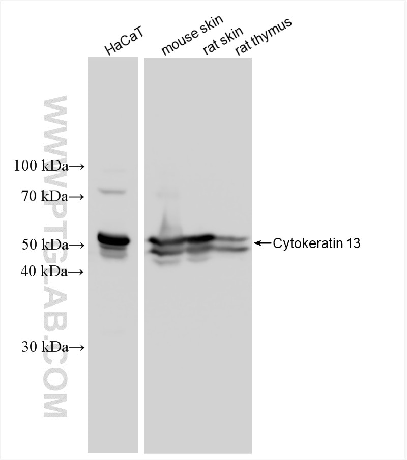
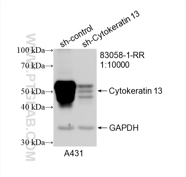
| Positive WB detected in | HaCaT cells, A431 cells, mouse skin tissue, rat skin tissue, rat thymus tissue |
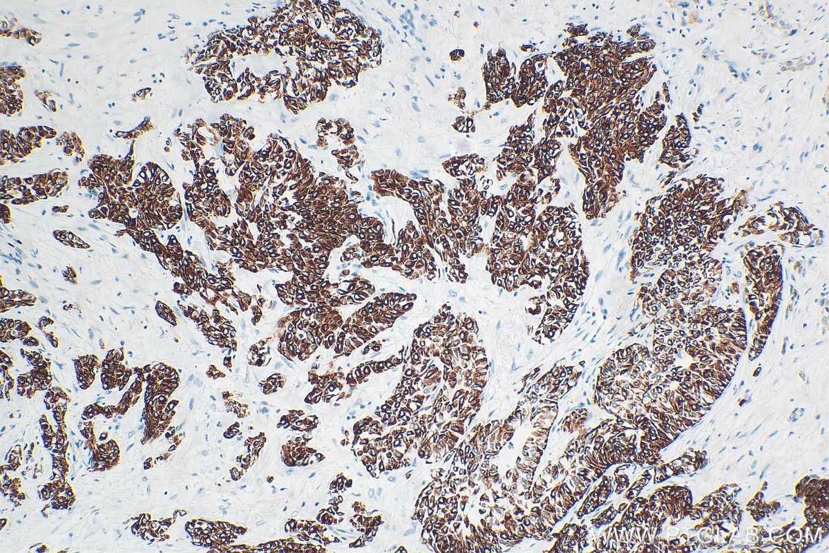
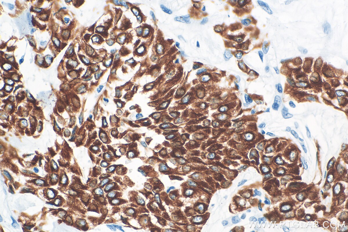
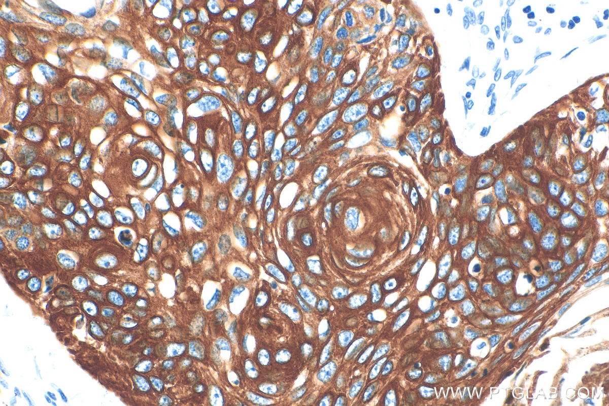
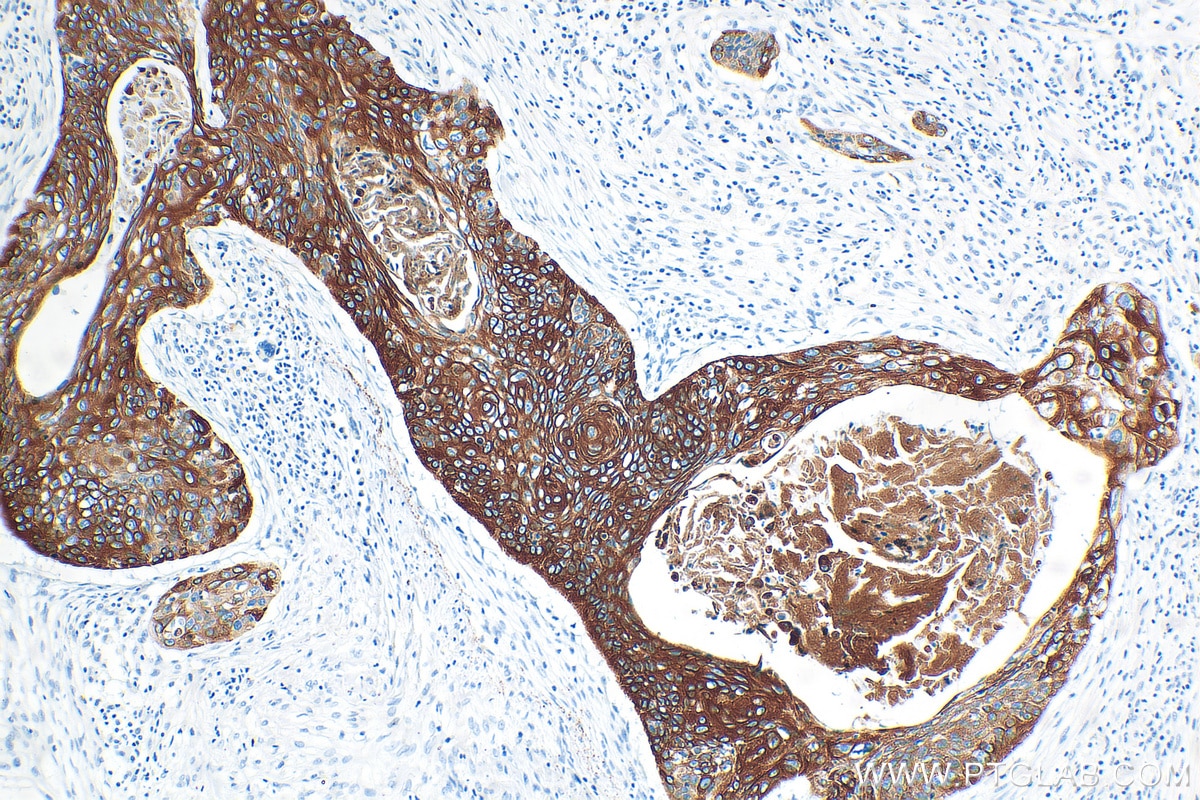
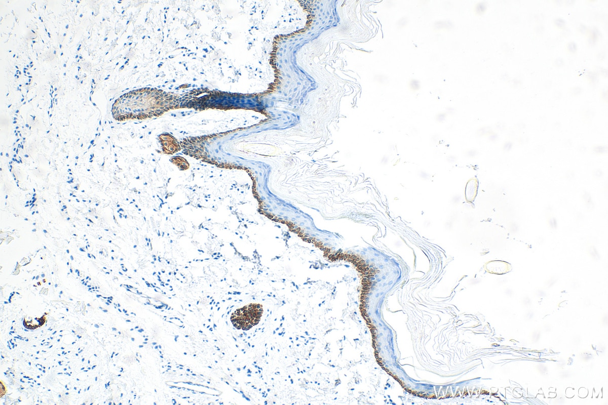
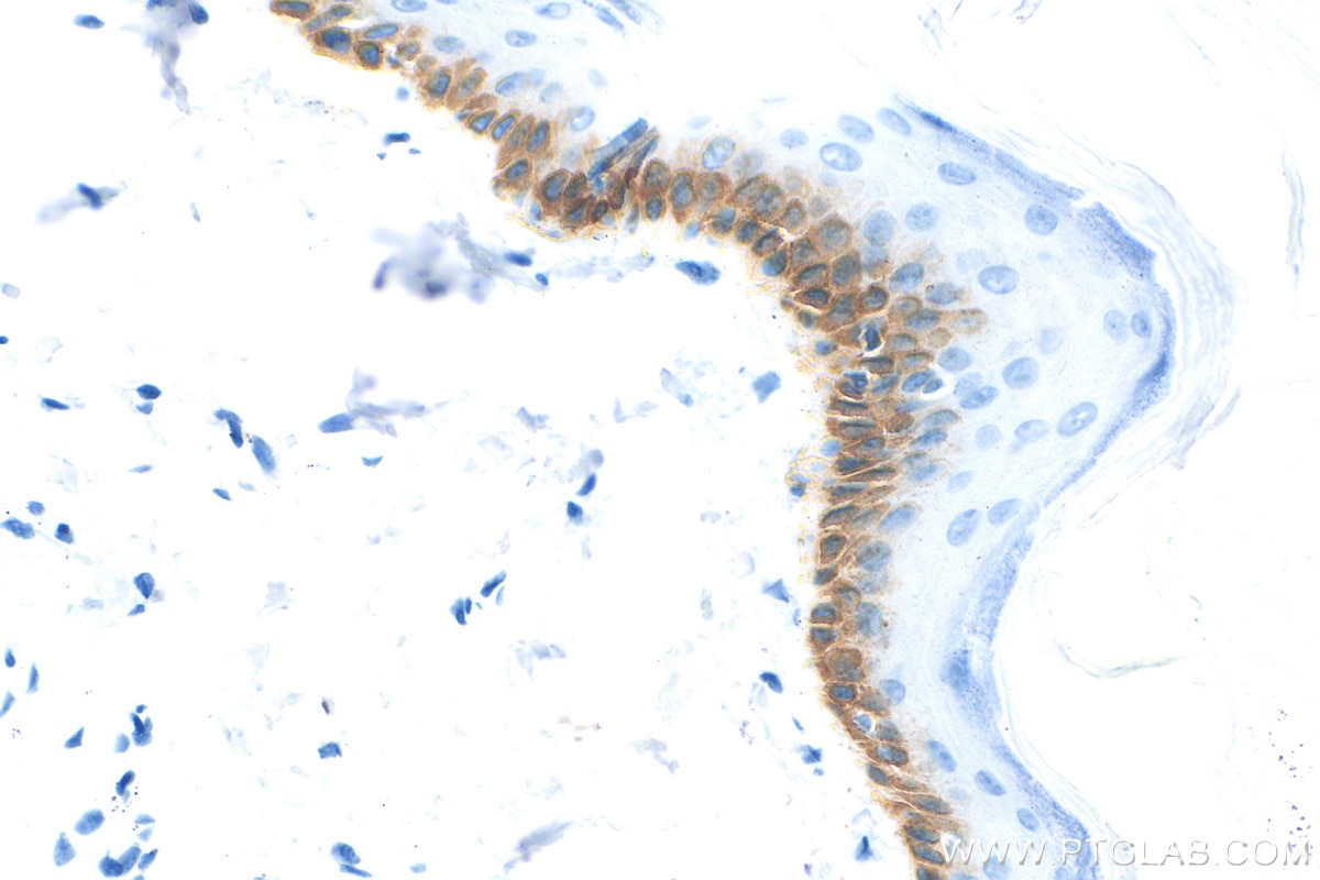
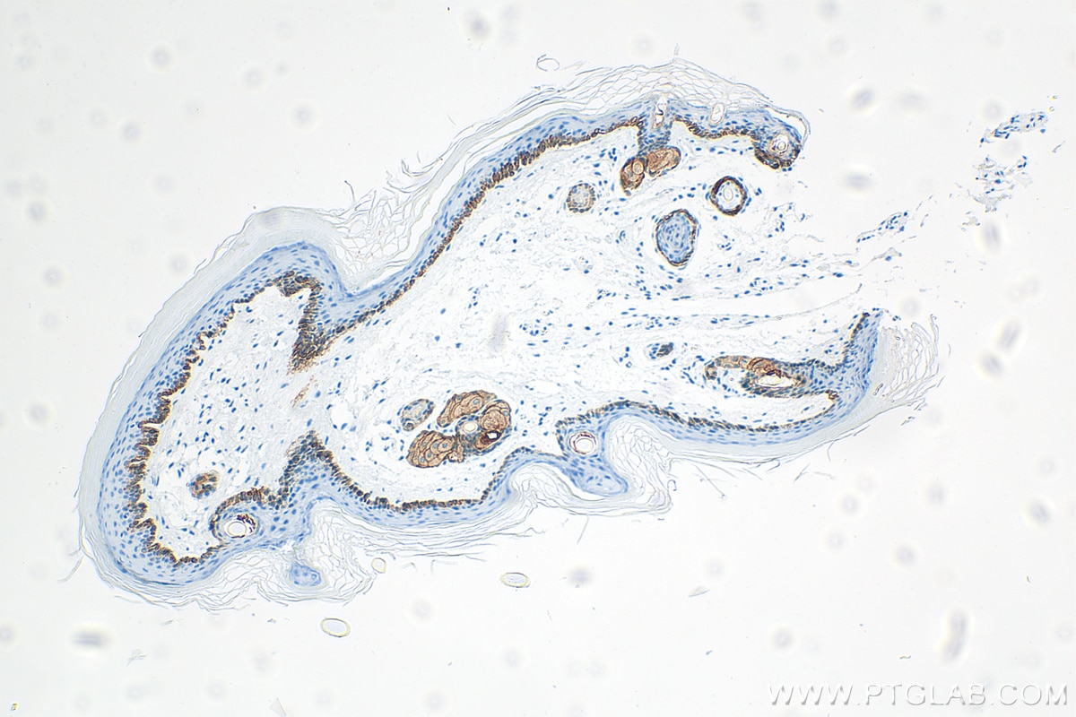
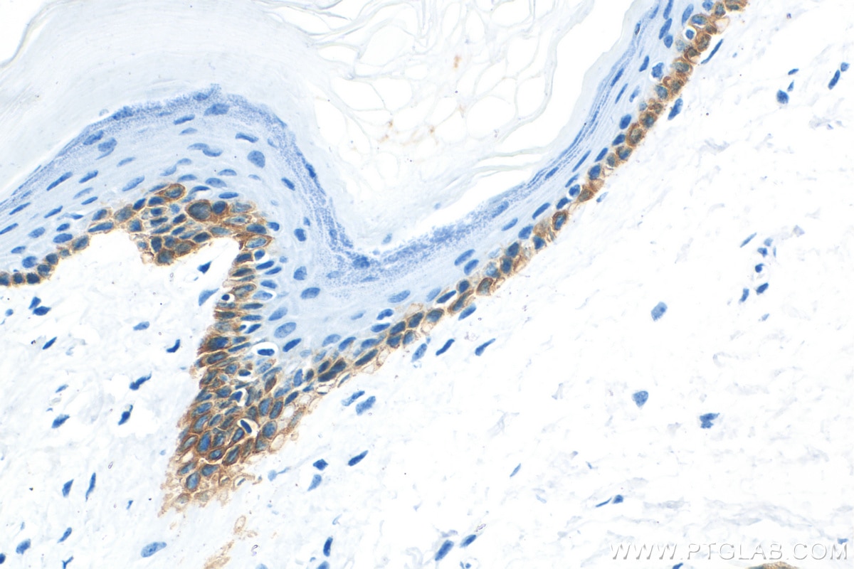
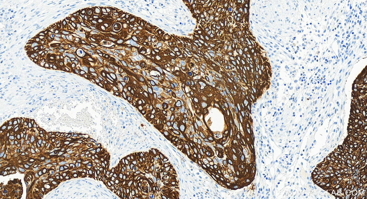
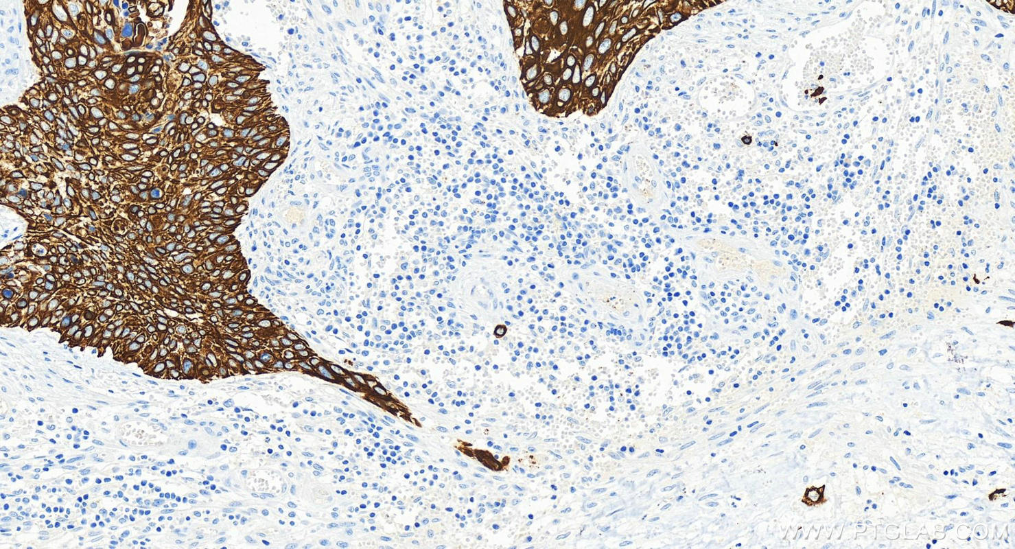
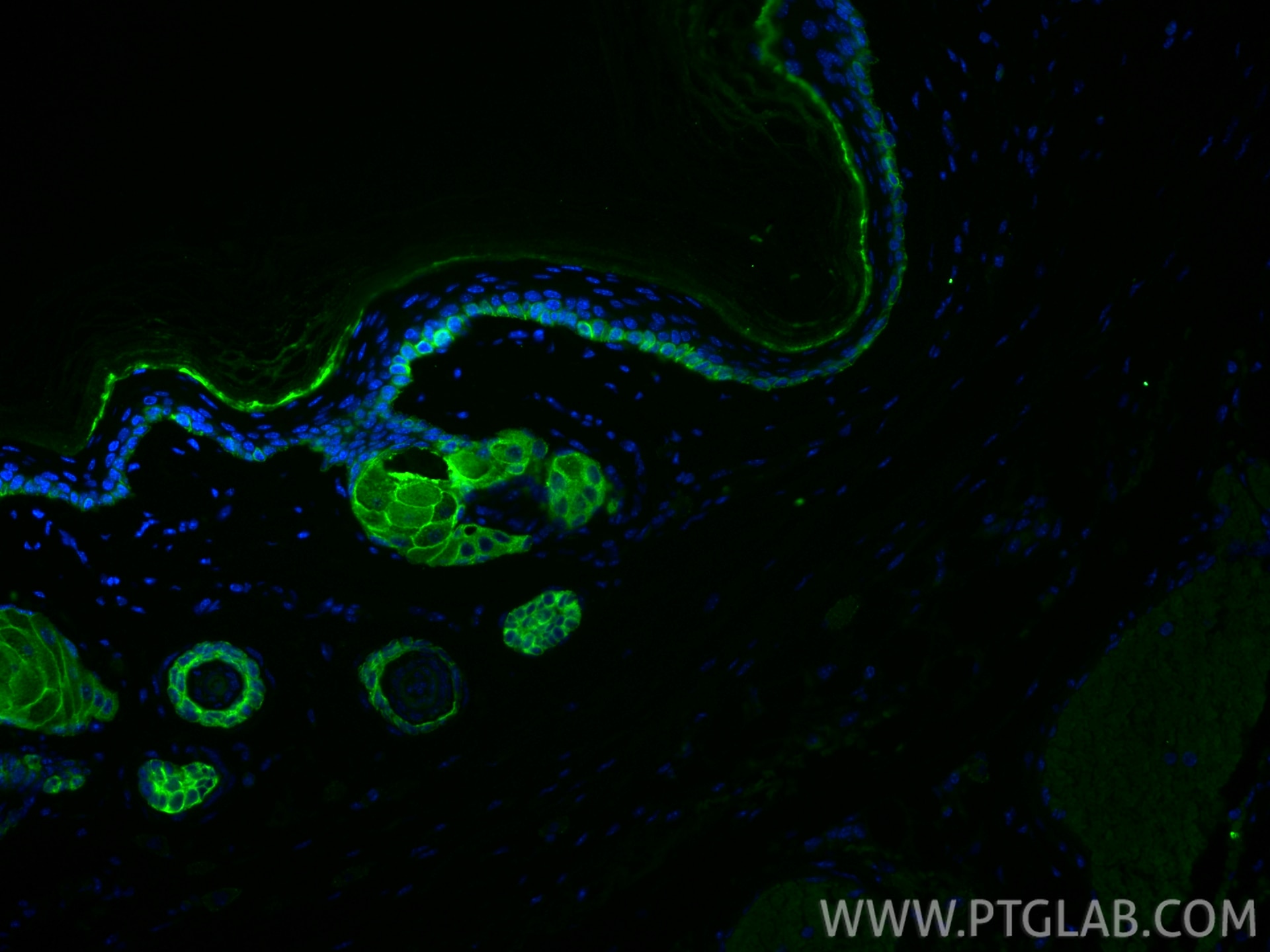
| Positive IHC detected in | human urothelial carcinoma tissue, human cervical squamous cancer tissue, human oesophagus cancer tissue, mouse skin tissue, rat skin tissue Note: suggested antigen retrieval with TE buffer pH 9.0; (*) Alternatively, antigen retrieval may be performed with citrate buffer pH 6.0 |
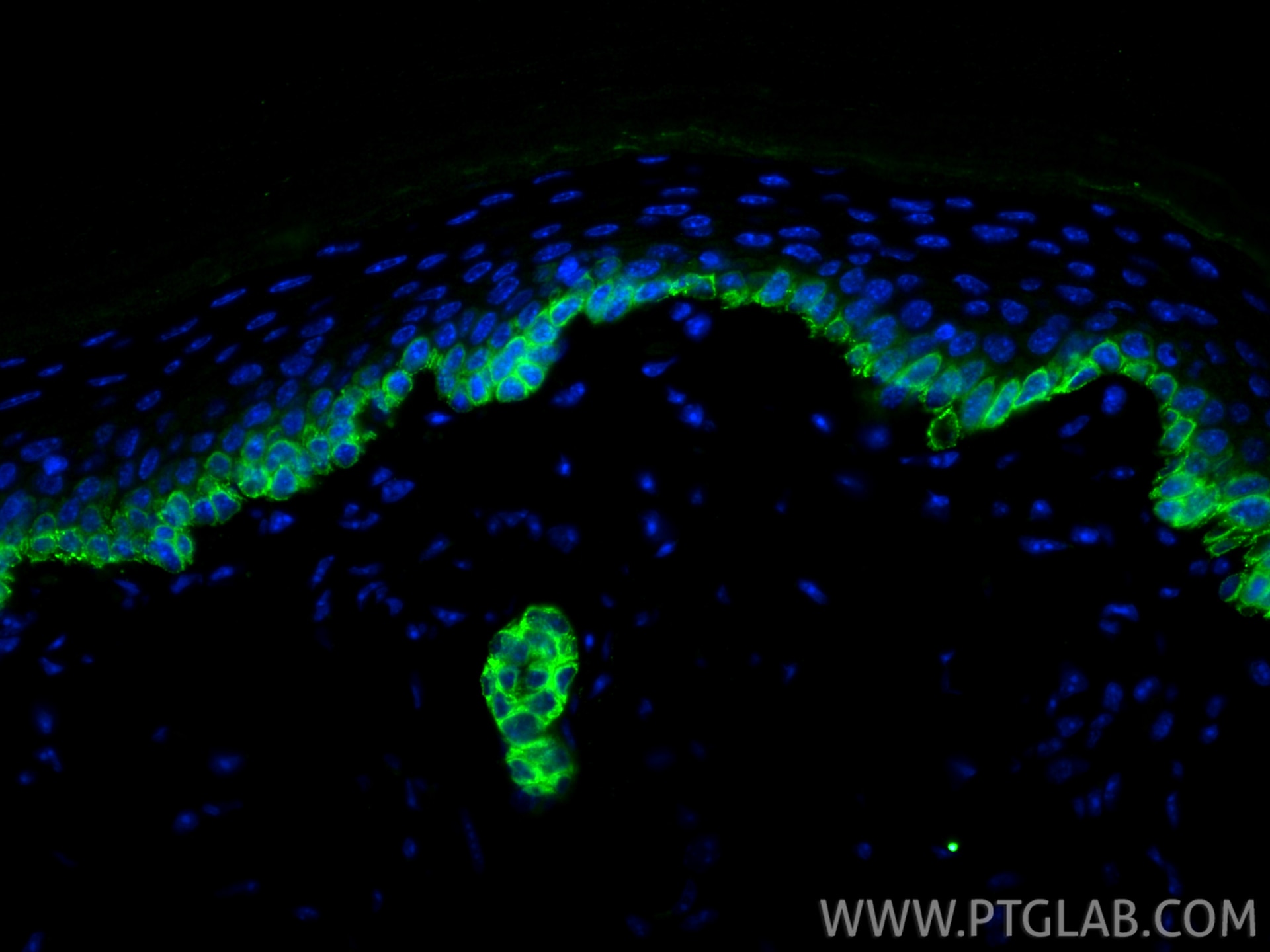
| Positive IF-P detected in | mouse skin tissue |
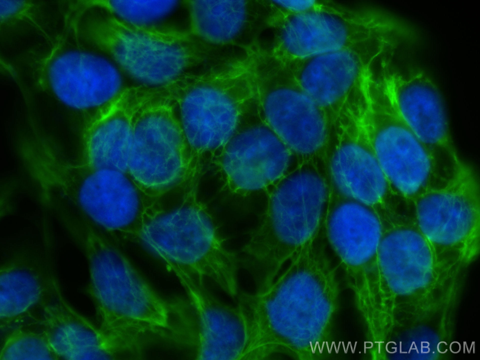
| Positive IF/ICC detected in | HaCaT cells |
Recommended dilution
| Application | Dilution |
|---|---|
| Western Blot (WB) | WB : 1:2000-1:16000 |
| Immunohistochemistry (IHC) | IHC : 1:500-1:2000 |
| Immunofluorescence (IF)-P | IF-P : 1:50-1:500 |
| Immunofluorescence (IF)/ICC | IF/ICC : 1:200-1:800 |
| It is recommended that this reagent should be titrated in each testing system to obtain optimal results. | |
| Sample-dependent, Check data in validation data gallery. | |
Product Information
83058-1-RR targets Cytokeratin 13 in WB, IHC, IF/ICC, IF-P, ELISA applications and shows reactivity with human, mouse, rat samples.
| Tested Reactivity | human, mouse, rat |
| Host / Isotype | Rabbit / IgG |
| Class | Recombinant |
| Type | Antibody |
| Immunogen |
CatNo: Ag0217 Product name: Recombinant human KRT13 protein Source: e coli.-derived, PGEX-4T Tag: GST Domain: 183-458 aa of BC002661 Sequence: ARLAVDDFRLKYENELALRQSVEADINGLRRVLDELTLSKTDLEMQIESLNEELAYMKKNHEEEMKEFSNQVVGQVNVEMDATPGIDLTRVLAEMREQYEAMAERNRRDAEEWFHAKSAELNKEVSTNTAMIQTSKTEITELRRTLQGLEIELQSQLSMKAGLENTVAETECRYALQLQQIQGLISSIEAQLSELRSEMECQNQEYKMLLDIKTRLEQEIATYRSLLEGQDAKMIGFPSSAGSVSPRSTSVTTTSSASVTTTSNASGRRTSDVRRP Predict reactive species |
| Full Name | keratin 13 |
| Calculated Molecular Weight | 50 kDa |
| Observed Molecular Weight | 50 kDa |
| GenBank Accession Number | BC002661 |
| Gene Symbol | Cytokeratin 13 |
| Gene ID (NCBI) | 3860 |
| RRID | AB_3670785 |
| Conjugate | Unconjugated |
| Form | Liquid |
| Purification Method | Protein A purification |
| UNIPROT ID | P13646 |
| Storage Buffer | PBS with 0.02% sodium azide and 50% glycerol, pH 7.3. |
| Storage Conditions | Store at -20°C. Stable for one year after shipment. Aliquoting is unnecessary for -20oC storage. 20ul sizes contain 0.1% BSA. |
Background Information
Keratin 13 is a member of the keratin family. The keratins are intermediate filament proteins responsible for the structural integrity of epithelial cells and are subdivided into cytokeratins and hair keratins. Most of the type I cytokeratins consist of acidic proteins which are arranged in pairs of heterotypic keratin chains. This type I cytokeratin is paired with keratin 4 and expressed in the suprabasal layers of non-cornified stratified epithelia. Mutations in keratin 13 gene and keratin 4 have been associated with the autosomal dominant disorder White Sponge Nevus. The type I cytokeratins are clustered in a region of chromosome 17q21.2.
Protocols
| Product Specific Protocols | |
|---|---|
| IF protocol for Cytokeratin 13 antibody 83058-1-RR | Download protocol |
| IHC protocol for Cytokeratin 13 antibody 83058-1-RR | Download protocol |
| WB protocol for Cytokeratin 13 antibody 83058-1-RR | Download protocol |
| Standard Protocols | |
|---|---|
| Click here to view our Standard Protocols |