Tested Applications
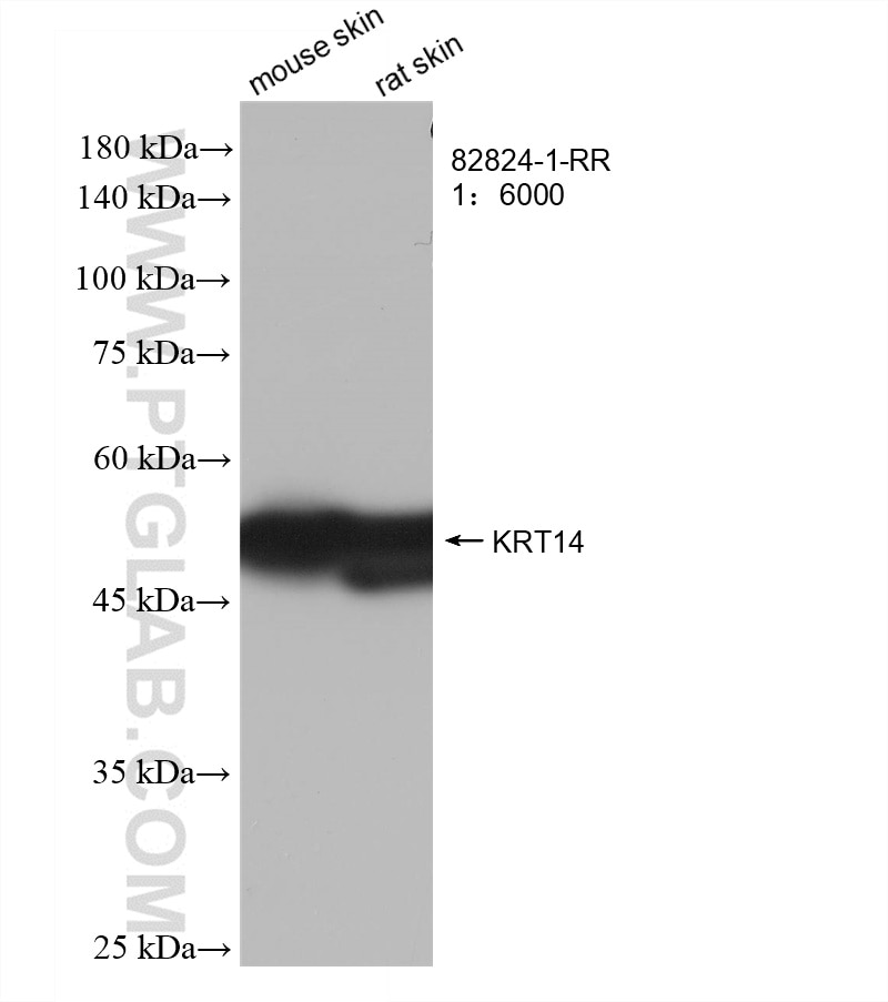
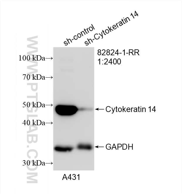
| Positive WB detected in | mouse skin tissue, A431 cells, rat skin tissue |
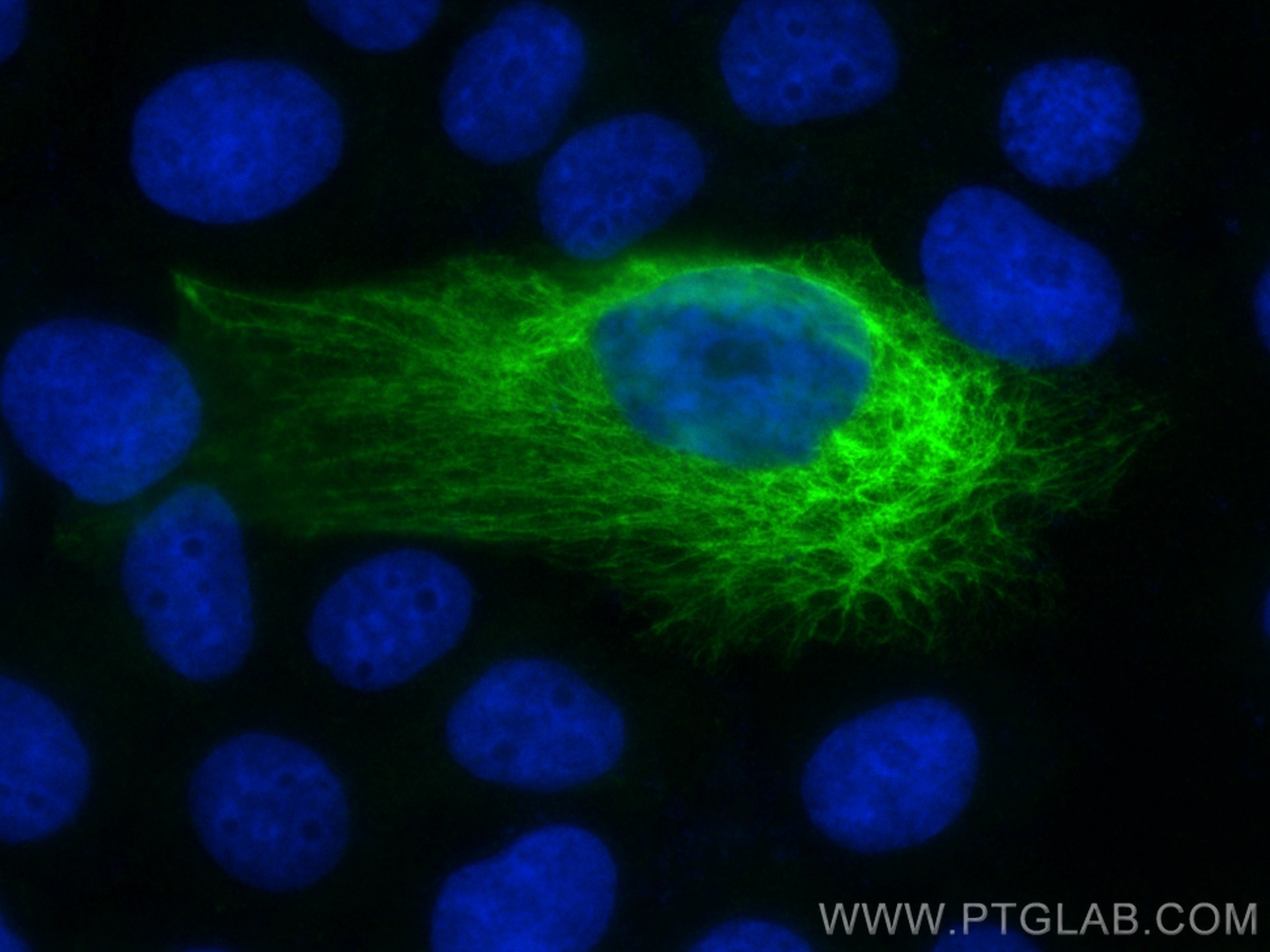
| Positive IF/ICC detected in | A431 cells |
Recommended dilution
| Application | Dilution |
|---|---|
| Western Blot (WB) | WB : 1:2000-1:12000 |
| Immunofluorescence (IF)/ICC | IF/ICC : 1:50-1:500 |
| It is recommended that this reagent should be titrated in each testing system to obtain optimal results. | |
| Sample-dependent, Check data in validation data gallery. | |
Product Information
82824-1-RR targets Cytokeratin 14 in WB, IF/ICC, ELISA applications and shows reactivity with human, mouse, rat samples.
| Tested Reactivity | human, mouse, rat |
| Host / Isotype | Rabbit / IgG |
| Class | Recombinant |
| Type | Antibody |
| Immunogen |
CatNo: Ag17559 Product name: Recombinant human KRT14 protein Source: e coli.-derived, PGEX-4T Tag: GST Domain: 426-472 aa of BC002690 Sequence: LSSSQFSSGSQSSRDVTSSSRQIRTKVMDVHDGKVVSTHEQVLRTKN Predict reactive species |
| Full Name | keratin 14 |
| Calculated Molecular Weight | 472 aa, 52 kDa |
| Observed Molecular Weight | 52 kDa |
| GenBank Accession Number | BC002690 |
| Gene Symbol | Cytokeratin 14 |
| Gene ID (NCBI) | 3861 |
| RRID | AB_3086548 |
| Conjugate | Unconjugated |
| Form | Liquid |
| Purification Method | Protein A purification |
| UNIPROT ID | P02533 |
| Storage Buffer | PBS with 0.02% sodium azide and 50% glycerol, pH 7.3. |
| Storage Conditions | Store at -20°C. Stable for one year after shipment. Aliquoting is unnecessary for -20oC storage. 20ul sizes contain 0.1% BSA. |
Background Information
Cytokeratin 14, one of about 20 different cytokeratin isotypes of human cells, is the intermediate filament protein characteristic of epithelial cells. Cytokeratin 14 is expressed in the basal compartment of all stratified squamous epithelia. In various kinds of human tumors, the appearance and increasing expression of Cytokeratin 14 were strikingly associated with higher grade and stage of carcinoma, with varying degrees of unfavorable prognosis. In lung squamous cell carcinoma(LSCC), Cytokeratin 14 was expressed in the tumor cell nests showing stromal invasion with fibrosis and lymph node metastases, indicating that Cytokeratin 14 involved in proliferation and metastasis of LSCC. Cytokeratin 14 expression is sometimes used in diagnosis of myoepithelioma1 and intraductal vs. invasive ductal carcinoma of the breast.
Protocols
| Product Specific Protocols | |
|---|---|
| IF protocol for Cytokeratin 14 antibody 82824-1-RR | Download protocol |
| WB protocol for Cytokeratin 14 antibody 82824-1-RR | Download protocol |
| Standard Protocols | |
|---|---|
| Click here to view our Standard Protocols |