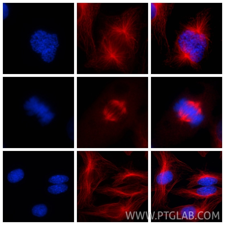
fixed SKOV-3 cells using Alpha Tubulin antibody (66031-1-Ig, Clone: 1E4C11 ) at dilution of 1:1000 and CoraLite®594-Conjugated AffiniPure Goat Anti-Mouse IgG(H+L), Nucleus was labelled in blue with DAPI.")
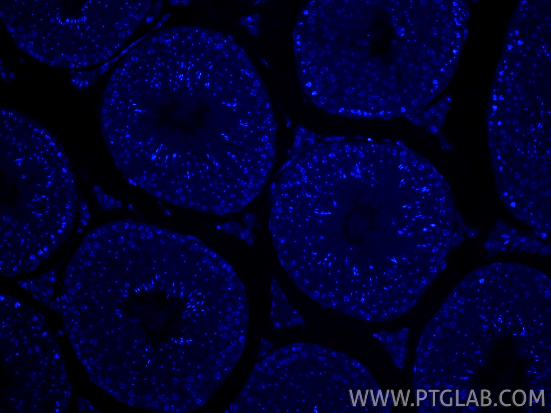
 fixed paraffin-embedded mouse testis tissue using DAPI. Heat mediated antigen retrieval with Tris-EDTA buffer (pH 9.0).")
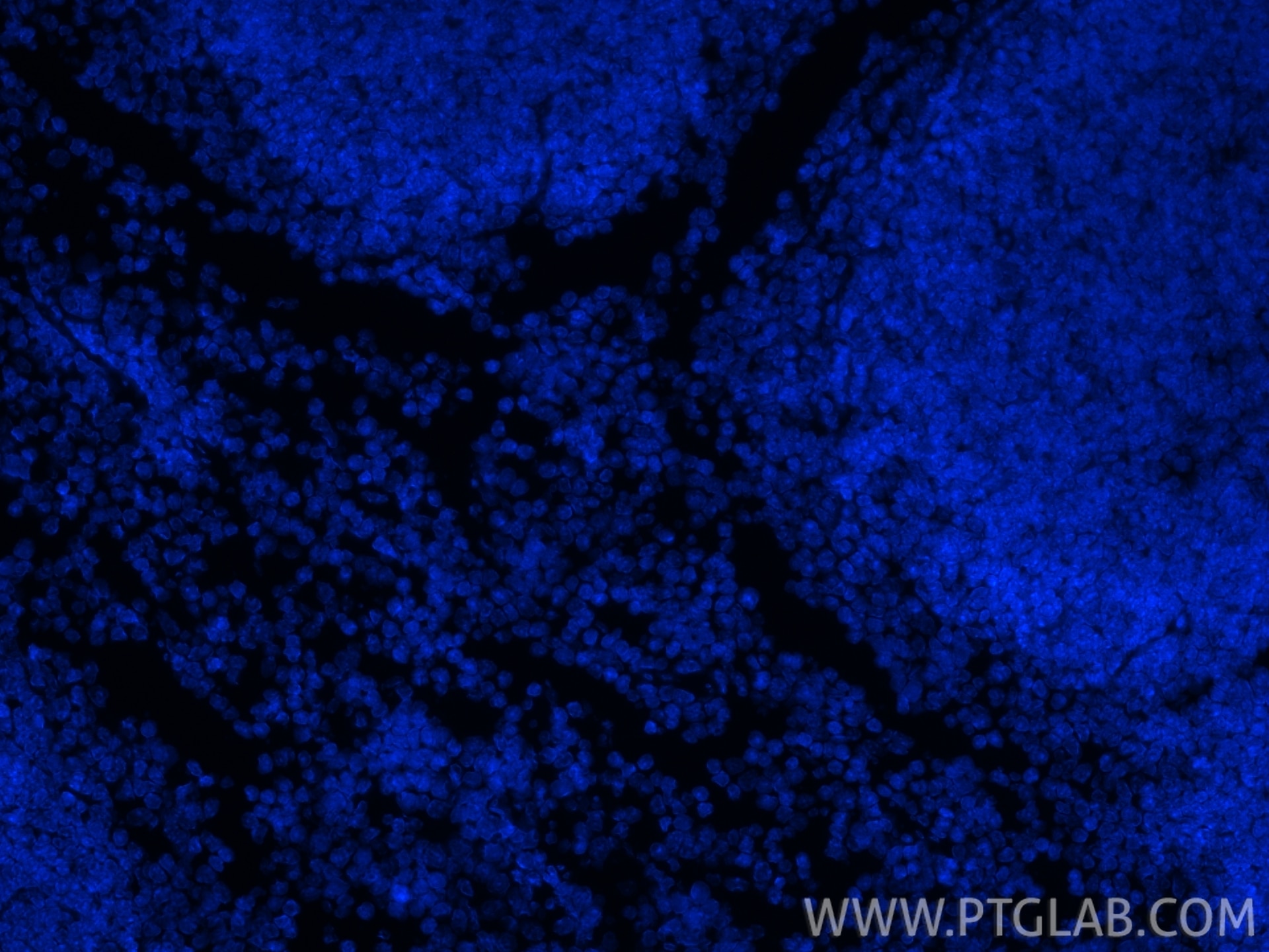
 fixed paraffin-embedded mouse spleen tissue using DAPI. Heat mediated antigen retrieval with Tris-EDTA buffer (pH 9.0).")
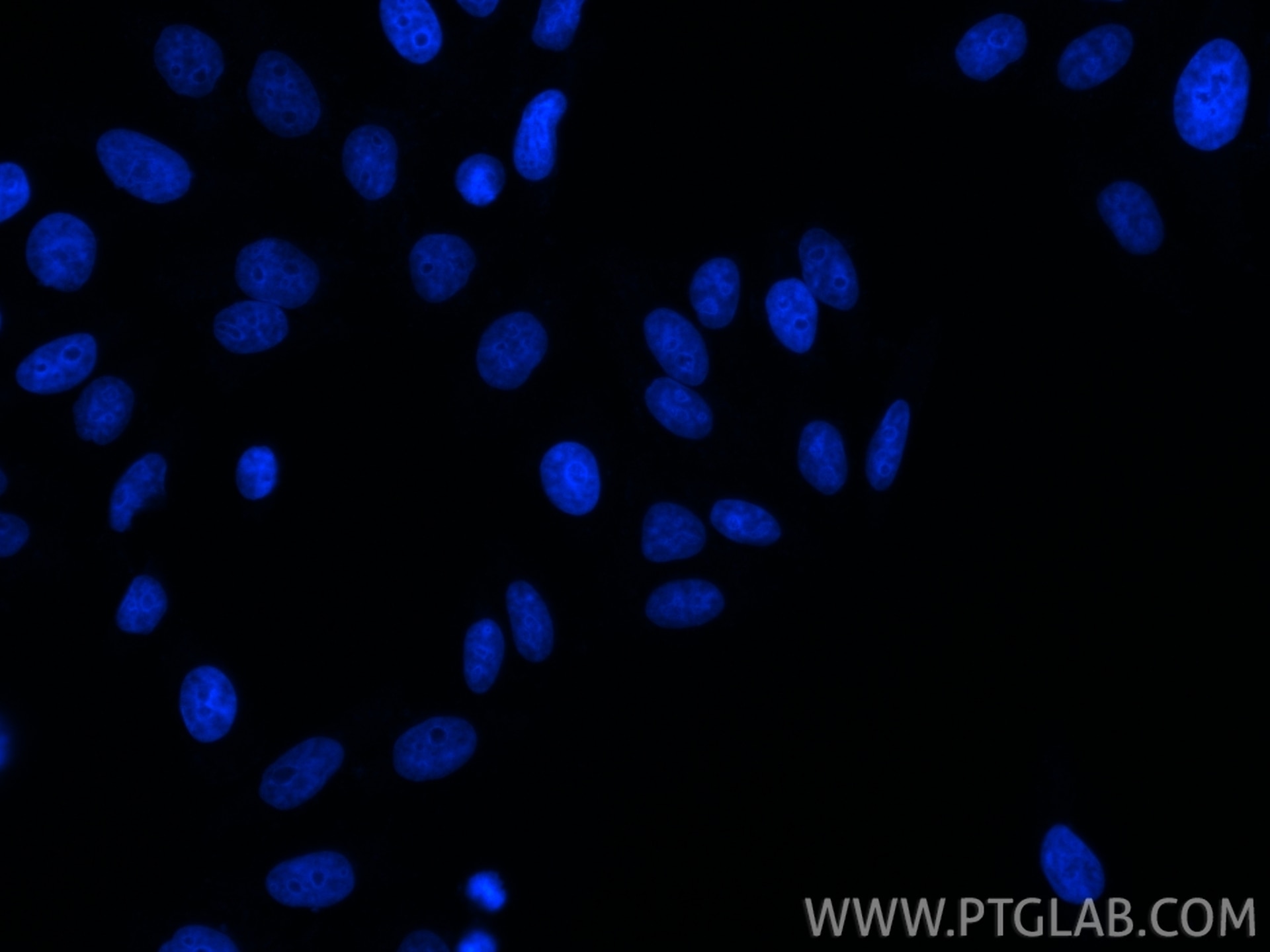
 fixed HepG2 cells using DAPI.")
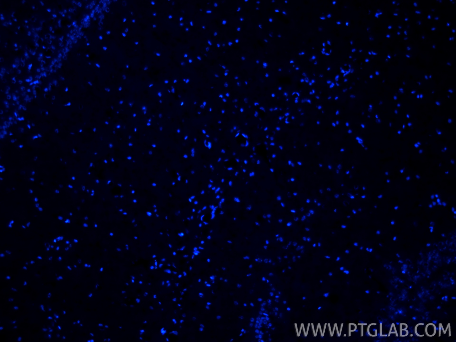
 fixed frozen OCT-embedded mouse brain tissue using DAPI (PR30021).")
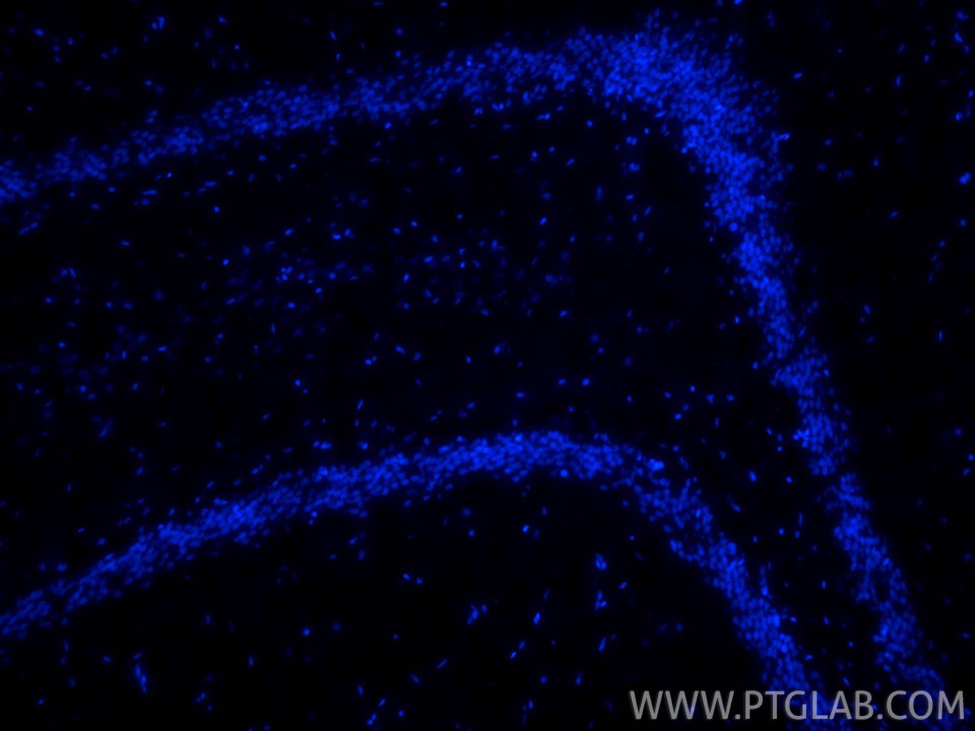
 fixed frozen OCT-embedded rat brain tissue using DAPI (PR30021).")
. Cells were fixed with 4% PFA.")
Product Information
DAPI, also known as DAPI dihydrochloride (4',6-diamidino-2-phenylindole), is a commonly used blue fluorescent nucleic acid stain. It binds to the minor groove of AT-rich base pair sequences in double-stranded DNA, and its blue fluorescence is enhanced more than 20-fold upon binding to DNA. It is frequently used for nuclear staining of fixed cells and tissue sections, which can be observed under a fluorescence microscope after staining.
When DAPI binds to DNA, its maximum excitation wavelength is 358 nm, and its maximum emission wavelength is 461 nm. Although DAPI is not membrane-permeable to live cells under normal conditions, it can enter live cells at elevated concentrations. Upon excitation, DAPI emits blue fluorescence, allowing it to be combined with other fluorochromes such as green, orange, and red ones to achieve multiplex fluorescent labeling.
This product is a ready-to-use DAPI aqueous solution, which can be directly applied to nuclear immunofluorescence staining of fixed cells or tissue sections, as well as flow cytometry.
Product Name | DAPI Staining Solution (Ready-to-use) |
Alternative Name | 4',6-diamidino-2-phenylindole |
Molecular Formula | C16H15N5·2HCl |
Molecular Weight | 350.25 |
CAS No. | 28718-90-3 |
Storage
Store at 2–8°C away from light. Valid for one year from the date of receipt.
Usage
Immunofluorescence Staining:
1. Staining: If other fluorescent staining is to be performed, immunofluorescence staining may be conducted first, followed by DAPI staining as the final step. If no other fluorescent staining is required, DAPI staining can be performed directly.
2. Add an appropriate volume of DAPI working solution to fixed adherent cells or tissue sections to fully immerse the samples, and incubate at room temperature for 8-10 minutes. For fixed suspension cells, add at least 3 times the volume of the staining solution relative to the sample volume, and mix thoroughly.
3. Stain at room temperature for 8-10 minutes.
4. Aspirate the DAPI staining solution, and wash with PBS 2-3 times, for 3-5 minutes each time.
5. Observe under a fluorescence microscope at Ex/Em = 358 nm/461 nm.
Flow Cytometry:
1. Collect the cultured cells into EP tubes, centrifuge at 300-400 × g for 5 minutes at room temperature, and discard the supernatant. Add 1 mL of PBS and pipette gently to ensure complete cell resuspension, and repeat this process three times.
2. Add 0.5 mL of 4% PFA or absolute ethanol to each EP tube containing 1 × 10⁶ cells, fix at room temperature for 15 minutes, centrifuge at 400–600 × g for 5 minutes at room temperature, and discard the supernatant.
3. Add 1 mL of PBS to the EP tube, pipette gently to ensure complete cell resuspension, centrifuge at 400–600 × g for 5 minutes, and discard the supernatant.
4. Repeat Step 3 three times.
5. Add 0.2 mL of DAPI working solution to each EP tube containing 1 × 10⁶ cells, and pipette gently to ensure complete cell resuspension. Incubate at room temperature for 10-15 minutes, protected from light.
6. Analyze the samples on a flow cytometer.
Notes
Cited in Article as
PR30021, DAPI Staining Solution (Ready-to-use), Proteintech, IL, USA
Documentation
| SDS |
|---|
| DAPI Staining Solution (Ready-to-use) SDS |
| Datasheet |
|---|
| DAPI Staining Solution (Ready-to-use) Datasheet |
Publications
| Application | Title |
|---|---|
J Transl Med Lipidomics reveals biomarkers of the efficacy of first-line ICI therapy combined with chemotherapy in NSCLC | |
J Ethnopharmacol Demethylzeylasteral protects against renal interstitial fibrosis by attenuating mitochondrial complex I-mediated oxidative stress | |
Front Biosci (Landmark Ed) Co-Highly Expressed SLC17A9 and KCNH1 as Potential Prognostic Biomarkers and Therapeutic Targets in Clear Cell Renal Cell Carcinoma | |
Front Bioeng Biotechnol An injectable curcumin-loaded hydrogel for neuroprotective treatment promote nerve tissue repair in rat severe spinal cord injury | |
Int Immunopharmacol H₂O₂ preconditioning enhances the transplantation-mediated therapeutic effect of bone marrow-derived mesenchymal stem cells after traumatic brain injury | |
Adv Healthc Mater Multifunctional Sr2+/Zn2+ Co-Doped Mesoporous Silica Nanoparticles in Injectable Hydrogel for Ameliorating Osteoporotic Osseointegration |