Tested Applications
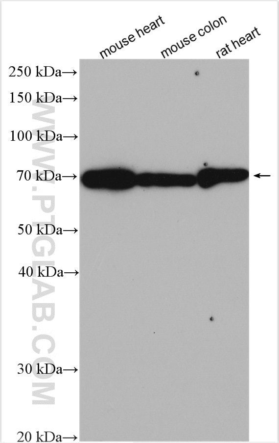
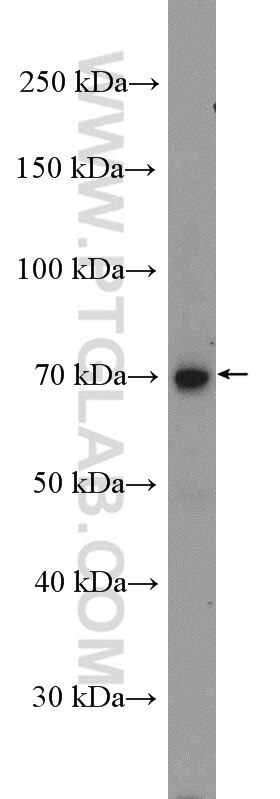
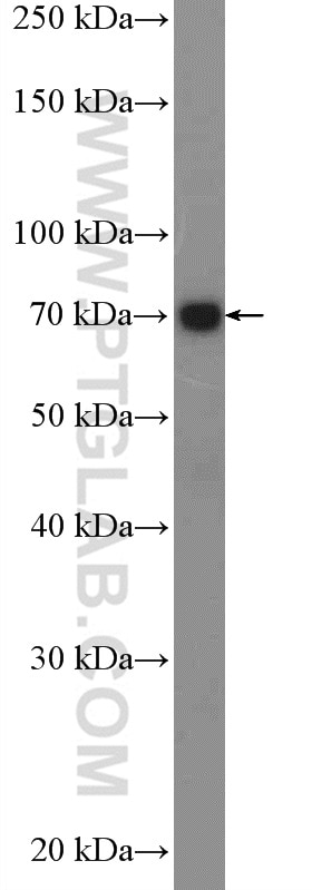
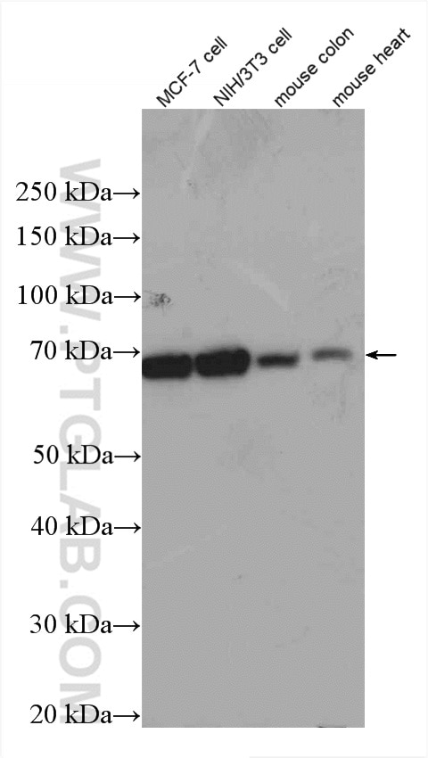
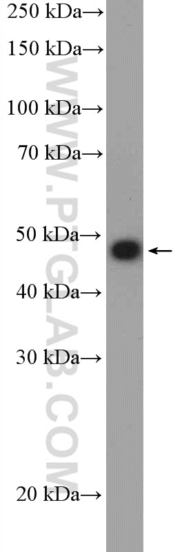
| Positive WB detected in | mouse heart tissue, MCF-7 cells, BxPC-3 cells, mouse colon tissue, rat heart tissue, NIH/3T3 cells |
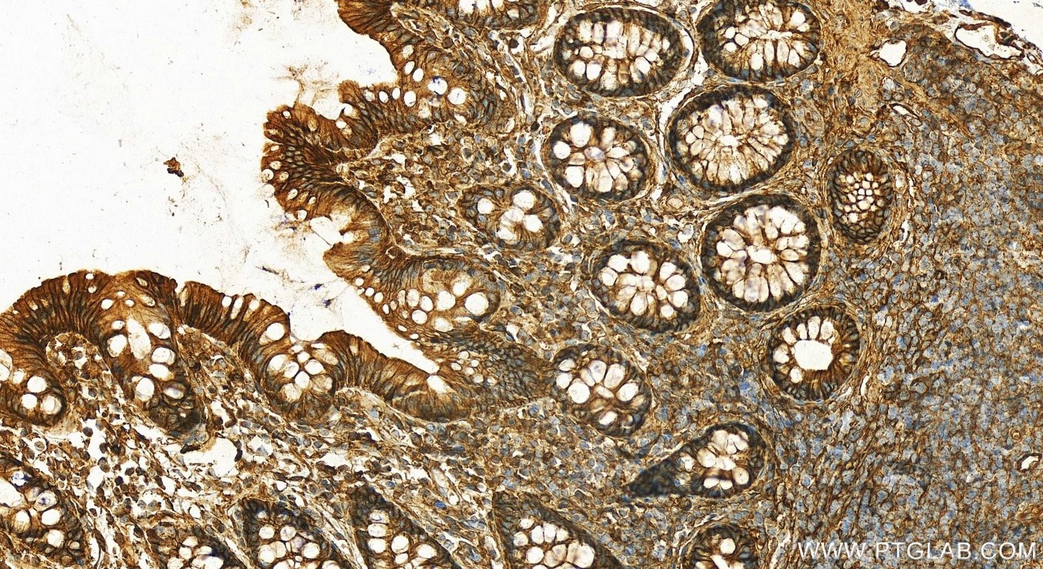
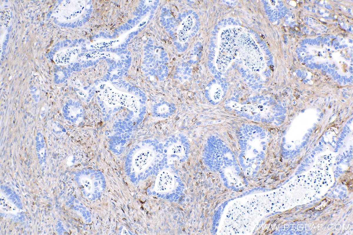
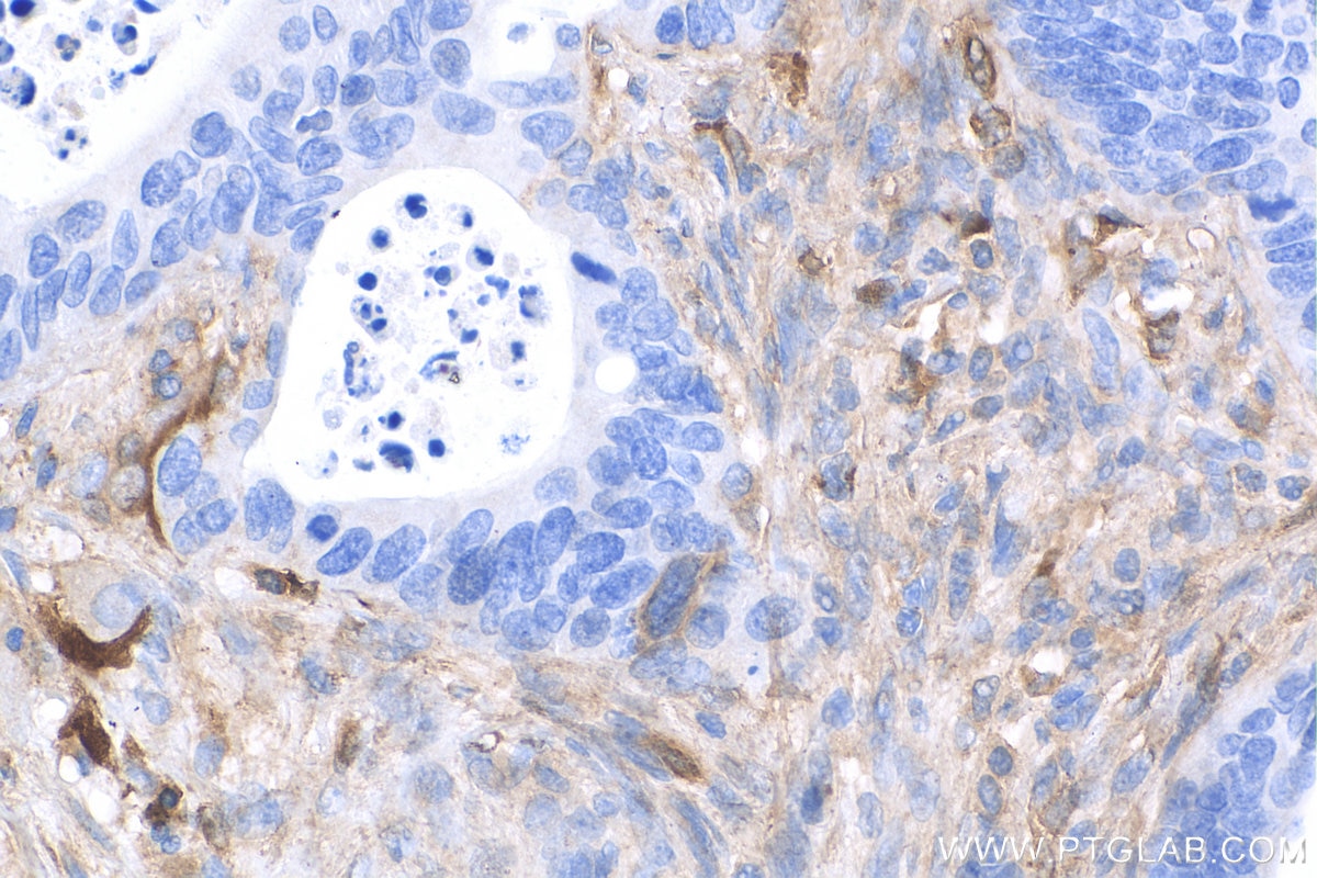
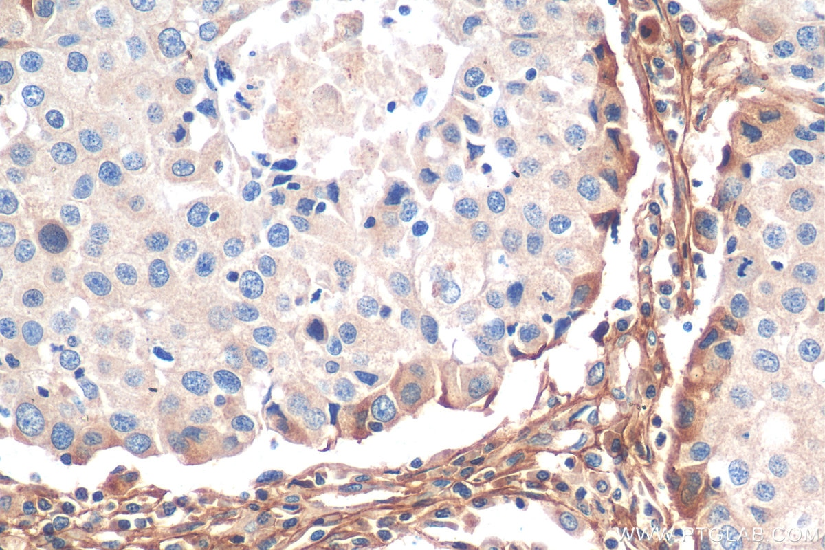
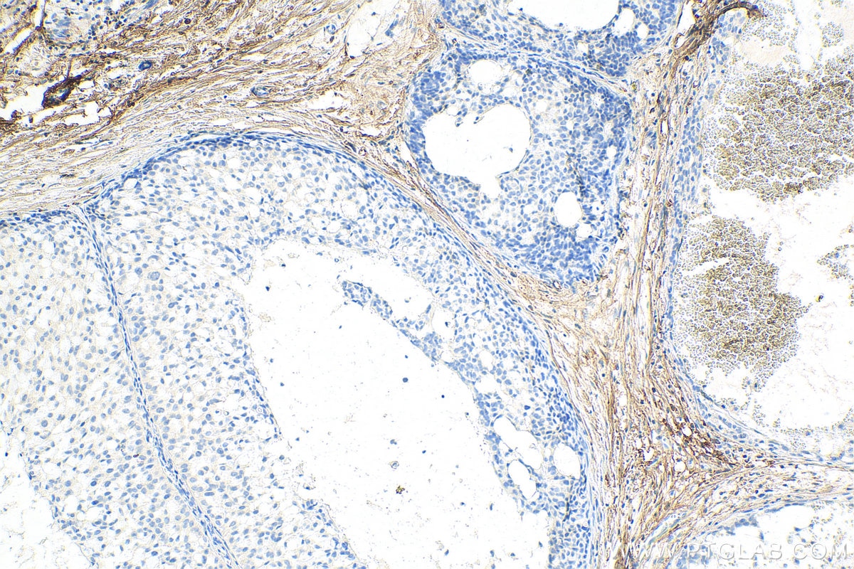
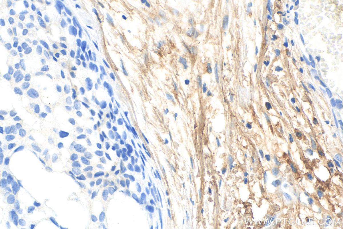
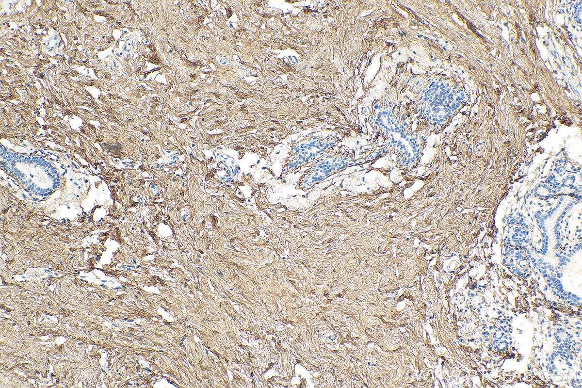
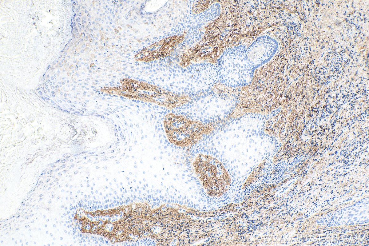
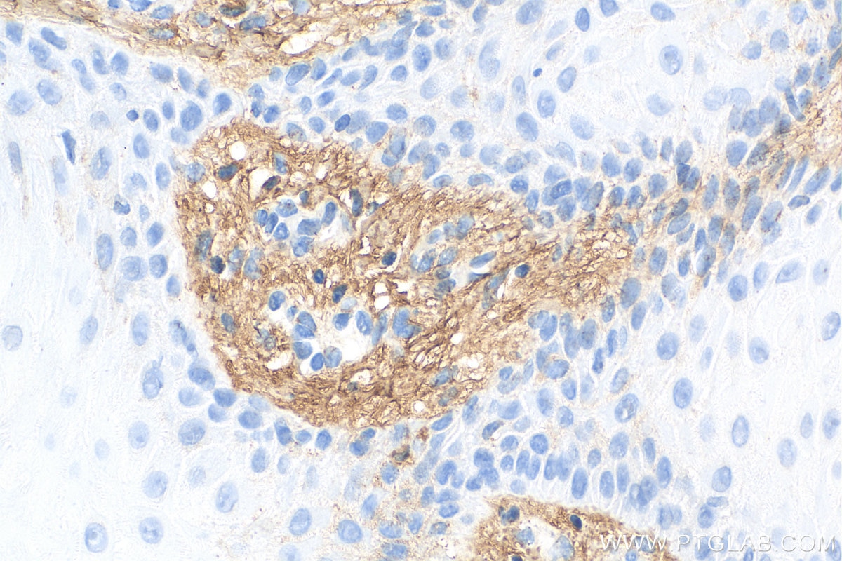
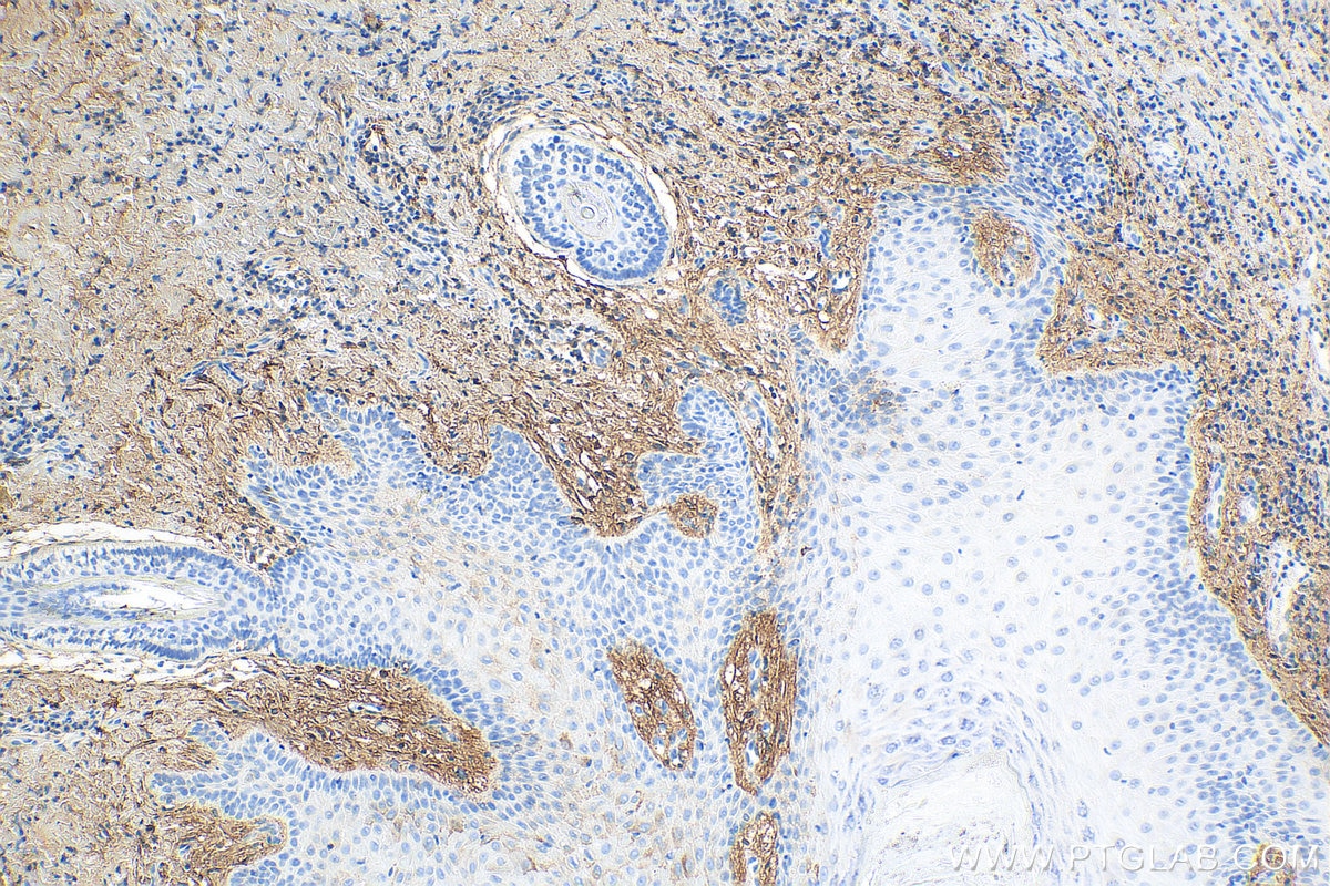
| Positive IHC detected in | human skin cancer tissue, human breast cancer tissue, human breast hyperplasia tissue, human colon cancer tissue Note: suggested antigen retrieval with TE buffer pH 9.0; (*) Alternatively, antigen retrieval may be performed with citrate buffer pH 6.0 |
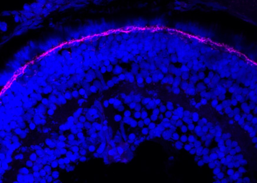
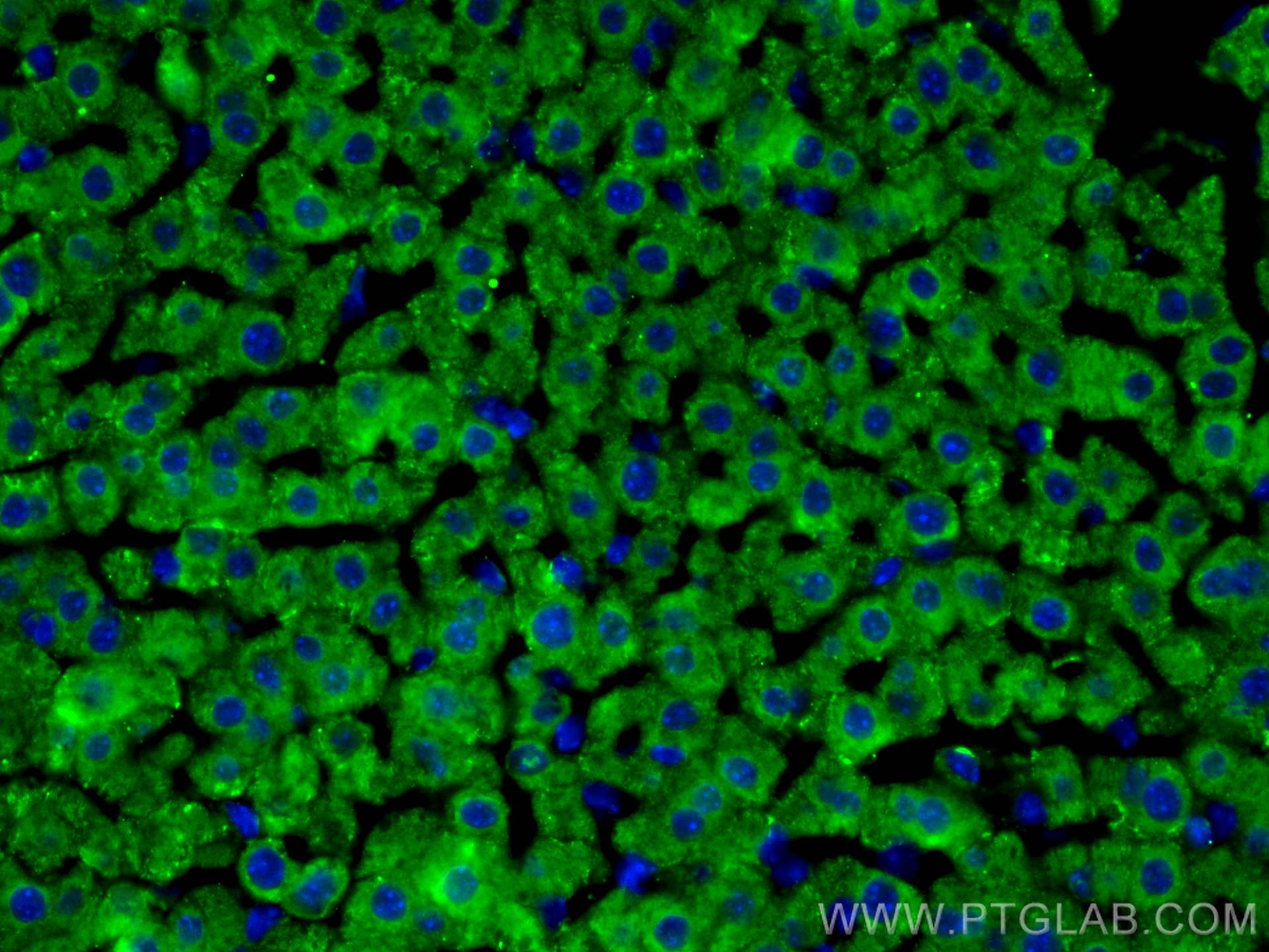
| Positive IF-P detected in | zebrafish retina, mouse liver tissue |
| Positive IF/ICC detected in | BxPC-3 cells |
Recommended dilution
| Application | Dilution |
|---|---|
| Western Blot (WB) | WB : 1:1000-1:4000 |
| Immunohistochemistry (IHC) | IHC : 1:1000-1:4000 |
| Immunofluorescence (IF)-P | IF-P : 1:50-1:500 |
| Immunofluorescence (IF)/ICC | IF/ICC : 1:50-1:500 |
| It is recommended that this reagent should be titrated in each testing system to obtain optimal results. | |
| Sample-dependent, Check data in validation data gallery. | |
Published Applications
| KD/KO | See 3 publications below |
| WB | See 27 publications below |
| IHC | See 7 publications below |
| IF | See 17 publications below |
Product Information
14667-1-AP targets Decorin in WB, IHC, IF/ICC, IF-P, ELISA applications and shows reactivity with human, mouse, rat, zabrafish samples.
| Tested Reactivity | human, mouse, rat, zabrafish |
| Cited Reactivity | human, mouse, rat, hamster |
| Host / Isotype | Rabbit / IgG |
| Class | Polyclonal |
| Type | Antibody |
| Immunogen |
CatNo: Ag6328 Product name: Recombinant human DCN protein Source: e coli.-derived, PGEX-4T Tag: GST Domain: 1-359 aa of BC005322 Sequence: MKATIILLLLAQVSWAGPFQQRGLFDFMLEDEASGIGPEVPDDRDFEPSLGPVCPFRCQCHLRVVQCSDLGLDKVPKDLPPDTTLLDLQNNKITEIKDGDFKNLKNLHALILVNNKISKVSPGAFTPLVKLERLYLSKNQLKELPEKMPKTLQELRAHENEITKVRKVTFNGLNQMIVIELGTNPLKSSGIENGAFQGMKKLSYIRIADTNITSIPQGLPPSLTELHLDGNKISRVDAASLKGLNNLAKLGLSFNSISAVDNGSLANTPHLRELHLDNNKLTRVPGGLAEHKYIQVVYLHNNNISVVGSSDFCPPGHNTKKASYSGVSLFSNPVQYWEIQPSTFRCVYVRSAIQLGNYK Predict reactive species |
| Full Name | decorin |
| Calculated Molecular Weight | 359 aa, 40 kDa |
| Observed Molecular Weight | 70 kDa, 45-48 kDa |
| GenBank Accession Number | BC005322 |
| Gene Symbol | Decorin |
| Gene ID (NCBI) | 1634 |
| RRID | AB_2090265 |
| Conjugate | Unconjugated |
| Form | Liquid |
| Purification Method | Antigen affinity purification |
| UNIPROT ID | P07585 |
| Storage Buffer | PBS with 0.02% sodium azide and 50% glycerol, pH 7.3. |
| Storage Conditions | Store at -20°C. Stable for one year after shipment. Aliquoting is unnecessary for -20oC storage. 20ul sizes contain 0.1% BSA. |
Background Information
Decorin is a member of the small leucine-rich proteoglycan family of proteins. Decorin interacts with type I collagen fibrils, thereby influencing the kinetics of fibril formation and the distance between adjacent collagen fibrils. The binding of this protein to multiple cell surface receptors mediates its role in tumor suppression, including a stimulatory effect on autophagy and inflammation and an inhibitory effect on angiogenesis and tumorigenesis. Decorin(~ 40 kDa) also binds glycosaminoglycan chains to form big molecules.
Protocols
| Product Specific Protocols | |
|---|---|
| IF protocol for Decorin antibody 14667-1-AP | Download protocol |
| IHC protocol for Decorin antibody 14667-1-AP | Download protocol |
| WB protocol for Decorin antibody 14667-1-AP | Download protocol |
| Standard Protocols | |
|---|---|
| Click here to view our Standard Protocols |
Publications
| Species | Application | Title |
|---|---|---|
Nat Commun The estrogen response in fibroblasts promotes ovarian metastases of gastric cancer | ||
Breast Cancer Res Single-cell RNA reveals a tumorigenic microenvironment in the interface zone of human breast tumors | ||
Front Immunol Development of a prognostic model for hepatocellular carcinoma based on microvascular invasion characteristic genes by spatial transcriptomics sequencing | ||
PLoS Genet Impaired proteoglycan glycosylation, elevated TGF-β signaling, and abnormal osteoblast differentiation as the basis for bone fragility in a mouse model for gerodermia osteodysplastica. | ||
Reviews
The reviews below have been submitted by verified Proteintech customers who received an incentive for providing their feedback.
FH Sai Sindhura (Verified Customer) (01-08-2026) | Decorin is good for IHC
|
FH Kenzo (Verified Customer) (08-29-2023) | Antibody is working pretty well for IF on mouse tissue sections.
|
FH Huai-Chin (Verified Customer) (10-24-2022) | Good antibody with only single band. Actual band size is higher than the calculated protein size which may indicated some protein modification.
|
FH balawant (Verified Customer) (07-25-2022) | I have this antibody for my research it is working great in both tissue lysate as well as in cell lysate.
|
FH Iram (Verified Customer) (06-13-2022) | Excellent antibody for western blotting
|
FH Balawant (Verified Customer) (05-08-2022) | I have used this antibody. This antibody is excellent, and I used it for western blotting. The dilution of 1:1000 is functioning well.
|
FH Susmita (Verified Customer) (09-17-2020) | Antibody is working very good for WB
|