Tested Applications
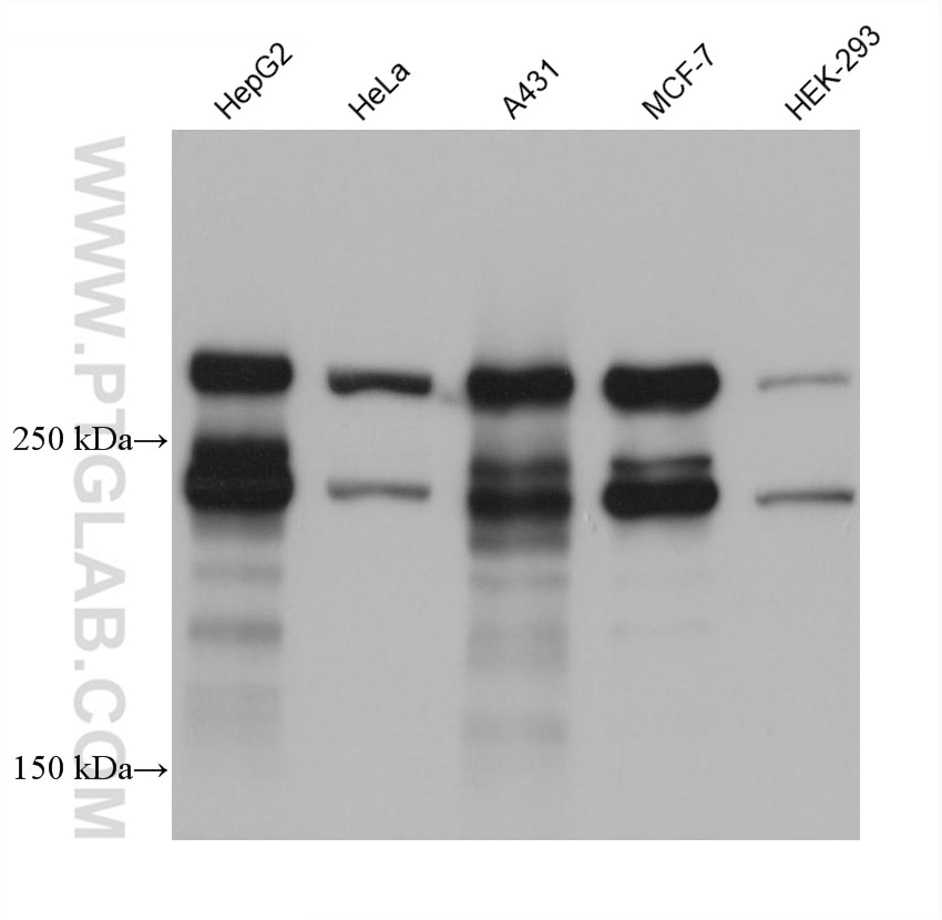
| Positive WB detected in | HepG2 cells, HeLa cells, A431 cells, MCF-7 cells, HEK-293 cells |
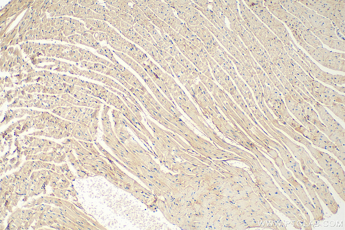
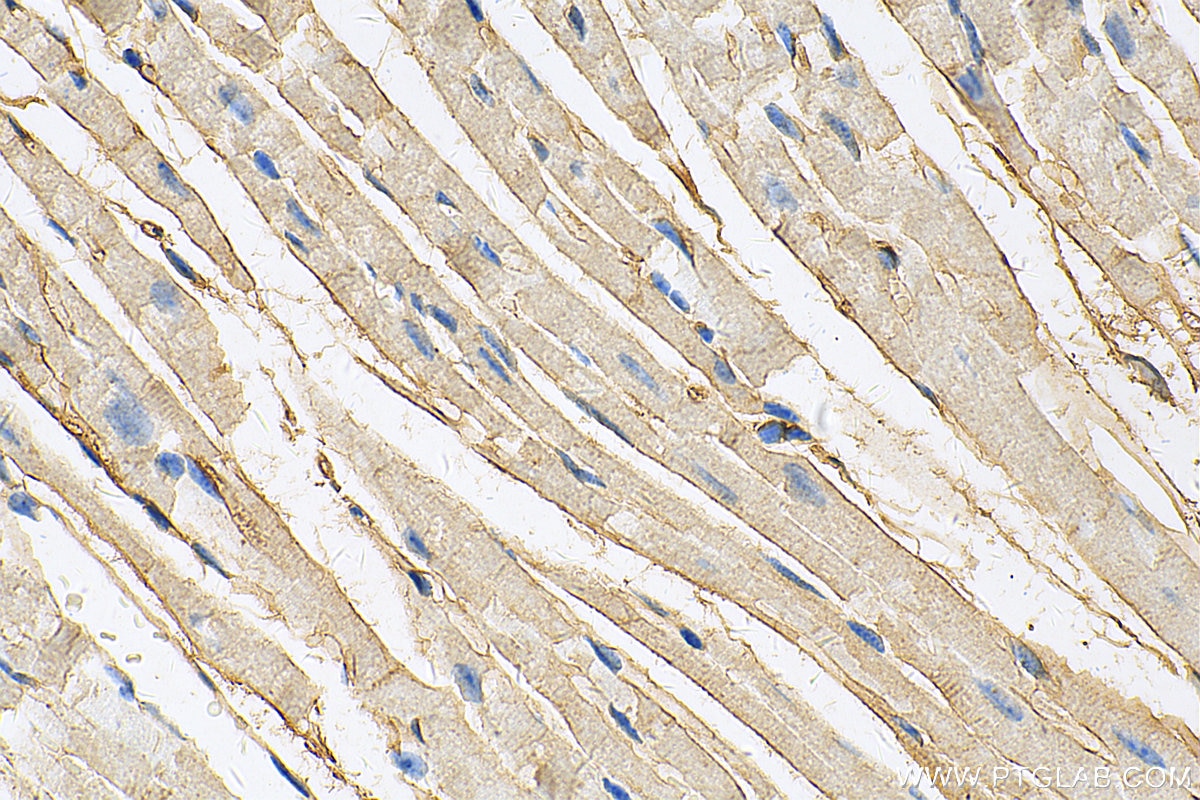
| Positive IHC detected in | mouse heart tissue Note: suggested antigen retrieval with TE buffer pH 9.0; (*) Alternatively, antigen retrieval may be performed with citrate buffer pH 6.0 |
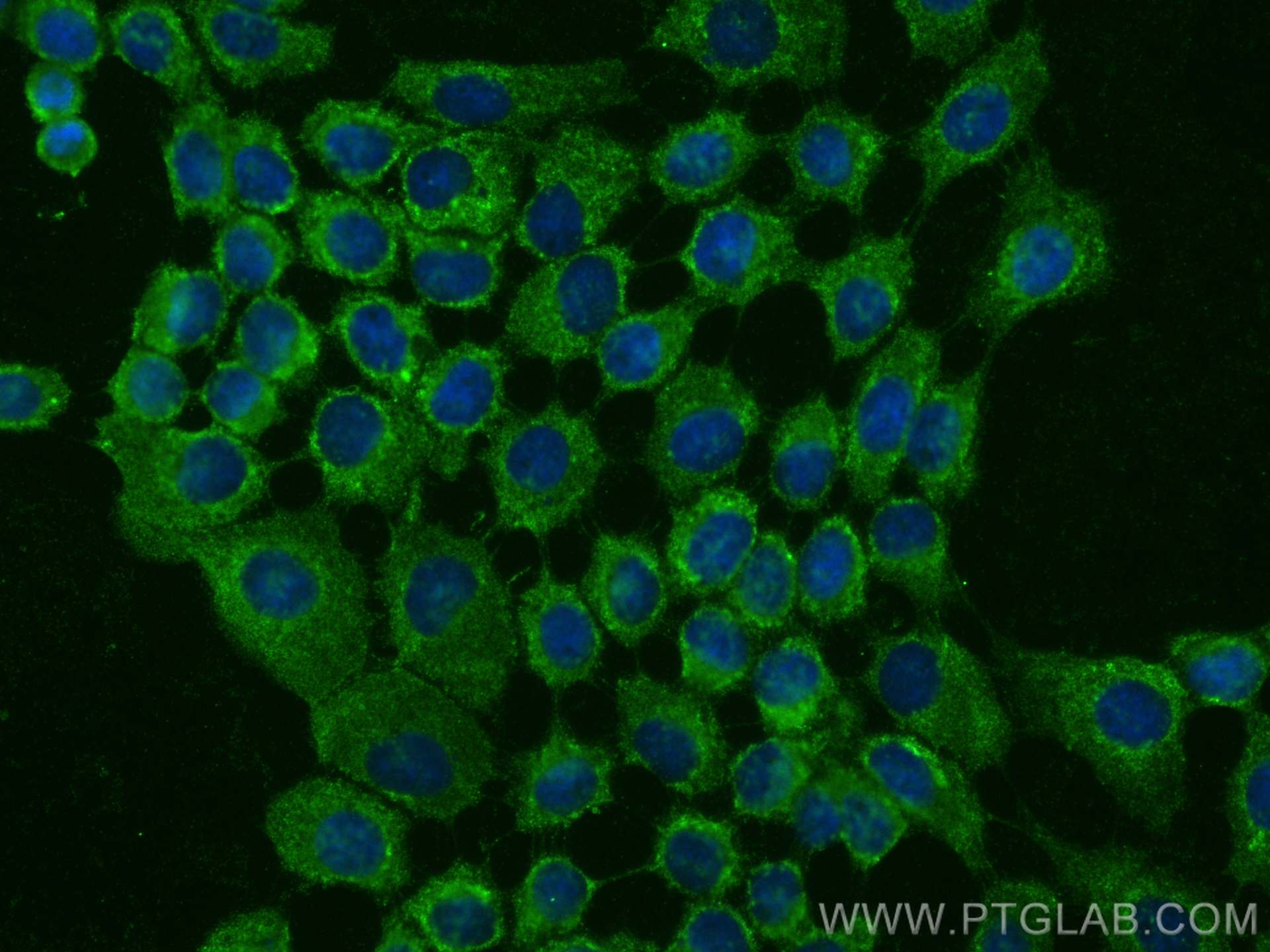
| Positive IF/ICC detected in | A431 cells |
Recommended dilution
| Application | Dilution |
|---|---|
| Western Blot (WB) | WB : 1:5000-1:50000 |
| Immunohistochemistry (IHC) | IHC : 1:1000-1:4000 |
| Immunofluorescence (IF)/ICC | IF/ICC : 1:500-1:2000 |
| It is recommended that this reagent should be titrated in each testing system to obtain optimal results. | |
| Sample-dependent, Check data in validation data gallery. | |
Product Information
68364-1-Ig targets Desmoplakin in WB, IHC, IF/ICC, ELISA applications and shows reactivity with human, mouse samples.
| Tested Reactivity | human, mouse |
| Host / Isotype | Mouse / IgG1 |
| Class | Monoclonal |
| Type | Antibody |
| Immunogen |
CatNo: Ag30499 Product name: Recombinant human DSP protein Source: e coli.-derived, PET28a Tag: 6*His Domain: 1-350 aa of BC140802 Sequence: MSCNGGSHPRINTLGRMIRAESGPDLRYEVTSGGGGTSRMYYSRRGVITDQNSDGYCQTGTMSRHQNQNTIQELLQNCSDCLMRAELIVQPELKYGDGIQLTRSRELDECFAQANDQMEILDSLIREMRQMGQPCDAYQKRLLQLQEQMRALYKAISVPRVRRASSKGGGGYTCQSGSGWDEFTKHVTSECLGWMRQQRAEMDMVAWGVDLASVEQHINSHRGIHNSIGDYRWQLDKIKADLREKSAIYQLEEEYENLLKASFERMDHLRQLQNIIQATSREIMWINDCEEEELLYDWSDKNTNIAQKQEAFSIRMSQLEVKEKELNKLKQESDQLVLNQHPASDKIEAY Predict reactive species |
| Full Name | desmoplakin |
| Calculated Molecular Weight | 2871 aa, 332 kDa |
| Observed Molecular Weight | 240-330 kDa |
| GenBank Accession Number | BC140802 |
| Gene Symbol | DSP |
| Gene ID (NCBI) | 1832 |
| RRID | AB_3085085 |
| Conjugate | Unconjugated |
| Form | Liquid |
| Purification Method | Protein G purification |
| UNIPROT ID | P15924 |
| Storage Buffer | PBS with 0.02% sodium azide and 50% glycerol, pH 7.3. |
| Storage Conditions | Store at -20°C. Stable for one year after shipment. Aliquoting is unnecessary for -20oC storage. 20ul sizes contain 0.1% BSA. |
Background Information
The desmoplakin (DP) is the most abundant desmosomal proteins and is located at the cytoplasmic portion of the desmosomes which are specialized cell-cell adhesion structures abundant in tissues such as muscle and epidermis that are subjected to mechanical stress. Desmoplakin 1 and 2 (DP 1 and 2) are two splice variants sharing common C-terminal and N-terminal globular head and tail domains, but differ in the length of the rod domain that links them. This antibody detects both DP1 (280-330 kDa) and DP2 (240-260 kDa). (PMID: 16467215, 11781569)
Protocols
| Product Specific Protocols | |
|---|---|
| IF protocol for Desmoplakin antibody 68364-1-Ig | Download protocol |
| IHC protocol for Desmoplakin antibody 68364-1-Ig | Download protocol |
| WB protocol for Desmoplakin antibody 68364-1-Ig | Download protocol |
| Standard Protocols | |
|---|---|
| Click here to view our Standard Protocols |
Reviews
The reviews below have been submitted by verified Proteintech customers who received an incentive for providing their feedback.
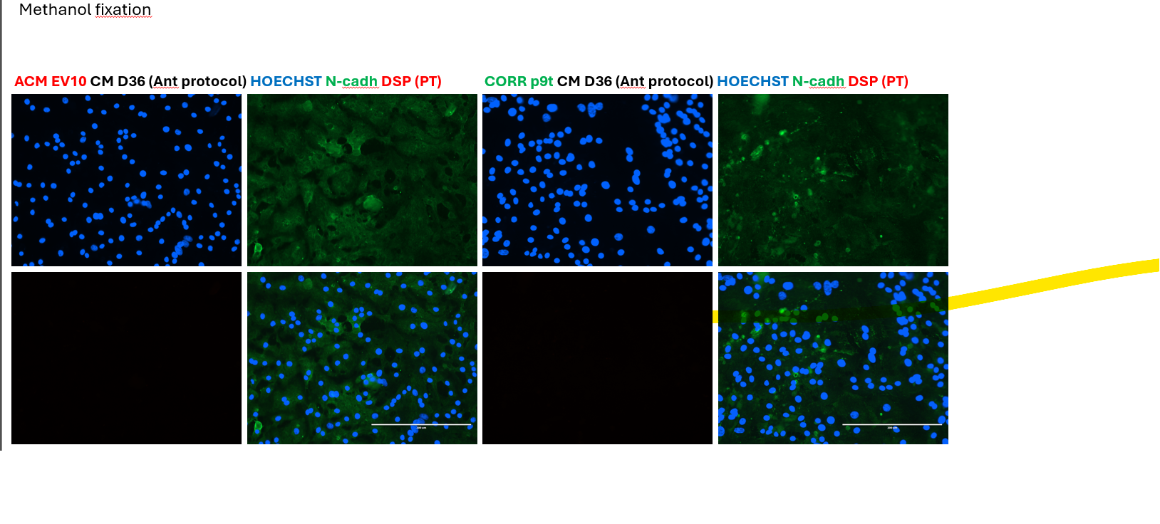
FH Kiara (Verified Customer) (07-28-2025) | Although validated by ProteinTech for IF, we did not observe a reliable signal for DSP in methanol fixed iPSC-cardiomyocytes.
 |