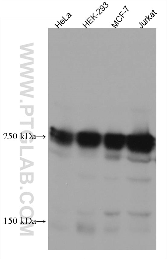
Various lysates were subjected to SDS PAGE followed by western blot with 67199-1-Ig (EIF4G1 antibody) at dilution of 1:10000 incubated at room temperature for 1.5 hours.
Various lysates were subjected to SDS PAGE followed by western blot with 67199-1-Ig (EIF4G1 antibody) at dilution of 1:10000 incubated at room temperature for 1.5 hours.
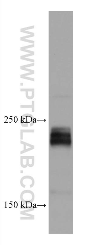
WB analysis of 4T1 using 67199-1-Ig
4T1 cells were subjected to SDS PAGE followed by western blot with 67199-1-Ig (EIF4G1 antibody) at dilution of 1:10000 incubated at room temperature for 1.5 hours.
4T1 cells were subjected to SDS PAGE followed by western blot with 67199-1-Ig (EIF4G1 antibody) at dilution of 1:10000 incubated at room temperature for 1.5 hours.
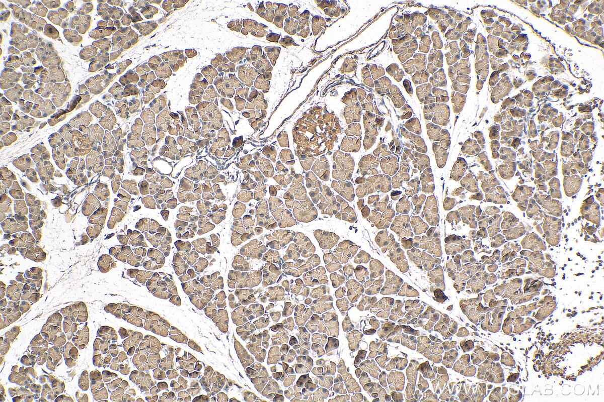
IHC staining of mouse pancreas using 67199-1-Ig
Immunohistochemical analysis of paraffin-embedded mouse pancreas tissue slide using 67199-1-Ig (EIF4G1 antibody) at dilution of 1:200 (under 10x lens). Heat mediated antigen retrieval with Tris-EDTA buffer (pH 9.0).
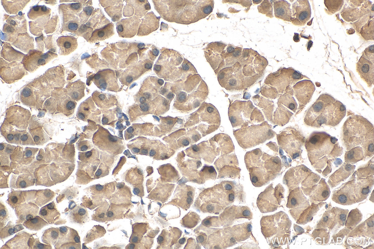
Immunohistochemical analysis of paraffin-embedded mouse pancreas tissue slide using 67199-1-Ig (EIF4G1 antibody) at dilution of 1:200 (under 40x lens). Heat mediated antigen retrieval with Tris-EDTA buffer (pH 9.0).
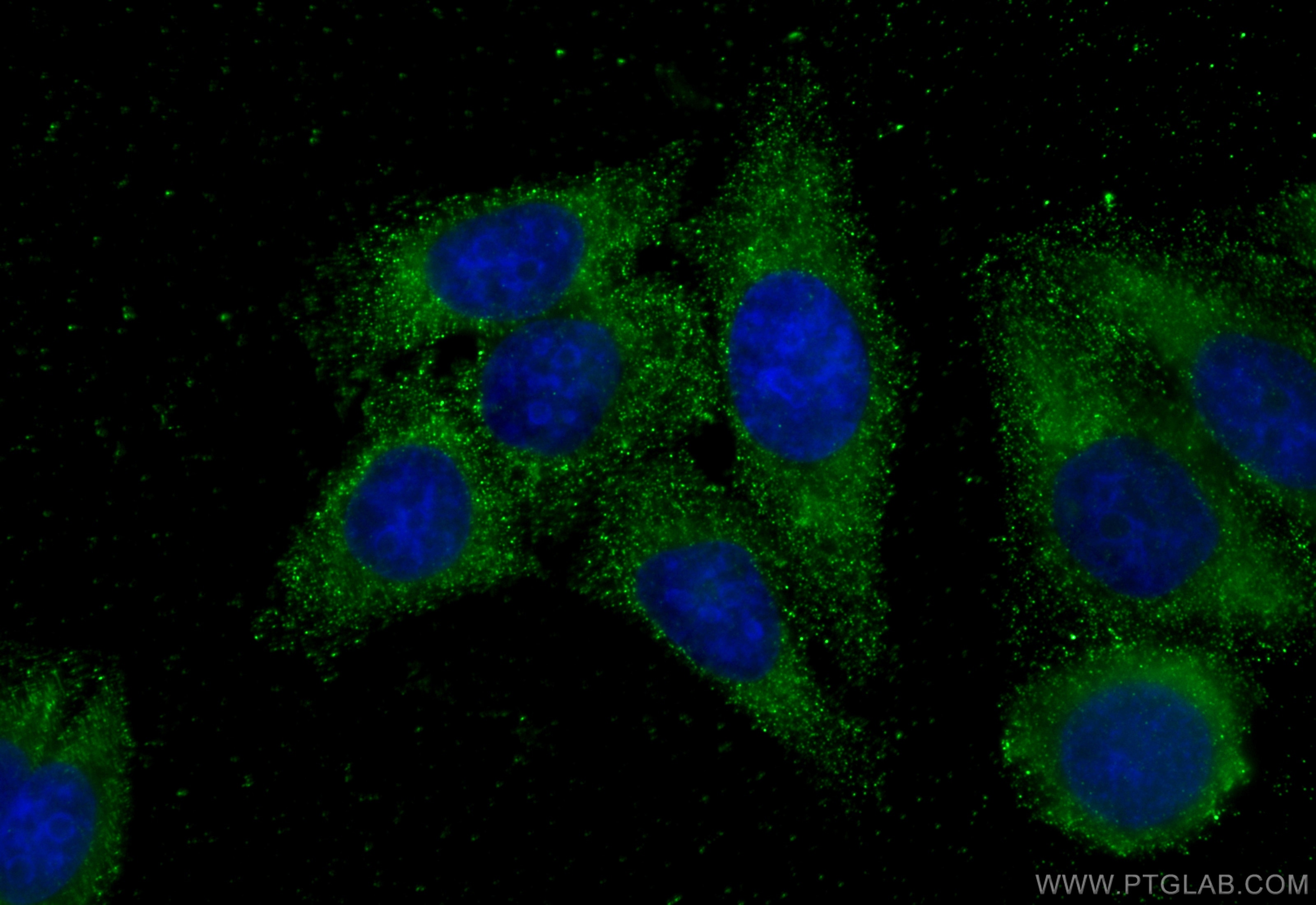
IF Staining of HepG2 using 67199-1-Ig
Immunofluorescent analysis of (-20°C Ethanol) fixed HepG2 cells using EIF4G1 antibody (67199-1-Ig, Clone: 2B10G8 ) at dilution of 1:400 and CoraLite®488-Conjugated Goat Anti-Mouse IgG(H+L) (SA00013-1).
Immunofluorescent analysis of (-20°C Ethanol) fixed HepG2 cells using EIF4G1 antibody (67199-1-Ig, Clone: 2B10G8 ) at dilution of 1:400 and CoraLite®488-Conjugated Goat Anti-Mouse IgG(H+L) (SA00013-1).
The Proteintech guarantee covers Proteintech antibodies in any species and any application, including those not listed on the datasheet. If the antibody doesn’t perform, you can receive a hassle-free refund or credit note.
HeLa cells, 4T1 cells, HEK-293 cells, MCF-7 cells, Jurkat cells
Positive IHC detected in
mouse pancreas tissue Note: suggested antigen retrieval with TE buffer pH 9.0; (*) Alternatively, antigen retrieval may be performed with citrate buffer pH 6.0
Positive IF/ICC detected in
HepG2 cells
Recommended dilution
Application
Dilution
Western Blot (WB)
WB : 1:5000-1:50000
Immunohistochemistry (IHC)
IHC : 1:50-1:500
Immunofluorescence (IF)/ICC
IF/ICC : 1:200-1:800
It is recommended that this reagent should be titrated in each testing system to obtain optimal results.
Sample-dependent, Check data in validation data gallery.
PBS with 0.02% sodium azide and 50% glycerol, pH 7.3.
Storage Conditions
Store at -20°C. Stable for one year after shipment. Aliquoting is unnecessary for -20oC storage. 20ul sizes contain 0.1% BSA.
Background Information
Eukaryotic cellular messenger RNAs are posttranscriptionally modified by addition of an m(7)GTP moiety to the 5-prime terminus, referred to as a cap. Recognition of the cap structure and unwinding of mRNA secondary structure during the initiation phase of protein synthesis is catalyzed by initiation factors of the eIF4 group. EIF4G1, a subunit of eIF4 gamma, forms various complexes with the other eIF4 polypeptides [PMID: 7601469]. Mutations in the EIF4G1 gene, encoding a component of the eIF4F translation initiation complex, were recently reported as a possible cause for the autosomal dominant form of Parkinson's disease [PMID:22658323]. The calcualted molecular weight of EIF4G1 is 175 kDa, but modified EIF4G1 is about 220-240 kDa. (PMID: 18426977 )
DDX3 Regulates the Cap-Independent Translation of the Japanese Encephalitis Virus via Its Interactions with PABP1 and the Untranslated Regions of the Viral Genome
Various lysates were subjected to SDS PAGE followed by western blot with 67199-1-Ig (EIF4G1 antibody) at dilution of 1:10000 incubated at room temperature for 1.5 hours.
WB analysis of 4T1 using 67199-1-Ig
4T1 cells were subjected to SDS PAGE followed by western blot with 67199-1-Ig (EIF4G1 antibody) at dilution of 1:10000 incubated at room temperature for 1.5 hours.
IHC Figures
IHC staining of mouse pancreas using 67199-1-Ig
Immunohistochemical analysis of paraffin-embedded mouse pancreas tissue slide using 67199-1-Ig (EIF4G1 antibody) at dilution of 1:200 (under 10x lens). Heat mediated antigen retrieval with Tris-EDTA buffer (pH 9.0).
IHC staining of mouse pancreas using 67199-1-Ig
Immunohistochemical analysis of paraffin-embedded mouse pancreas tissue slide using 67199-1-Ig (EIF4G1 antibody) at dilution of 1:200 (under 40x lens). Heat mediated antigen retrieval with Tris-EDTA buffer (pH 9.0).
IF/ICC Figures
IF Staining of HepG2 using 67199-1-Ig
Immunofluorescent analysis of (-20°C Ethanol) fixed HepG2 cells using EIF4G1 antibody (67199-1-Ig, Clone: 2B10G8 ) at dilution of 1:400 and CoraLite®488-Conjugated Goat Anti-Mouse IgG(H+L) (SA00013-1).
The species listed in Tested Reactivity are in-house verified and applicable species. For unlisted species, please refer to the homology analysis of the immunogen sequence and related species. For rabbit polyclonal antibodies, homology >70% is recommended. For mouse monoclonal antibodies and rabbit recombinant antibodies, homology >90% is recommended. Generally, the higher the homology, the greater the applicability. However, there will be certain differences in protein expression in different species, tissues or cells. Therefore, the homology analysis results are for reference only and do not serve as a guarantee.
At Proteintech, we pride ourselves on our antibody quality, customer service and transparency. As such, we are comparing our antibodies with other vendors, enabling easy identification and comparisons of key data to help you choose the suitable antibody for your needs.
We have selected the top cited antibodies from these vendors for you to compare.
 at dilution of 1:10000 incubated at room temperature for 1.5 hours.")
 at dilution of 1:10000 incubated at room temperature for 1.5 hours.")
 at dilution of 1:200 (under 10x lens). Heat mediated antigen retrieval with Tris-EDTA buffer (pH 9.0).")
 at dilution of 1:200 (under 40x lens). Heat mediated antigen retrieval with Tris-EDTA buffer (pH 9.0).")
 fixed HepG2 cells using EIF4G1 antibody (67199-1-Ig, Clone: 2B10G8 ) at dilution of 1:400 and CoraLite®488-Conjugated Goat Anti-Mouse IgG(H+L) (SA00013-1).")