Tested Applications
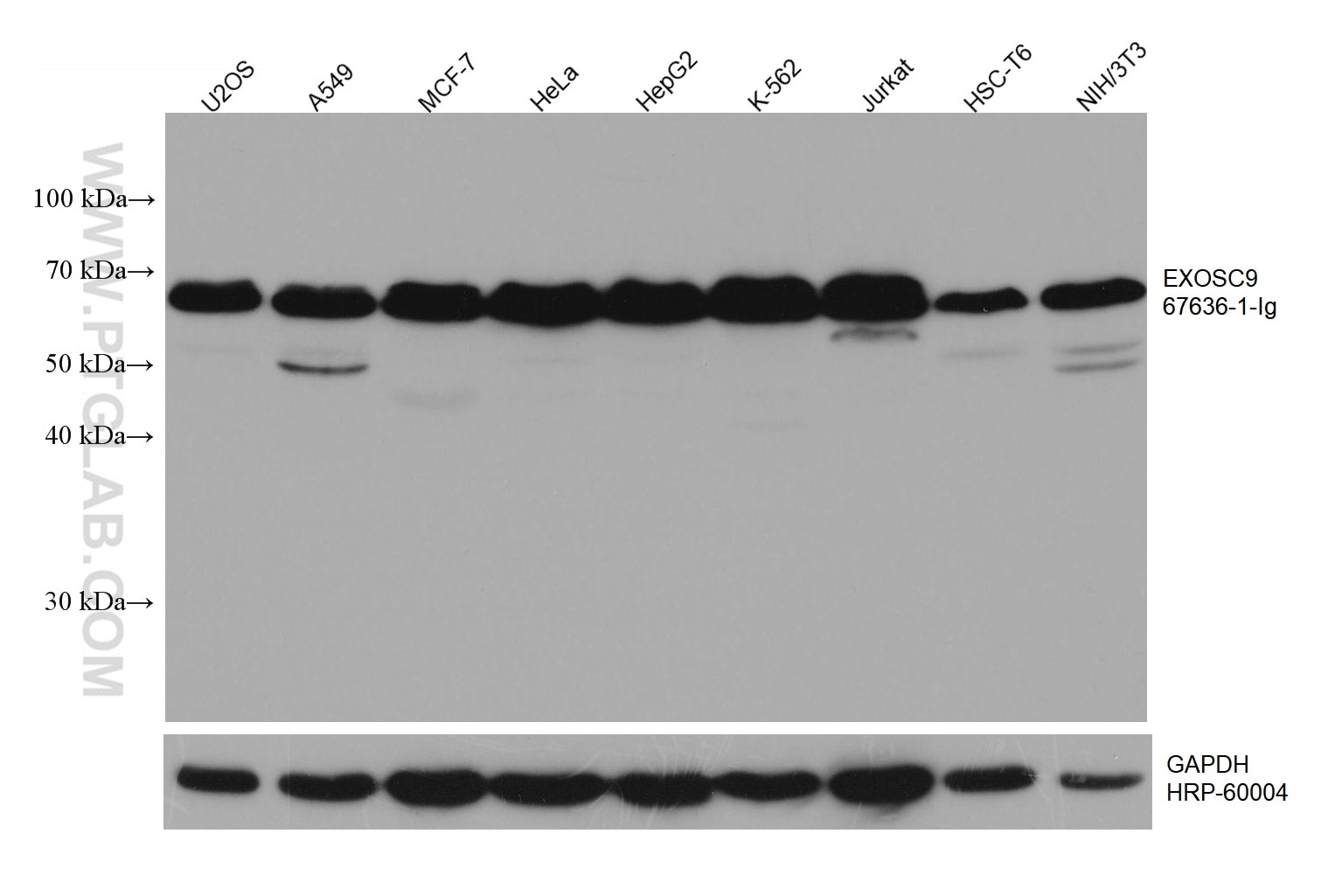
| Positive WB detected in | U2OS cells, HeLa cells, A549 cells, MCF-7 cells, HepG2 cells, K-562 cells, Jurkat cells, HSC-T6 cells, NIH/3T3 cells |
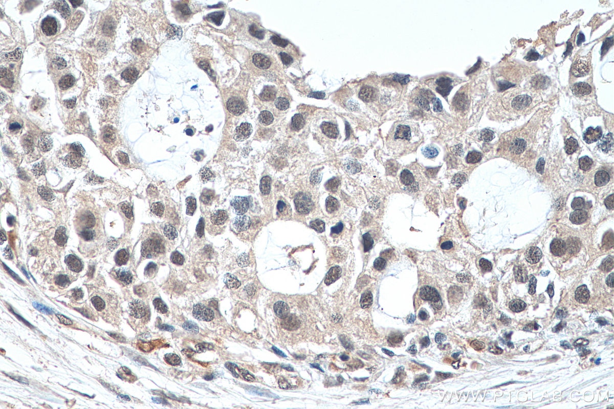
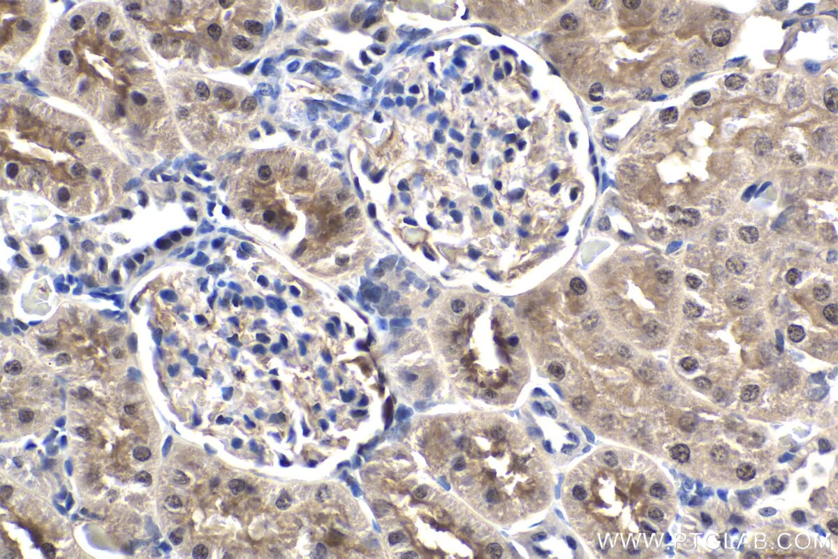
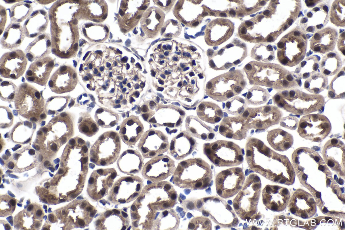
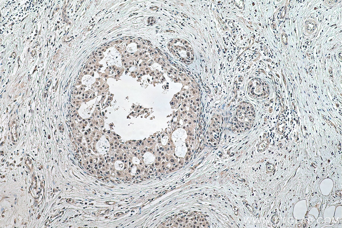
| Positive IHC detected in | human breast cancer tissue, mouse kidney tissue, rat kidney tissue Note: suggested antigen retrieval with TE buffer pH 9.0; (*) Alternatively, antigen retrieval may be performed with citrate buffer pH 6.0 |
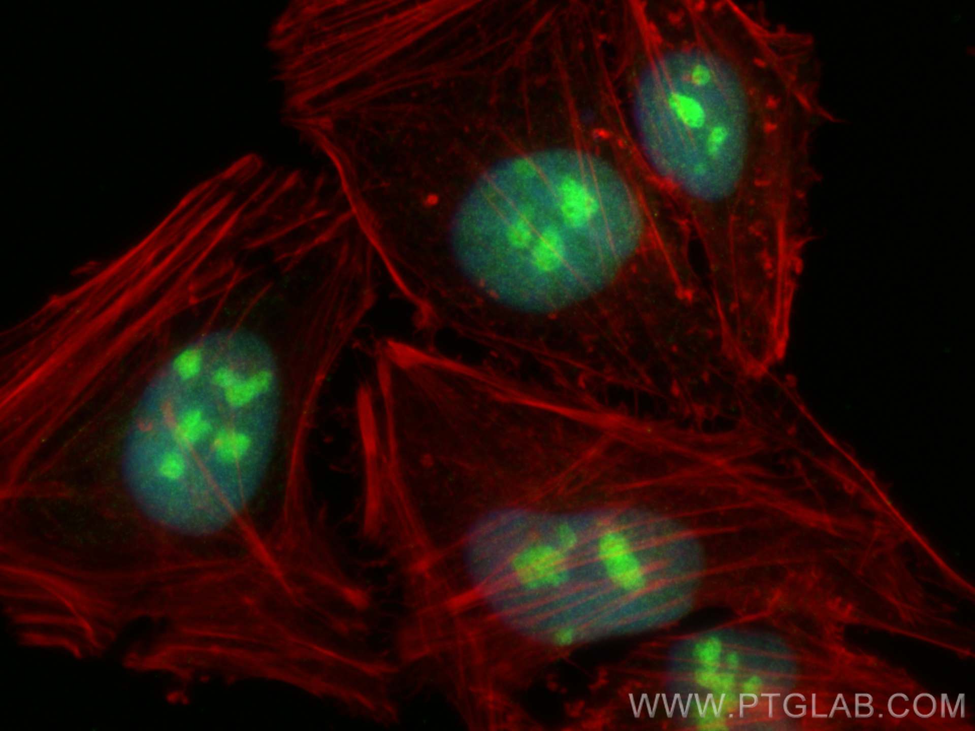
| Positive IF/ICC detected in | HeLa cells |
Recommended dilution
| Application | Dilution |
|---|---|
| Western Blot (WB) | WB : 1:5000-1:50000 |
| Immunohistochemistry (IHC) | IHC : 1:750-1:3000 |
| Immunofluorescence (IF)/ICC | IF/ICC : 1:1000-1:4000 |
| It is recommended that this reagent should be titrated in each testing system to obtain optimal results. | |
| Sample-dependent, Check data in validation data gallery. | |
Published Applications
| WB | See 1 publications below |
Product Information
67636-1-Ig targets EXOSC9 in WB, IHC, IF/ICC, ELISA applications and shows reactivity with Human, Mouse, Rat samples.
| Tested Reactivity | Human, Mouse, Rat |
| Cited Reactivity | human |
| Host / Isotype | Mouse / IgG2a |
| Class | Monoclonal |
| Type | Antibody |
| Immunogen |
CatNo: Ag19783 Product name: Recombinant human EXOSC9 protein Source: e coli.-derived, PET28a Tag: 6*His Domain: 1-351 aa of BC142978 Sequence: MKETPLSNCERRFLLRAIEEKKRLDGRQTYDYRNIRISFGTDYGCCIVELGKTRVLGQVSCELVSPKLNRATEGILFFNLELSQMAAPAFEPGRQSDLLVKLNRLMERCLRNSKCIDTESLCVVAGEKVWQIRVDLHLLNHDGNIIDAASIAAIVALCHFRRPDVSVQGDEVTLYTPEERDPVPLSIHHMPICVSFAFFQQGAYLLVDPNEREERVMDGLLVIAMNKHREICTIQSSGGIMLLKDQVLRCSKIAGVKVAEITELILKALENDQKVRKEGGKFGFAESIANQRITAFKMEKAPIDTSDVEEKAEEIIAEAEPPSEVVSTPVLWTPGTAQIGEGVENSWGDLE Predict reactive species |
| Full Name | exosome component 9 |
| Calculated Molecular Weight | 456 aa, 51 kDa |
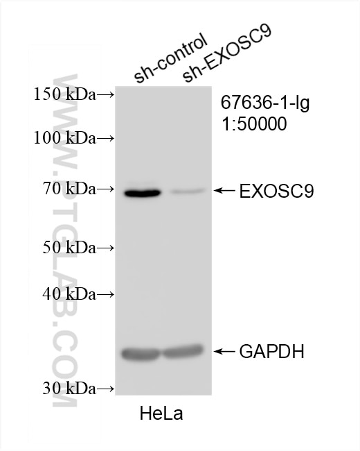
| Observed Molecular Weight | 65-70 kDa |
| GenBank Accession Number | BC142978 |
| Gene Symbol | EXOSC9 |
| Gene ID (NCBI) | 5393 |
| RRID | AB_2882837 |
| Conjugate | Unconjugated |
| Form | Liquid |
| Purification Method | Protein A purification |
| UNIPROT ID | Q06265 |
| Storage Buffer | PBS with 0.02% sodium azide and 50% glycerol, pH 7.3. |
| Storage Conditions | Store at -20°C. Stable for one year after shipment. Aliquoting is unnecessary for -20oC storage. 20ul sizes contain 0.1% BSA. |
Background Information
EXOSC9, also named as PMSCL1 and PM/Scl-75, is a key subunit of the exosome complex. EXOSC9 maintains self-renewal in a cell-autonomous manner. EXOSC9 has 4 isoforms with MW 49 kDa, 51 kDa, 39 kDa and 41 kDa. It's polymyositis/scleroderma auto-antigen 75 kDa. The MW of EXOSC9 is migrated to 60-75 kDa for modification in WB detection.
Protocols
| Product Specific Protocols | |
|---|---|
| IF protocol for EXOSC9 antibody 67636-1-Ig | Download protocol |
| IHC protocol for EXOSC9 antibody 67636-1-Ig | Download protocol |
| WB protocol for EXOSC9 antibody 67636-1-Ig | Download protocol |
| Standard Protocols | |
|---|---|
| Click here to view our Standard Protocols |
Reviews
The reviews below have been submitted by verified Proteintech customers who received an incentive for providing their feedback.
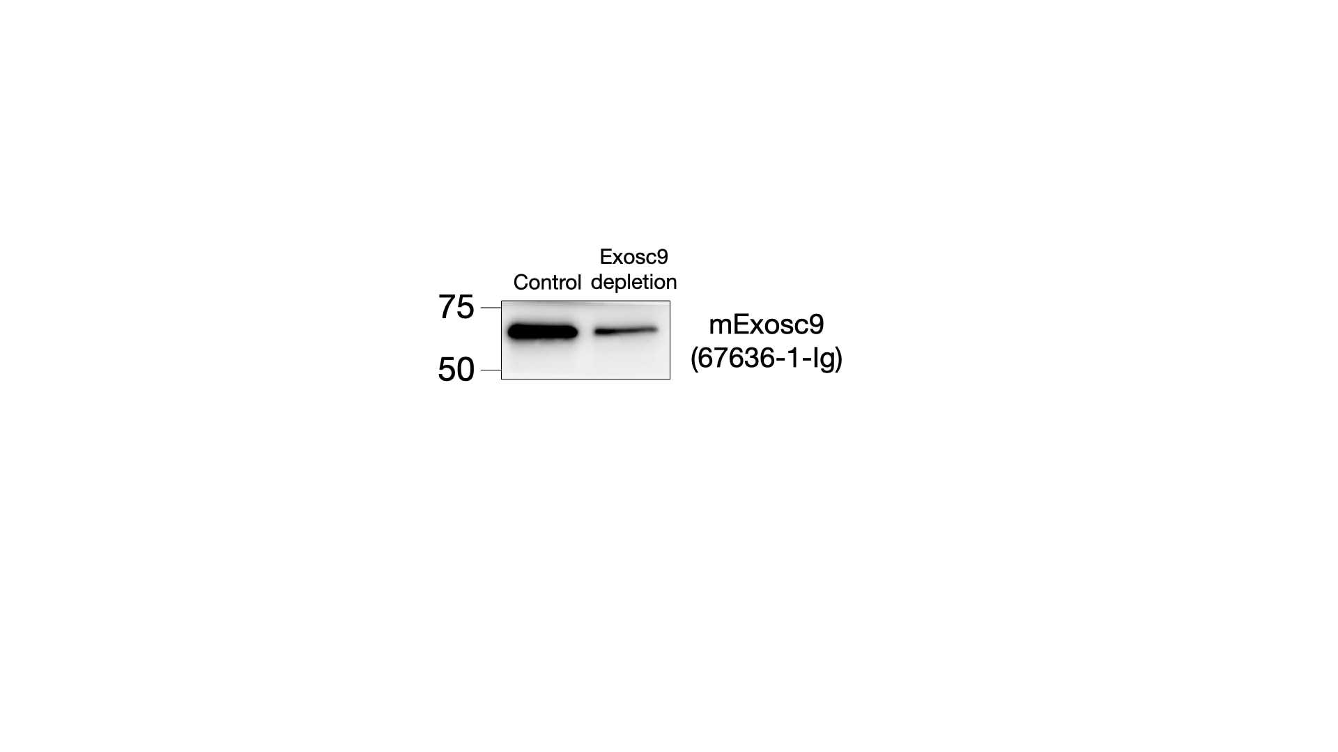
FH Nikolaus (Verified Customer) (07-14-2022) | Antibodies were incubated ON at 4C. Exosc9 depletion was used as a control to check the specificity of the band.
 |