Tested Applications
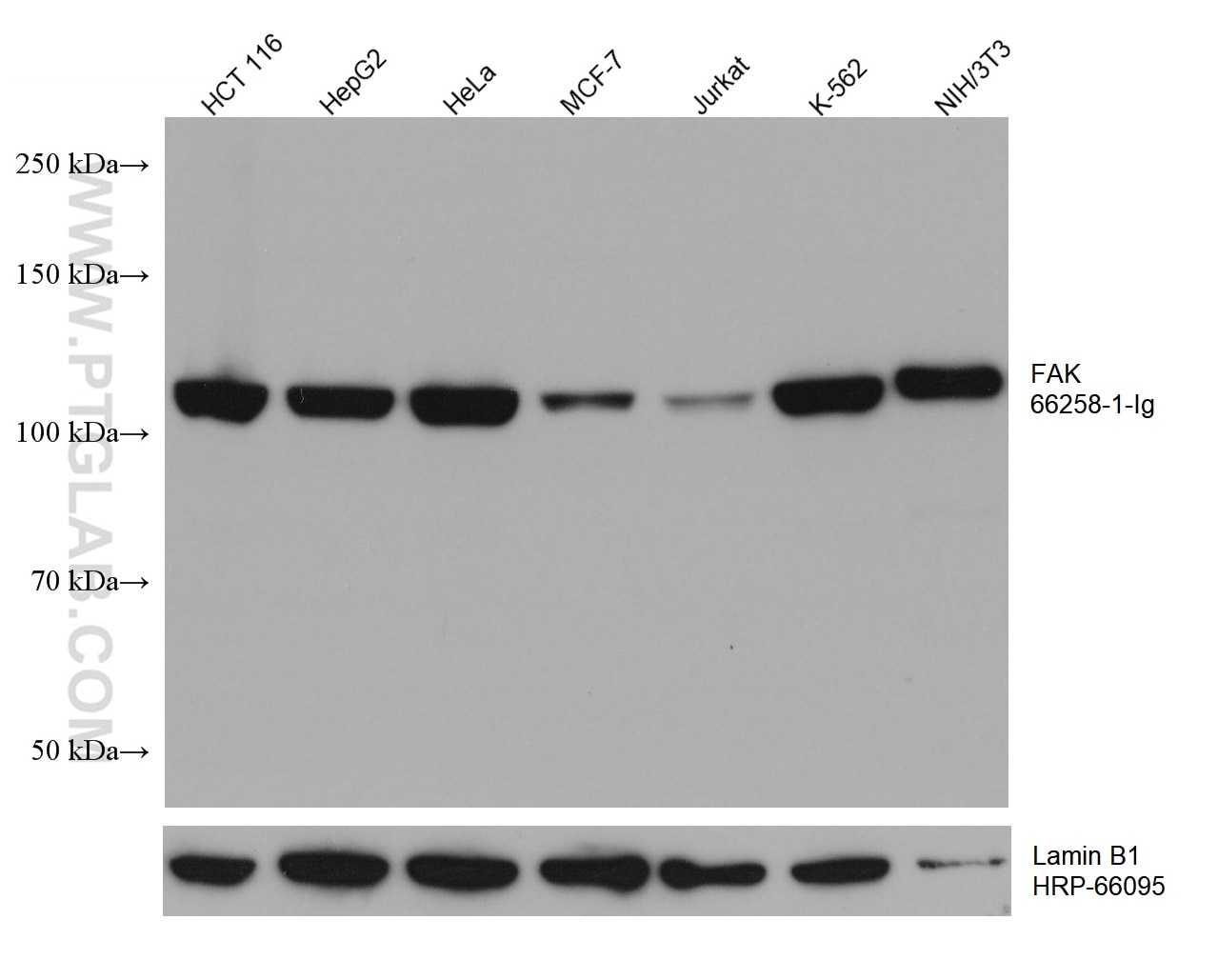
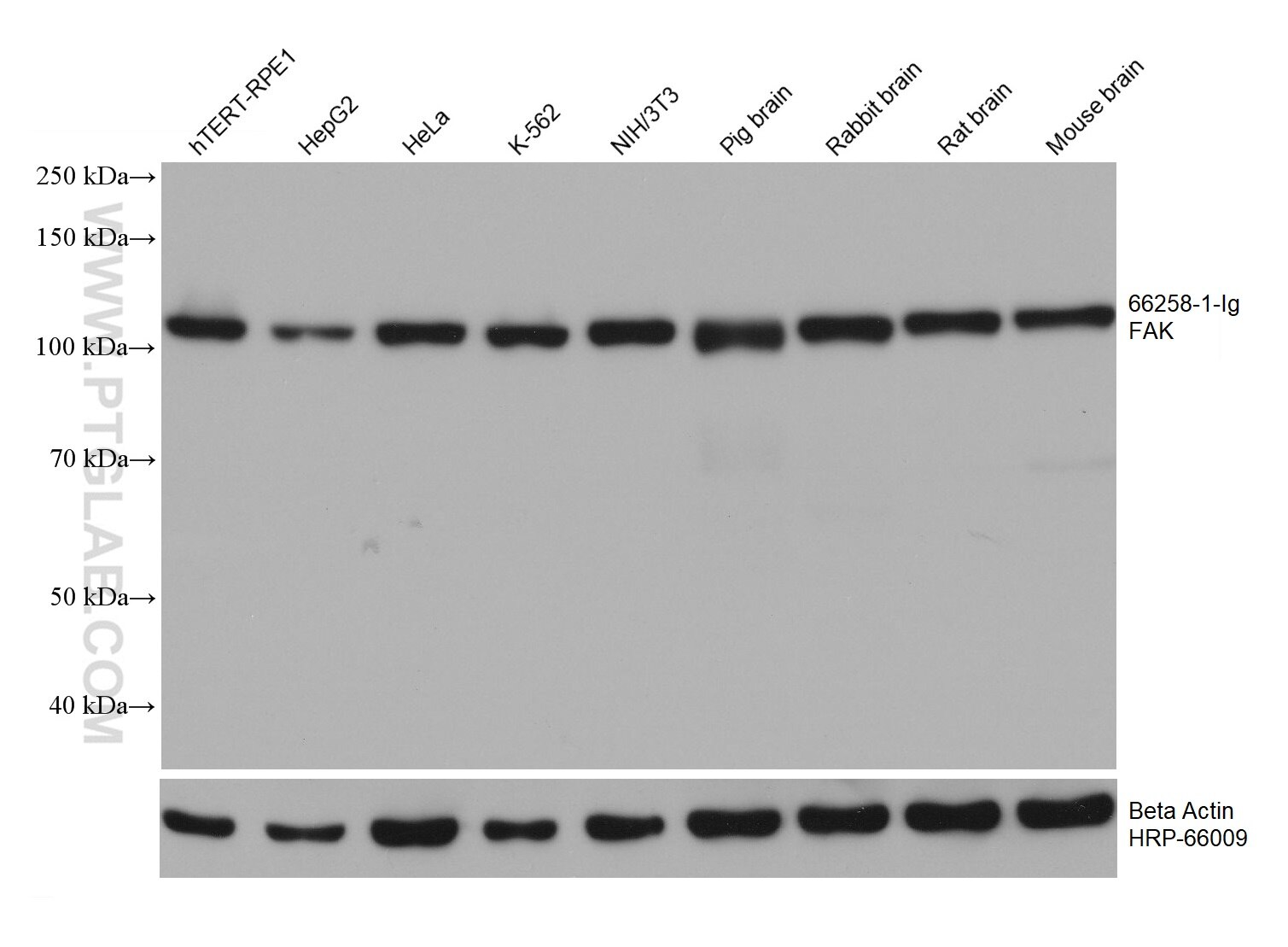
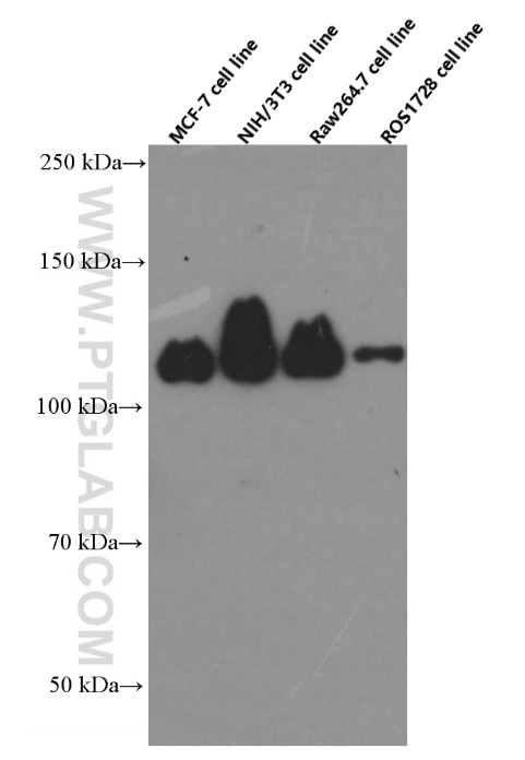
| Positive WB detected in | hTERT-RPE1 cells, human testis tissue, MCF-7 cells, HCT 116 cells, NIH/3T3 cells, RAW 264.7 cells, ROS1728 cells, HepG2 cells, HeLa cells, Jurkat cells, K-562 cells, pig brain tissue, rabbit brain tissue, rat brain tissue, mouse brain tissue |
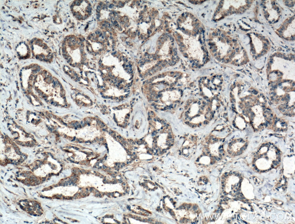
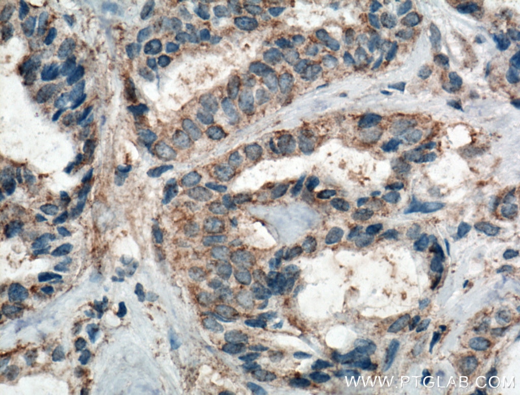
| Positive IHC detected in | human breast cancer tissue Note: suggested antigen retrieval with TE buffer pH 9.0; (*) Alternatively, antigen retrieval may be performed with citrate buffer pH 6.0 |
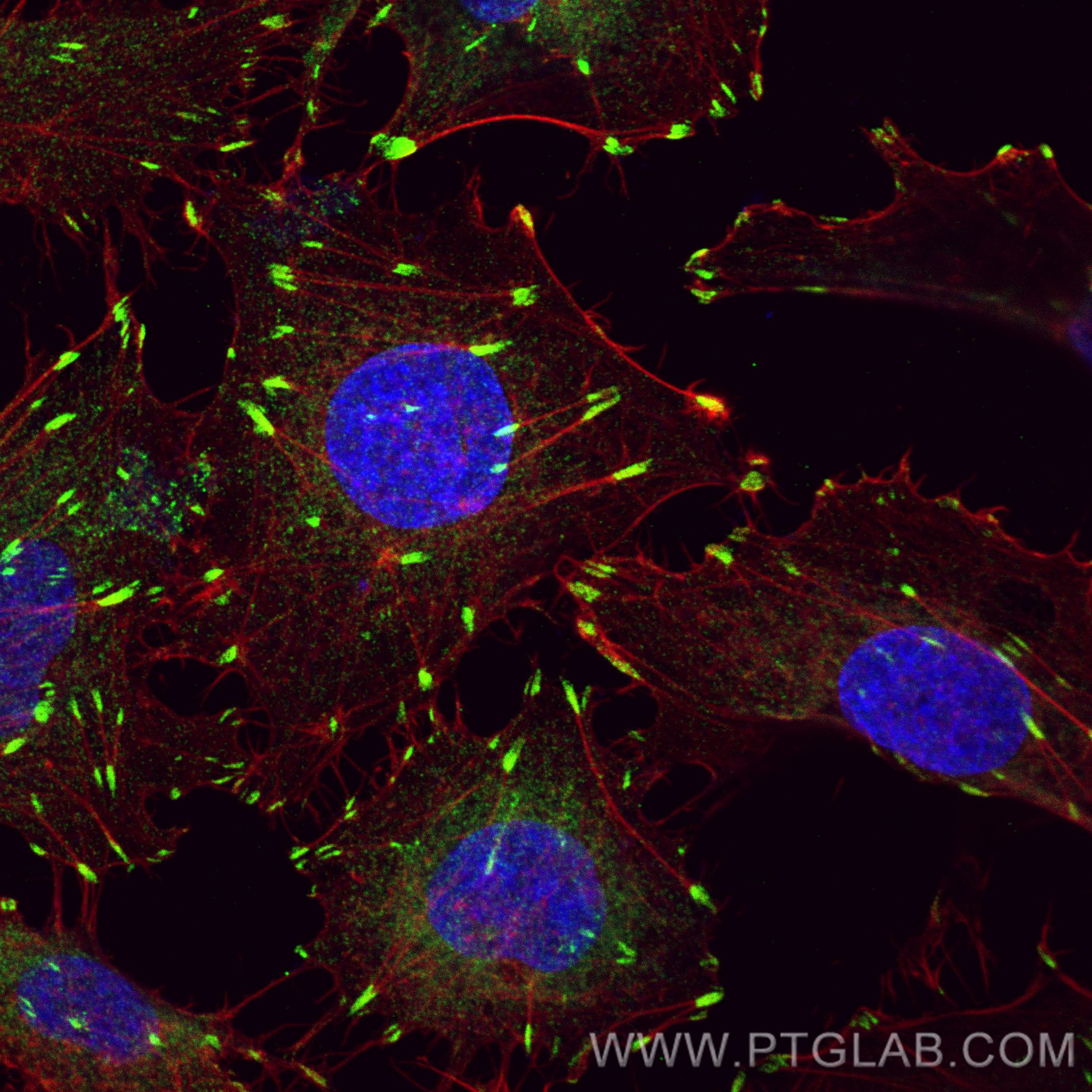
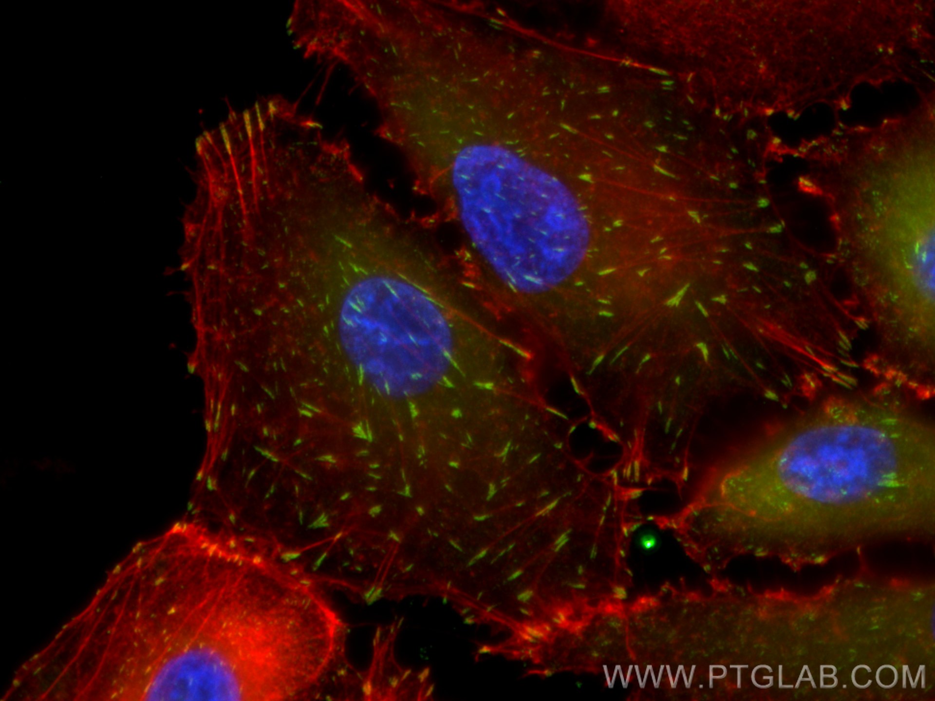
| Positive IF/ICC detected in | HUVEC cells, A549 cells |
Recommended dilution
| Application | Dilution |
|---|---|
| Western Blot (WB) | WB : 1:5000-1:50000 |
| Immunohistochemistry (IHC) | IHC : 1:50-1:500 |
| Immunofluorescence (IF)/ICC | IF/ICC : 1:200-1:800 |
| It is recommended that this reagent should be titrated in each testing system to obtain optimal results. | |
| Sample-dependent, Check data in validation data gallery. | |
Published Applications
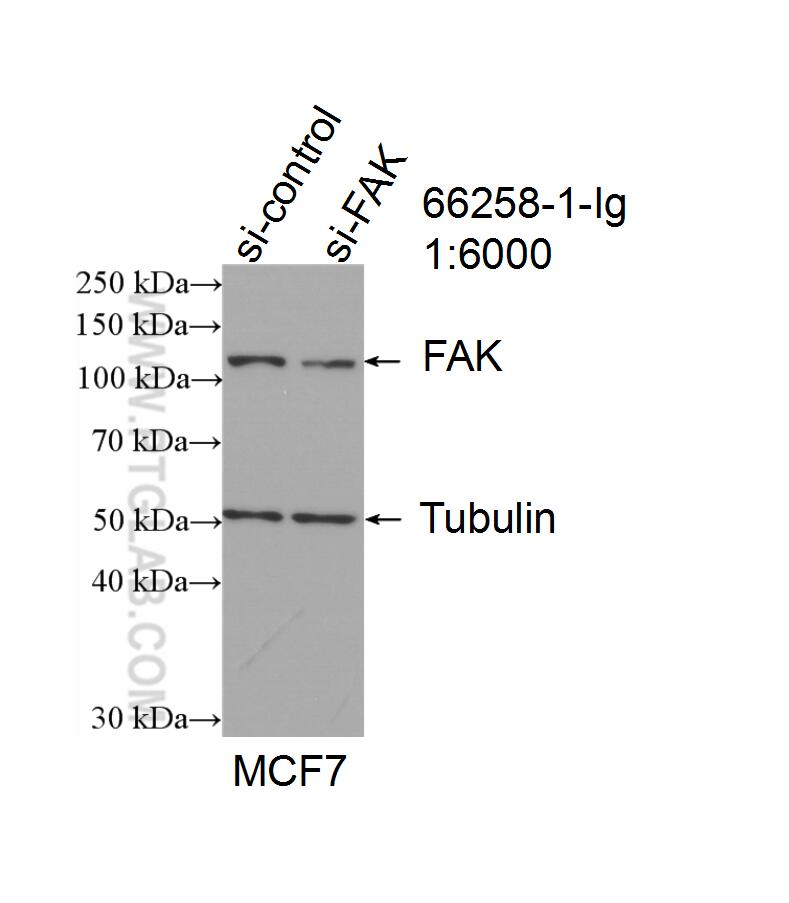
| KD/KO | See 2 publications below |
| WB | See 54 publications below |
| IHC | See 5 publications below |
| IF | See 21 publications below |
| IP | See 1 publications below |
| CoIP | See 1 publications below |
Product Information
66258-1-Ig targets FAK in WB, IHC, IF/ICC, IP, CoIP, ELISA applications and shows reactivity with human, mouse, rat samples.
| Tested Reactivity | human, mouse, rat |
| Cited Reactivity | human, mouse, rat |
| Host / Isotype | Mouse / IgG2c |
| Class | Monoclonal |
| Type | Antibody |
| Immunogen |
CatNo: Ag17966 Product name: Recombinant human FAK protein Source: e coli.-derived, PET28a Tag: 6*His Domain: 381-678 aa of BC028733 Sequence: ITAMAGSIYPGQASLLDQTDSWNHRPQEIAMWQPNVEDSTVLDLRGIGQVLPTHLMEERLIRQQQEMEEDQRWLEKEERFLKPDVRLSRGSIDREDGSLQGPIGNQHIYQPVGKPDPAAPPKKPPRPGAPGHLGSLASLSSPADSYNEGVKLKPQEISPPPTANLDRSNDKVYENVTGLVKAVIEMSSKIQPAPPEEYVPMVKEVGLALRTLLATVDETIPLLPASTHREIEMAQKLLNSDLGELINKMKLAQQYVMTSLQQEYKKQMLTAAHALAVDAKNLLDVIDQARLKMLGQTR Predict reactive species |
| Full Name | PTK2 protein tyrosine kinase 2 |
| Calculated Molecular Weight | 1052 aa, 119 kDa |
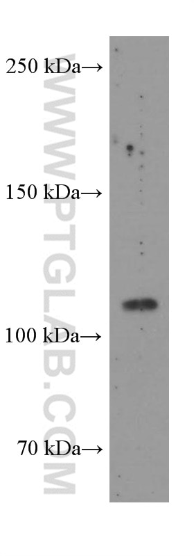
| Observed Molecular Weight | 110 kDa |
| GenBank Accession Number | BC028733 |
| Gene Symbol | FAK |
| Gene ID (NCBI) | 5747 |
| RRID | AB_2881646 |
| Conjugate | Unconjugated |
| Form | Liquid |
| Purification Method | Protein A purification |
| UNIPROT ID | Q05397 |
| Storage Buffer | PBS with 0.02% sodium azide and 50% glycerol, pH 7.3. |
| Storage Conditions | Store at -20°C. Stable for one year after shipment. Aliquoting is unnecessary for -20oC storage. 20ul sizes contain 0.1% BSA. |
Background Information
FAK (Focal adhesion kinase 1) is also named as FAK1, FADK, pp125FAK, FAK and belongs to the protein kinase superfamily. It is a critical tyrosine kinase that modulates cell adhesion, migration, proliferation and survival in response to extracellular signals (PMID:19664602). It also acts as a pivotal signal 'integrator', controlling and coordinating cellular responses that include cell migration, survival, proliferation and, epithelial tissue repair after DNA damage (PMID:20966971). This protein has some isoforms produced by alternative promoter usage and alternative splicing, and the range of the molecular weights are 100-120kD and 40-60kD.
Protocols
| Product Specific Protocols | |
|---|---|
| IF protocol for FAK antibody 66258-1-Ig | Download protocol |
| IHC protocol for FAK antibody 66258-1-Ig | Download protocol |
| WB protocol for FAK antibody 66258-1-Ig | Download protocol |
| Standard Protocols | |
|---|---|
| Click here to view our Standard Protocols |
Publications
| Species | Application | Title |
|---|---|---|
Acta Biomater Sodium alginate/collagen/stromal cell-derived factor-1 neural scaffold loaded with BMSCs promotes neurological function recovery after traumatic brain injury. | ||
Cancer Lett Direct contact between tumor cells and platelets initiates a FAK-dependent F3/TGF-β positive feedback loop that promotes tumor progression and EMT in osteosarcoma | ||
Oncogene MiR-19a/miR-96-mediated low expression of KIF26A suppresses metastasis by regulating FAK pathway in gastric cancer. | ||
Aging (Albany NY) TNFSF9 promotes metastasis of pancreatic cancer through Wnt/Snail signaling and M2 polarization of macrophages. |
Reviews
The reviews below have been submitted by verified Proteintech customers who received an incentive for providing their feedback.
FH Balawant (Verified Customer) (12-02-2022) | This antibody is working great both for mice colon sample as well as for colon epithelial cells
|