

Tested Applications
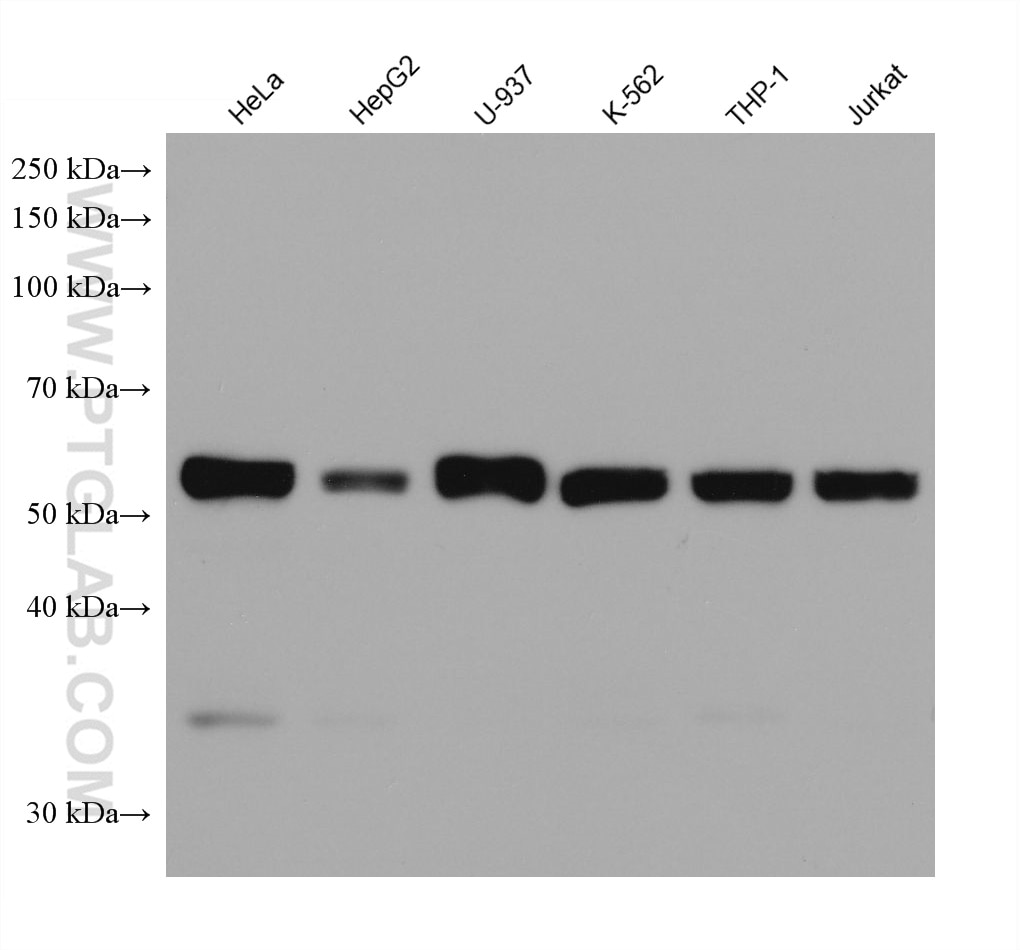
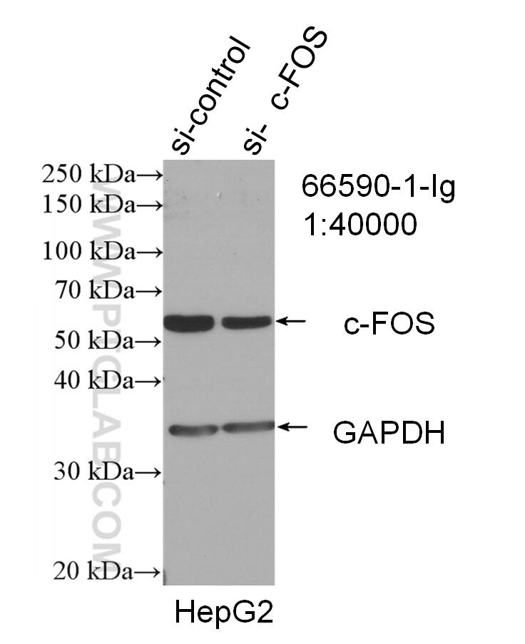
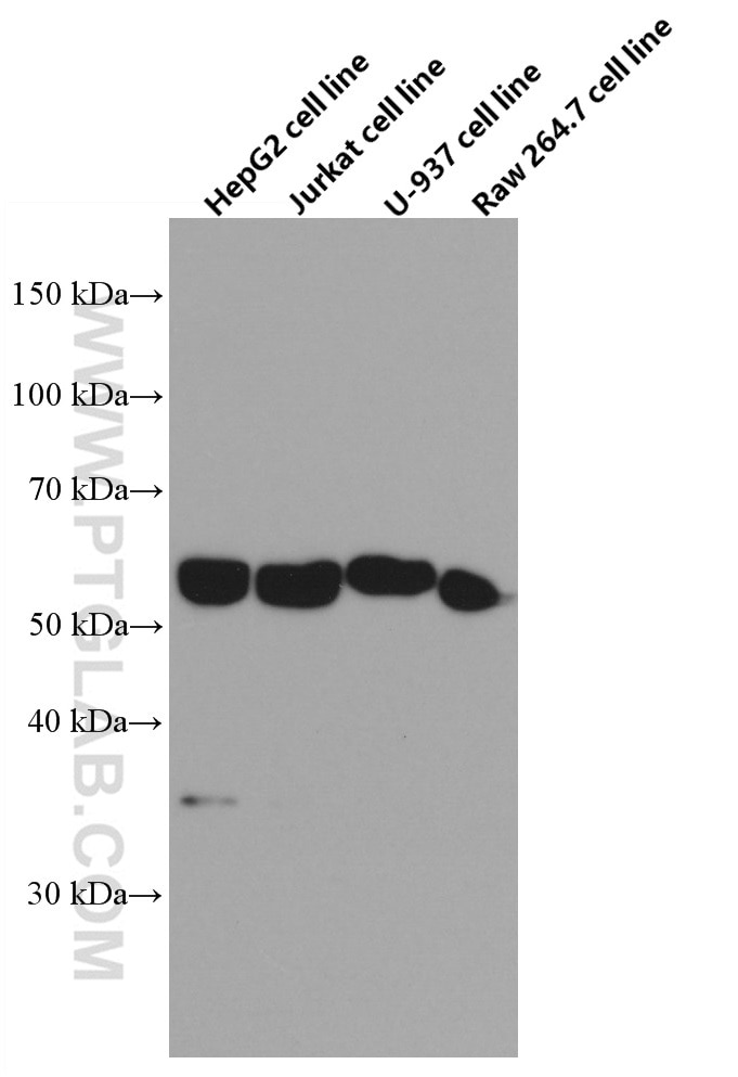
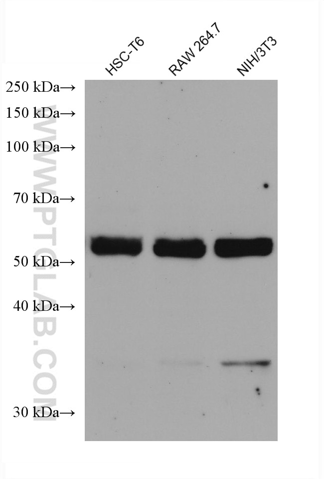
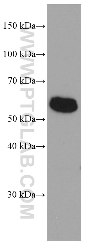
| Positive WB detected in | HeLa cells, HepG2 cells, HSC-T6 cells, Jurkat cells, U-937 cells, RAW 264.7 cells, K-562 cells, THP-1 cells, NIH/3T3 cells |
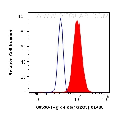
| Positive FC (Intra) detected in | HeLa cells |
c-Fos is an extremely short-lived intracellular protein that needs to be detected in fresh samples to prevent its degradation.
Recommended dilution
| Application | Dilution |
|---|---|
| Western Blot (WB) | WB : 1:5000-1:50000 |
| Flow Cytometry (FC) (INTRA) | FC (INTRA) : 0.5 ug per 10^6 cells in a 100 µl suspension |
| It is recommended that this reagent should be titrated in each testing system to obtain optimal results. | |
| Sample-dependent, Check data in validation data gallery. | |
Product Information
66590-1-Ig targets c-Fos in WB, IHC, FC (Intra), IP, CoIP, ELISA applications and shows reactivity with human, mouse, rat samples.
| Tested Reactivity | human, mouse, rat |
| Cited Reactivity | human, mouse, rat, rabbit |
| Host / Isotype | Mouse / IgG1 |
| Class | Monoclonal |
| Type | Antibody |
| Immunogen |
CatNo: Ag24340 Product name: Recombinant human FOS protein Source: e coli.-derived, PGEX-4T Tag: GST Domain: 196-341 aa of BC004490 Sequence: ILAAHRPACKIPDDLGFPEEMSVASLDLTGGLPEVATPESEEAFTLPLLNDPEPKPSVEPVKSISSMELKTEPFDDFLFPASSRPSGSETARSVPDMDLSGSFYAADWEPLHSGSLGMGPMATELEPLCTPVVTCTPSCTAYTSSF Predict reactive species |
| Full Name | FOS |
| Calculated Molecular Weight | 41 kDa |
| Observed Molecular Weight | 55-60 kDa |
| GenBank Accession Number | BC004490 |
| Gene Symbol | c-Fos |
| Gene ID (NCBI) | 2353 |
| RRID | AB_2881950 |
| Conjugate | Unconjugated |
| Form | Liquid |
| Purification Method | Protein G purification |
| UNIPROT ID | P01100 |
| Storage Buffer | PBS with 0.02% sodium azide and 50% glycerol, pH 7.3. |
| Storage Conditions | Store at -20°C. Stable for one year after shipment. Aliquoting is unnecessary for -20oC storage. 20ul sizes contain 0.1% BSA. |
Background Information
c-Fos, also named as FOS and G0/G1 switch regulatory protein 7, is a 380 amino acid protein, which contains 1 bZIP (basic-leucine zipper) domain and belongs to the bZIP family. c-Fos is expressed at very low levels in quiescent cells. When cells are stimulated to reenter growth, c-Fos undergo 2 waves of expression, the first one peaks 7.5 minutes following FBS induction. At this stage, the c-Fos protein is localized endoplasmic reticulum. The second wave of expression occurs at about 20 minutes after induction and peaks at 1 hour. At this stage, the c-FOS protein becomes nuclear. c-Fos is a very short-lived intracellular protein, which is very easy to degrade. The calculated molecular weight of c-Fos is 40 kDa, but Phosphorylated c-Fos protein is about 60-65 kDa. It is involved in important cellular events, including cell proliferation, differentiation and survival; genes associated with hypoxia; and angiogenesis; which makes its dysregulation an important factor for cancer development. It can also induce a loss of cell polarity and epithelial-mesenchymal transition, leading to invasive and metastatic growth in mammary epithelial cells. Expression of c-Fos is an indirect marker of neuronal activity because c-Fos is often expressed when neurons fire action potentials. Upregulation of c-Fos mRNA in a neuron indicates recent activity.
Protocols
| Product Specific Protocols | |
|---|---|
| FC protocol for c-Fos antibody 66590-1-Ig | Download protocol |
| WB protocol for c-Fos antibody 66590-1-Ig | Download protocol |
| Standard Protocols | |
|---|---|
| Click here to view our Standard Protocols |
Publications
What published studies show
Anti-c-Fos (Cat# 66590-1-Ig) has 180 citations spanning over 90 journals, including Nature Communications, Advanced Materials, ACS Nano, EMBO Journal, Science Advances, eLife, Cell Reports, and Phytomedicine. Key research fields include oncology, neuroscience (pain, neurodegeneration, anxiety), bone metabolism/osteoclastogenesis, immunology/inflammation, traditional Chinese medicine pharmacology, cardiology, and renal disease. Applications are predominantly Western blot, supplemented by IHC, IP, and CoIP.
| Species | Application | Title |
|---|---|---|
Adv Mater Noninvasive Optogenetics Realized by iPSC-Derived Tentacled Carrier in Alzheimer's Disease Treatment | ||
Mil Med Res Propionate and butyrate attenuate macrophage pyroptosis and osteoclastogenesis induced by CoCrMo alloy particles | ||
Bioact Mater Mantis shrimp saddle-mimetic amorphous calcium (zinc) phosphate/chitin scaffolds with superior mechanical properties and bioactivity for bone regeneration. | ||
Nat Commun Paraventricular hypothalamic RUVBL2 neurons suppress appetite by enhancing excitatory synaptic transmission in distinct neurocircuits | ||
Nat Commun Proteogenomics of different urothelial bladder cancer stages reveals distinct molecular features for papillary cancer and carcinoma in situ |
Reviews
What customers say
Users report mixed results with this c-Fos antibody. One reviewer rated it 4/5 for IF on brain slices at 1:750, though it failed on Adipo-cleared whole brain. Another gave 3/5, noting inconsistent nuclear vs. cytoplasmic staining in human FFPE tissue. A third reviewer awarded 5/5 for IHC on paraffinized mouse brain cortex at 1:1000, reporting strong, specific neuronal signal that increased after restraint stress. Overall, the antibody performs well in IHC and some IF applications but may show variable specificity depending on tissue type and protocol.
The reviews below have been submitted by verified Proteintech customers who received an incentive for providing their feedback.
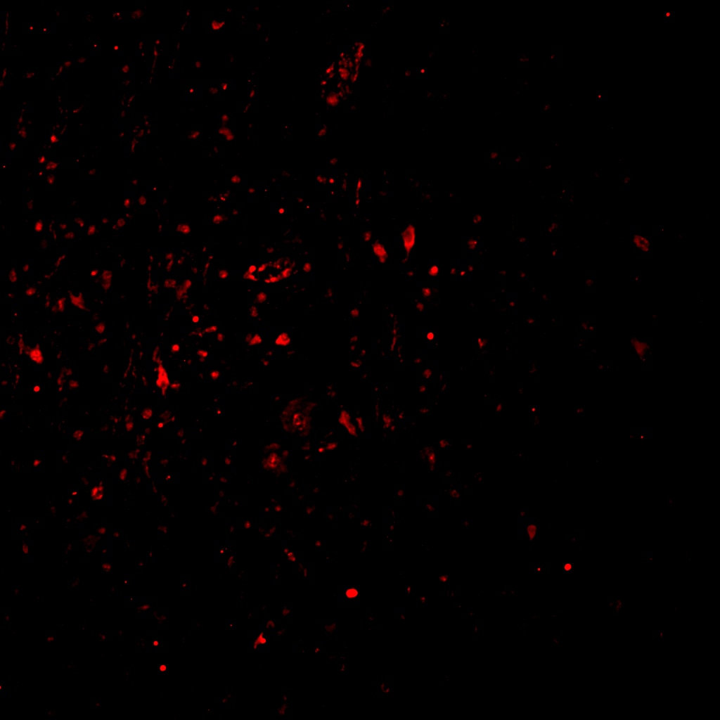
Pratiksha (Verified Customer) (03-28-2026) | The antibody worked on IF for brain slices at 1:750 but did not work for Adipo cleared whole brain.
|
Reyes (Verified Customer) (02-14-2025) | cFOS (a nuclear marker) did not perform as expected in my epileptic human FFPE tissue. It seems to appear in the nucleus in a few cells, but also a strong marking in the cyoplasm of others.
 |
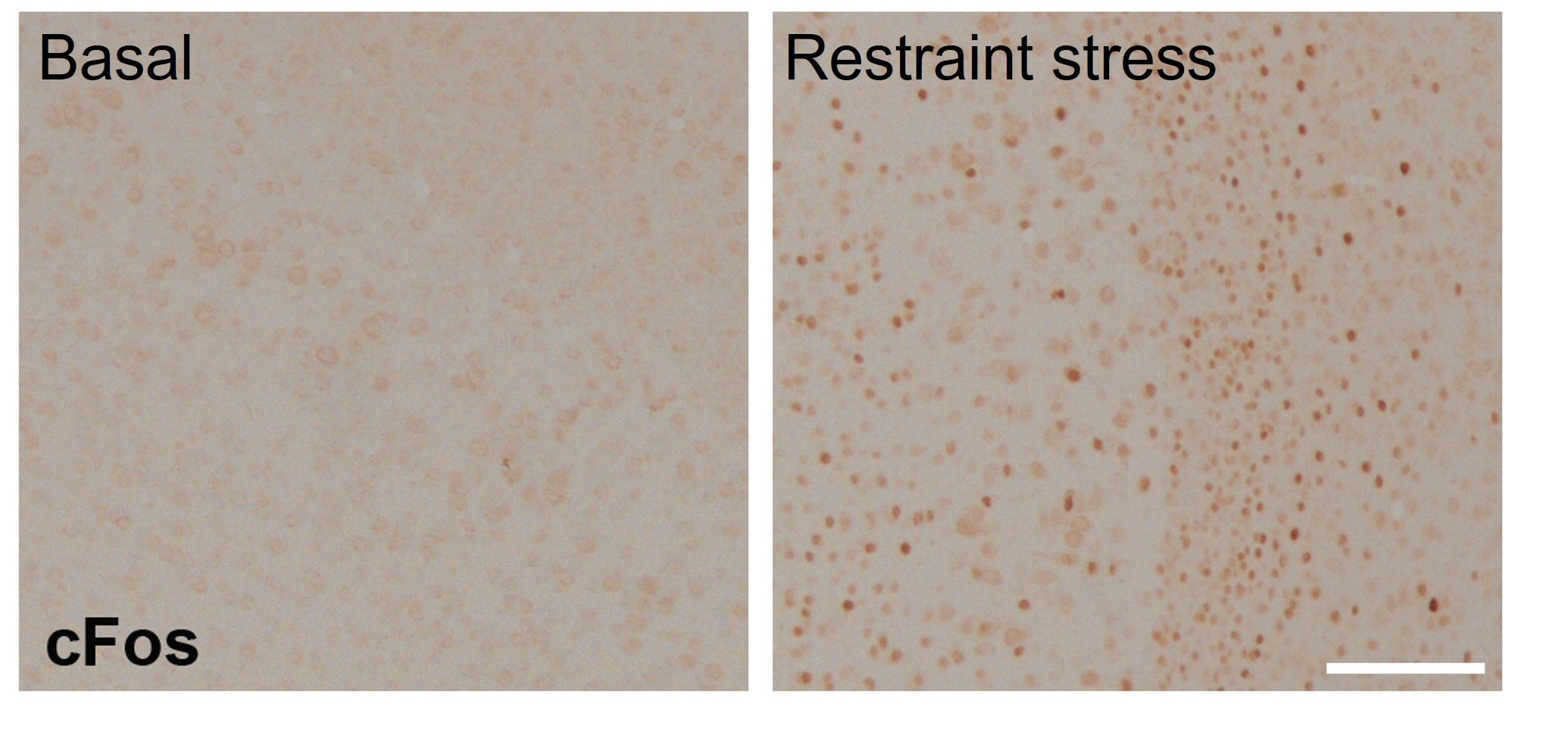
Tatyana (Verified Customer) (01-21-2023) | Suitable for IHC in the paraffinised brain sections of mice (cortex). Samples were fixed in 4% and standard IHC procedure with antigen retrieval and DAB detection was performed. Antibody was incubated at 1:1000 dilution overnight at 4C. Provided a good specific signal in neurons that increased after restraint stress.
 |







