Tested Applications
| Positive FC (Intra) detected in | HeLa cells |
Recommended dilution
| Application | Dilution |
|---|---|
| Flow Cytometry (FC) (INTRA) | FC (INTRA) : 0.80 ug per 10^6 cells in a 100 µl suspension |
| It is recommended that this reagent should be titrated in each testing system to obtain optimal results. | |
| Sample-dependent, Check data in validation data gallery. | |
Product Information
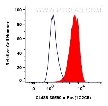
CL488-66590 targets c-Fos in FC (Intra) applications and shows reactivity with human, mouse, rat samples.
| Tested Reactivity | human, mouse, rat |
| Host / Isotype | Mouse / IgG1 |
| Class | Monoclonal |
| Type | Antibody |
| Immunogen |
CatNo: Ag24340 Product name: Recombinant human FOS protein Source: e coli.-derived, PGEX-4T Tag: GST Domain: 196-341 aa of BC004490 Sequence: ILAAHRPACKIPDDLGFPEEMSVASLDLTGGLPEVATPESEEAFTLPLLNDPEPKPSVEPVKSISSMELKTEPFDDFLFPASSRPSGSETARSVPDMDLSGSFYAADWEPLHSGSLGMGPMATELEPLCTPVVTCTPSCTAYTSSF Predict reactive species |
| Full Name | FOS |
| Calculated Molecular Weight | 41 kDa |
| Observed Molecular Weight | 55-60 kDa |
| GenBank Accession Number | BC004490 |
| Gene Symbol | c-Fos |
| Gene ID (NCBI) | 2353 |
| RRID | AB_2934481 |
| Conjugate | CoraLite® Plus 488 Fluorescent Dye |
| Excitation/Emission Maxima Wavelengths | 493 nm / 522 nm |
| Excitation Laser | Blue laser (488 nm) |
| Form | Liquid |
| Purification Method | Protein G purification |
| UNIPROT ID | P01100 |
| Storage Buffer | PBS with 50% glycerol, 0.05% Proclin300, 0.5% BSA, pH 7.3. |
| Storage Conditions | Store at -20°C. Avoid exposure to light. Stable for one year after shipment. Aliquoting is unnecessary for -20oC storage. |
Background Information
c-FOS, also named as Proto-oncogene c-Fos and G0/G1 switch regulatory protein 7, is a 380 amino acid protein, which contains 1 bZIP (basic-leucine zipper) domain and belongs to the bZIP family. FOS is expressed at very low levels in quiescent cells. When cells are stimulated to reenter growth, FOS undergo 2 waves of expression, the first one peaks 7.5 minutes following FBS induction. At this stage, the FOS protein is localized endoplasmic reticulum. The second wave of expression occurs at about 20 minutes after induction and peaks at 1 hour. At this stage, the FOS protein becomes nuclear. The calculated molecular weight of FOS is 40 kDa, but Phosphorylated FOS protein is about 60-65 kDa. It is involved in important cellular events, including cell proliferation, differentiation and survival; genes associated with hypoxia; and angiogenesis; which makes its dysregulation an important factor for cancer development. It can also induce a loss of cell polarity and epithelial-mesenchymal transition, leading to invasive and metastatic growth in mammary epithelial cells. Expression of c-fos is an indirect marker of neuronal activity because c-fos is often expressed when neurons fire action potentials. Upregulation of c-fos mRNA in a neuron indicates recent activity
Protocols
| Product Specific Protocols | |
|---|---|
| FC protocol for CL Plus 488 c-Fos antibody CL488-66590 | Download protocol |
| Standard Protocols | |
|---|---|
| Click here to view our Standard Protocols |
Reviews
The reviews below have been submitted by verified Proteintech customers who received an incentive for providing their feedback.
FH Lindsay (Verified Customer) (09-04-2025) | This pre-conjugated c-fos primary works very well in mouse brain tissue, and limits the time required for single or dual color IHC staining with secondaries. It may not be as stable as others, though, as we found the conjugate to stop working after fewer freeze-thaw cycles than the non-conjugated fos primaries from PT.
|