





Tested Applications
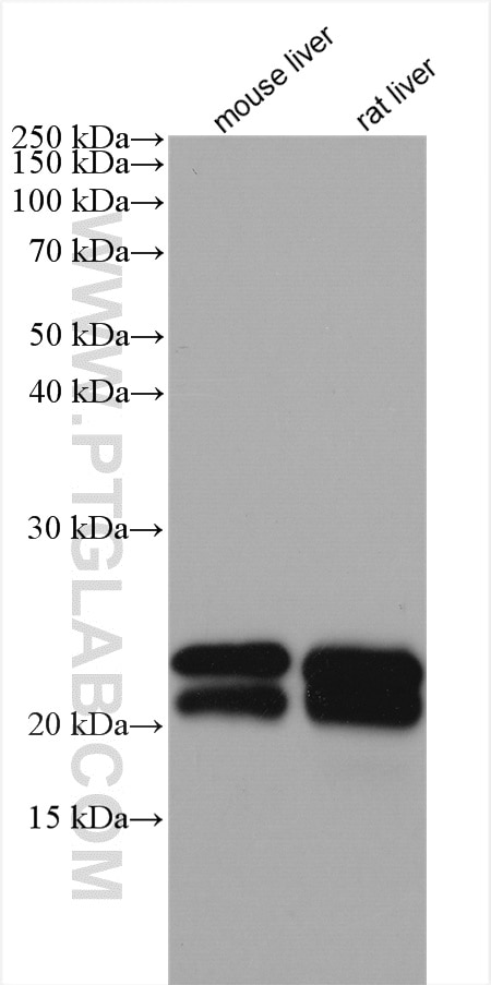
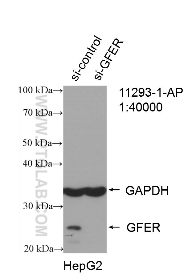
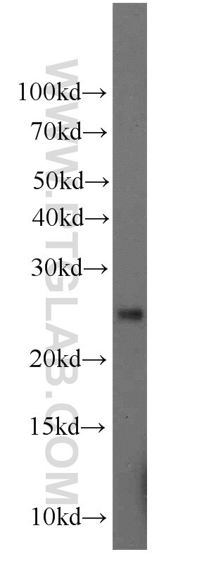
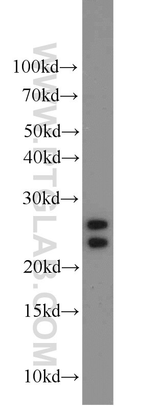
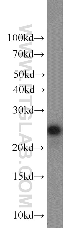
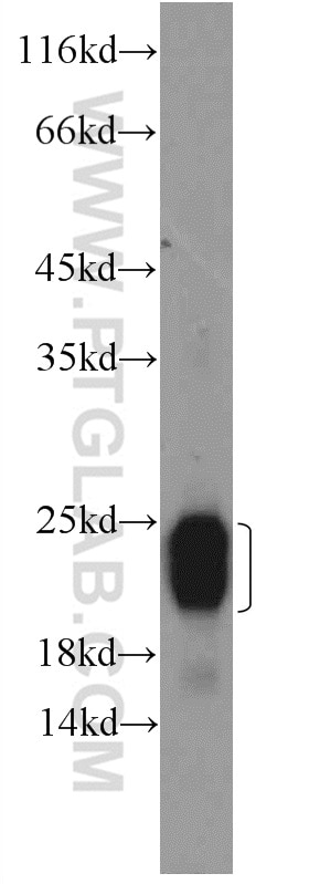
| Positive WB detected in | mouse liver tissue, A431 cells, A375 cells, rat liver tissue, HepG2 cells |
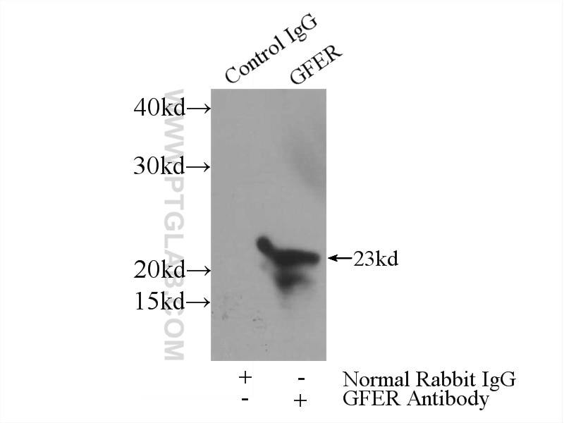
| Positive IP detected in | mouse liver tissue |
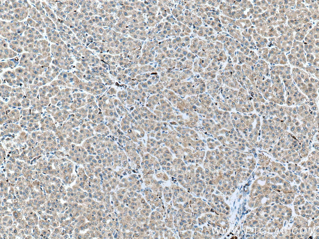
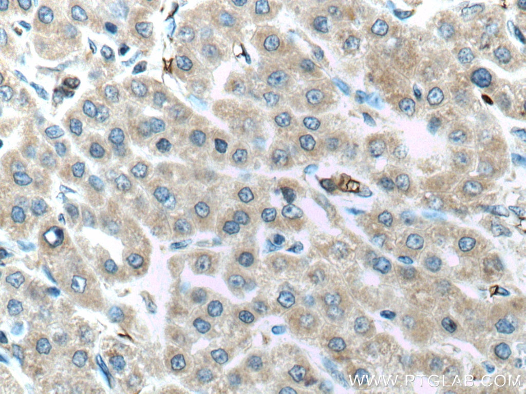
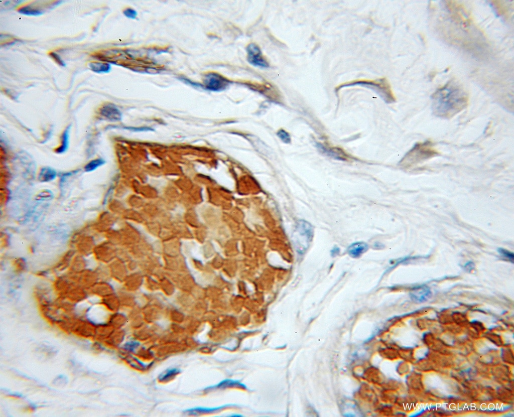
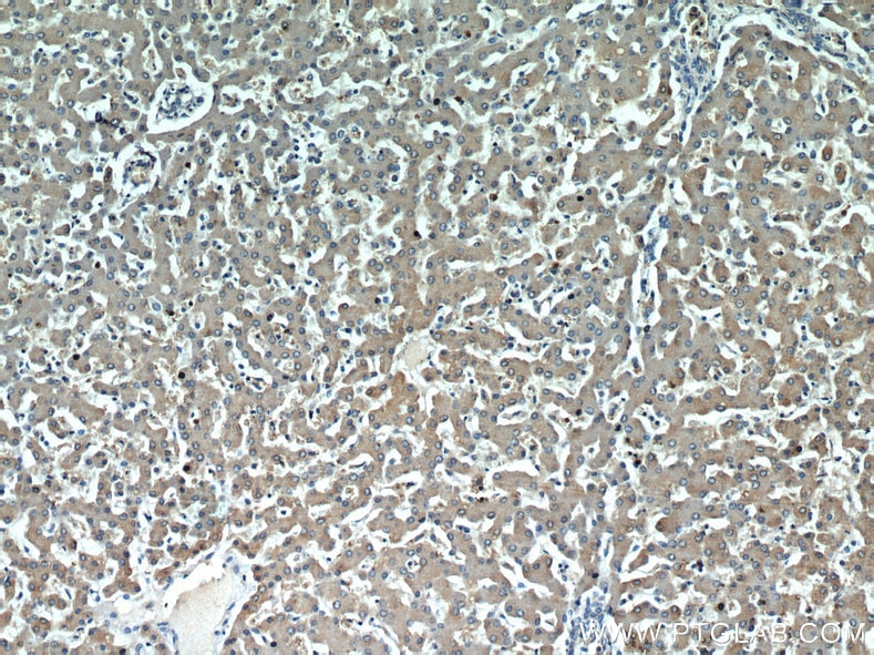
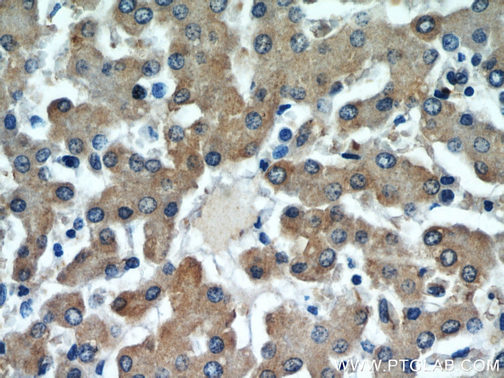
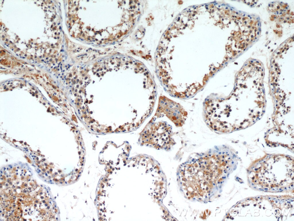
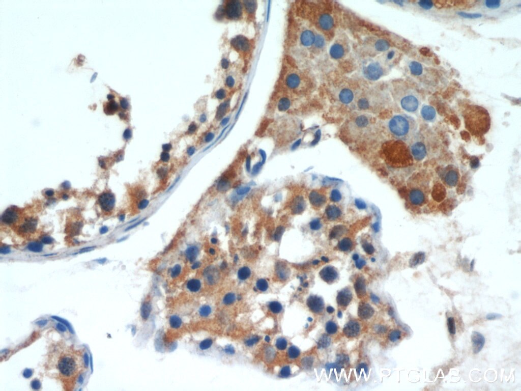
| Positive IHC detected in | human liver cancer tissue, mouse stomach tissue, rat stomach tissue, human liver tissue, human colon cancer tissue, human testis tissue, human stomach cancer tissue Note: suggested antigen retrieval with TE buffer pH 9.0; (*) Alternatively, antigen retrieval may be performed with citrate buffer pH 6.0 |
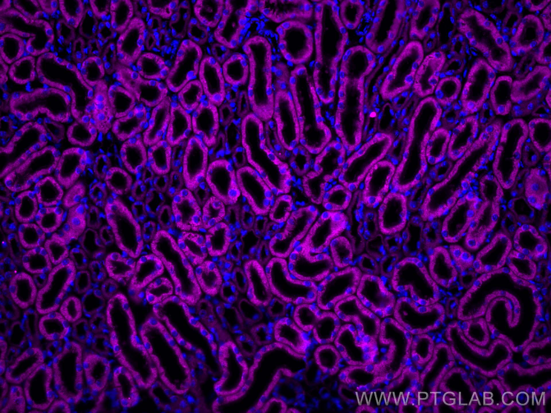
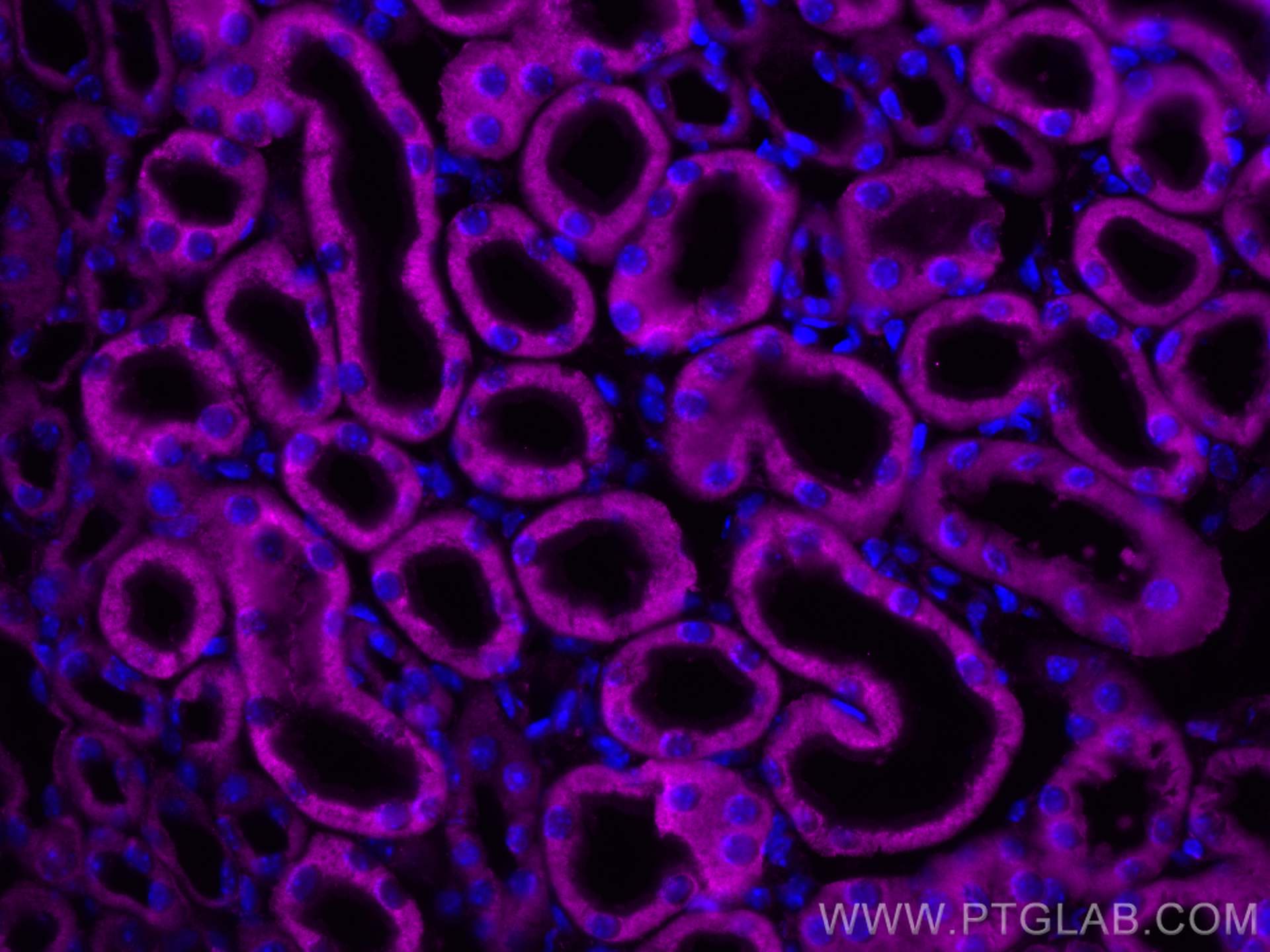
| Positive IF-P detected in | mouse kidney tissue |
| Positive FC (Intra) detected in | A431 cells, HepG2 cells |
Recommended dilution
| Application | Dilution |
|---|---|
| Western Blot (WB) | WB : 1:1000-1:8000 |
| Immunoprecipitation (IP) | IP : 0.5-4.0 ug for 1.0-3.0 mg of total protein lysate |
| Immunohistochemistry (IHC) | IHC : 1:50-1:500 |
| Immunofluorescence (IF)-P | IF-P : 1:50-1:500 |
| Flow Cytometry (FC) (INTRA) | FC (INTRA) : 0.25 ug per 10^6 cells in a 100 µl suspension |
| It is recommended that this reagent should be titrated in each testing system to obtain optimal results. | |
| Sample-dependent, Check data in validation data gallery. | |
Product Information
11293-1-AP targets ALR in WB, IHC, IF-P, FC (Intra), IP, ELISA applications and shows reactivity with human, mouse, rat samples.
| Tested Reactivity | human, mouse, rat |
| Cited Reactivity | human, mouse, rat |
| Host / Isotype | Rabbit / IgG |
| Class | Polyclonal |
| Type | Antibody |
| Immunogen |
CatNo: Ag1840 Product name: Recombinant human GFER protein Source: e coli.-derived, PGEX-4T Tag: GST Domain: 1-125 aa of BC028348 Sequence: MRTQQKRDTKFREDCPPDREELGRHSWAVLHTLAAYYPDLPTPEQQQDMAQFIHLFSKFYPCEECAEDLRKRLCRNHPDTRTRACFTQWLCHLHNEVNRKLGKPDFDCSKVDERWRDGWKDGSCD Predict reactive species |
| Full Name | growth factor, augmenter of liver regeneration |
| Calculated Molecular Weight | 15 kDa, 23 kDa |
| Observed Molecular Weight | 23-25 kDa |
| GenBank Accession Number | BC028348 |
| Gene Symbol | GFER |
| Gene ID (NCBI) | 2671 |
| RRID | AB_2109970 |
| Conjugate | Unconjugated |
| Form | Liquid |
| Purification Method | Antigen affinity purification |
| UNIPROT ID | P55789 |
| Storage Buffer | PBS with 0.02% sodium azide and 50% glycerol, pH 7.3. |
| Storage Conditions | Store at -20°C. Stable for one year after shipment. Aliquoting is unnecessary for -20oC storage. 20ul sizes contain 0.1% BSA. |
Background Information
GFER (FAD-linked sulfhydryl oxidase) is also named as ALR, HERV1, HPO. It plays an important role in the disulfide relay system (DRS) in human mitochondria. The GFER gene codes for 2 distinct isoforms that are probably synthesized from the same mRNA with the use of different initiation codons. The long isoform (205 amino acids, 23/21 kD) is located mainly in the mitochondrial intermembrane space and exists under nonreducing and nondenaturing conditions as a homodimer and a heterodimer. The shorter isoform (125 amino acids, 15 kD), which lacks 80 amino acids at its N terminus compared to the longer isoform, is present predominantly in the nucleus (PMID: 19409522, 24880092, 21152698).
Protocols
| Product Specific Protocols | |
|---|---|
| IF protocol for ALR antibody 11293-1-AP | Download protocol |
| IHC protocol for ALR antibody 11293-1-AP | Download protocol |
| IP protocol for ALR antibody 11293-1-AP | Download protocol |
| WB protocol for ALR antibody 11293-1-AP | Download protocol |
| Standard Protocols | |
|---|---|
| Click here to view our Standard Protocols |
Publications
What published studies show
The anti-ALR (Augmenter of Liver Regeneration) antibody (Cat# 11293-1-AP) has 32 citations across journals including Cell Stem Cell, Cancer Research, Hepatology, Redox Biology, Oncogene, FASEB Journal, eLife, and PLoS ONE. Research fields span mitochondrial biology, liver disease (fibrosis, steatosis, hepatocellular carcinoma), ferroptosis, redox homeostasis, neurodegeneration, kidney injury, iron homeostasis, and ischemia-reperfusion injury, with applications primarily in Western blot, IF, IHC, and IP.
| Species | Application | Title |
|---|---|---|
Cell Stem Cell Disrupting Mitochondrial Copper Distribution Inhibits Leukemic Stem Cell Self-Renewal.
| ||
Cancer Res GFER Represents a Target for Dual Disruption of Redox Homeostasis and Reactivation of the Immune Response in Pancreatic Adenocarcinoma.
| ||
Hepatology Hepatic stimulator substance resists hepatic ischemia/reperfusion injury by regulating Drp1 translocation and activation.
| ||
Redox Biol Lead aggravates Alzheimer's disease pathology via mitochondrial copper accumulation regulated by COX17 | ||
Adv Sci (Weinh) Single-Cell Transcriptomics Unravels Growth Factor Erv1-Like Mediated Ferroptosis as a Key Driver of Intestinal Epithelial Dysfunction in Ulcerative Colitis | ||
Oncogene Imbalance in ALR ubiquitination accelerates the progression of nonalcoholic steatohepatitis to hepatocellular carcinoma |
Reviews
What customers say
The single review for this antibody is highly negative (1/5). The reviewer used it for Western Blot at 1:1000 dilution on isolated rat liver mitochondria and whole cell lysates, reporting very poor signal intensity even after extended exposure times. They also observed a high number of non-specific bands. Attempts to improve results by adjusting PAGE, blotting, lysate, and reduction protocols, as well as overnight incubation, were unsuccessful. Overall, the reviewer expressed strong dissatisfaction with the antibody's performance.
The reviews below have been submitted by verified Proteintech customers who received an incentive for providing their feedback.
Yaschar (Verified Customer) (06-26-2018) | sample: 20 µg highly purified rat liver mitochondriaVery poor signal intensity even after 30(!) mintues of exposure (see image). Note that this is the best signal I was able to get.Barely any signal in different whole cell lysate.Even after changing the PAGE and Blotting protocols the signal does not get better, incubating over night does not help either.High number of unspecified bands, different lysate and reduction protocols did not help in this regard.I am extremely unsatisfied with this antibody.
 |




















