Filter:


Tested Applications
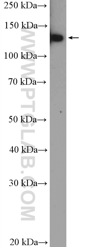
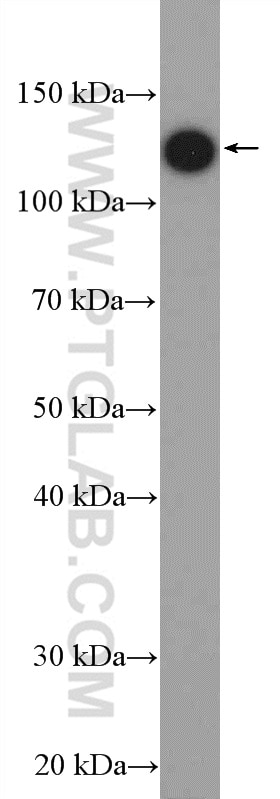
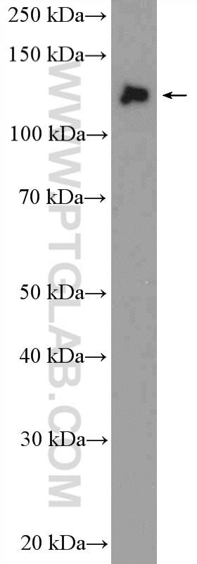
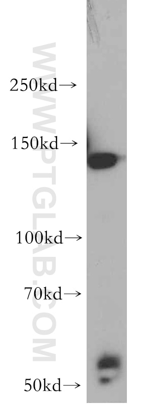
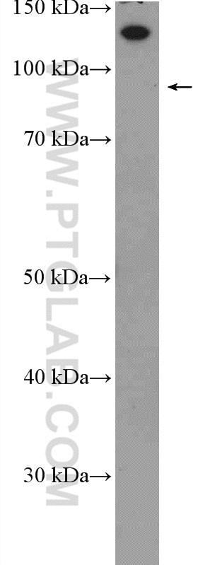
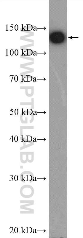
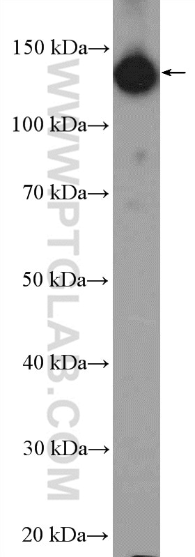
| Positive WB detected in | HeLa cells, mouse brain tissue, HepG2 cells, rat brain tissue, Jurkat cells |
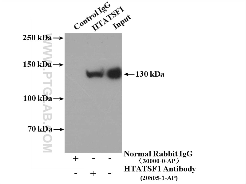
| Positive IP detected in | HepG2 cells |
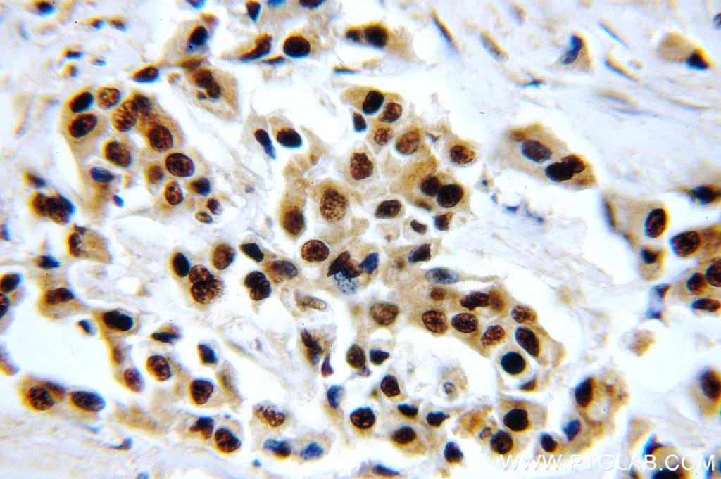
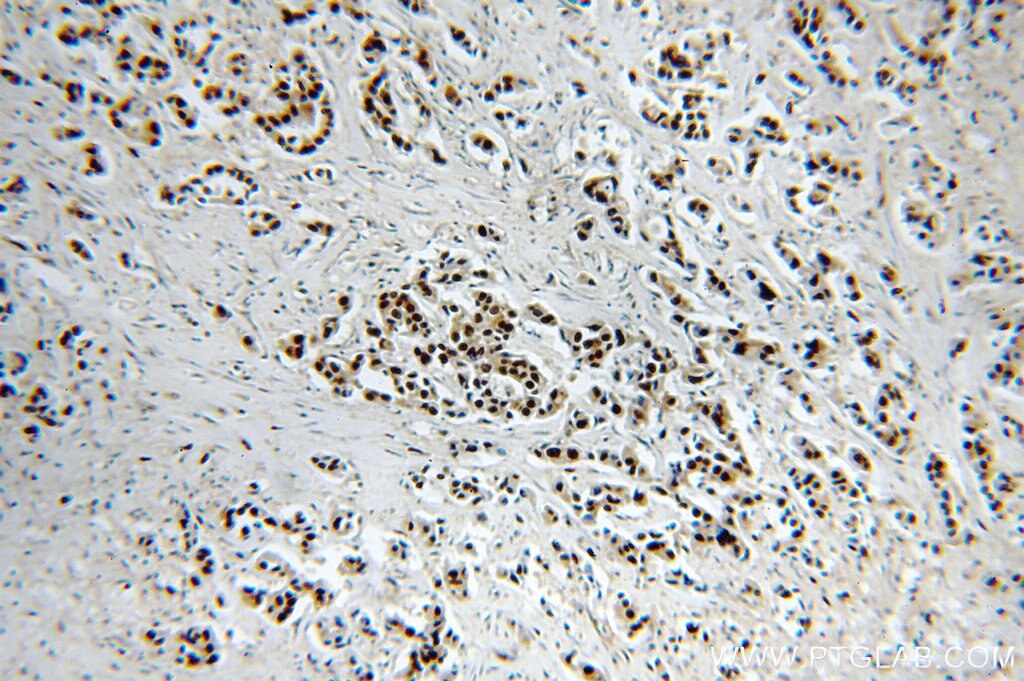
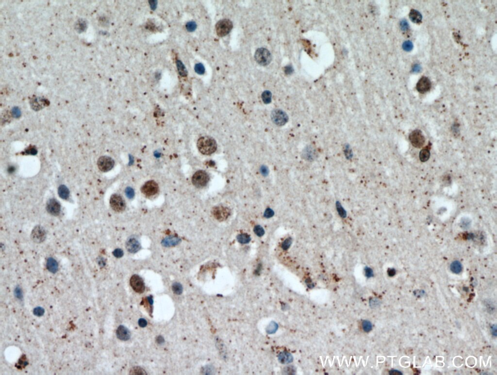
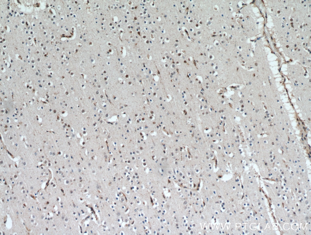
| Positive IHC detected in | human breast cancer tissue, human brain tissue Note: suggested antigen retrieval with TE buffer pH 9.0; (*) Alternatively, antigen retrieval may be performed with citrate buffer pH 6.0 |
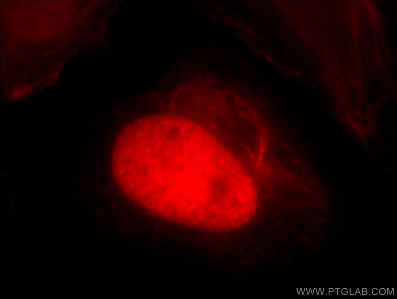
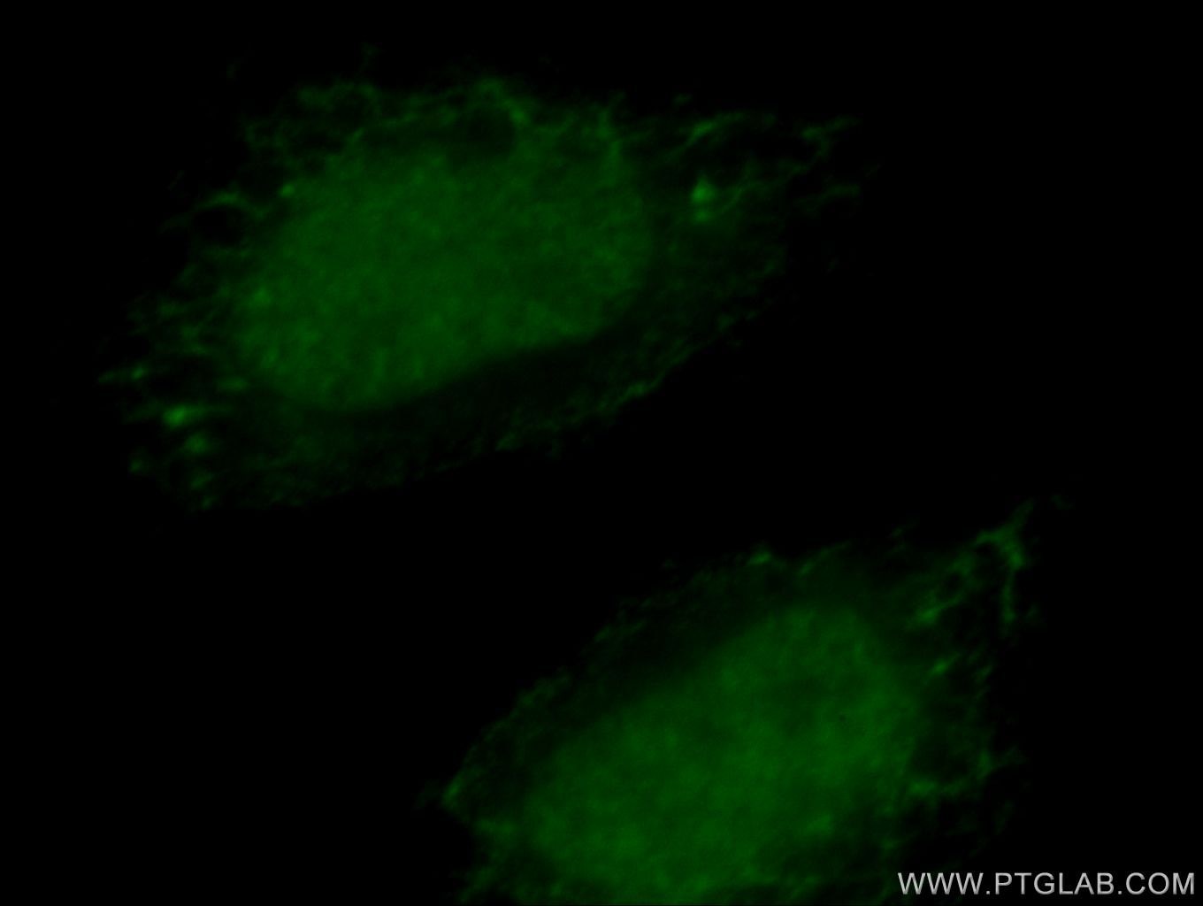
| Positive IF/ICC detected in | Hela cells |
Recommended dilution
| Application | Dilution |
|---|---|
| Western Blot (WB) | WB : 1:1000-1:4000 |
| Immunoprecipitation (IP) | IP : 0.5-4.0 ug for 1.0-3.0 mg of total protein lysate |
| Immunohistochemistry (IHC) | IHC : 1:20-1:200 |
| Immunofluorescence (IF)/ICC | IF/ICC : 1:10-1:100 |
| It is recommended that this reagent should be titrated in each testing system to obtain optimal results. | |
| Sample-dependent, Check data in validation data gallery. | |
Published Applications
| KD/KO | See 3 publications below |
| WB | See 4 publications below |
| IHC | See 1 publications below |
| IP | See 1 publications below |
| CoIP | See 1 publications below |
Product Information
20805-1-AP targets HTATSF1 in WB, IHC, IF/ICC, IP, CoIP, ELISA applications and shows reactivity with human, mouse, rat samples.
| Tested Reactivity | human, mouse, rat |
| Cited Reactivity | human, mouse |
| Host / Isotype | Rabbit / IgG |
| Class | Polyclonal |
| Type | Antibody |
| Immunogen |
CatNo: Ag14744 Product name: Recombinant human HTATSF1 protein Source: e coli.-derived, PGEX-4T Tag: GST Domain: 1-350 aa of BC009896 Sequence: MSGTNLDGNDEFDEQLRMQELYGDGKDGDTQTDAGGEPDSLGQQPTDTPYEWDLDKKAWFPKITEDFIATYQANYGFSNDGASSSTANVEDVHARTAEEPPQEKAPEPTDARKKGEKRKAESGWFHVEEDRNTNVYVSGLPPDITVDEFIQLMSKFGIIMRDPQTEEFKVKLYKDNQGNLKGDGLCCYLKRESVELALKLLDEDEIRGYKLHVEVAKFQLKGEYDASKKKKKCKDYKKKLSMQQKQLDWRPERRAGPSRMRHERVVIIKNMFHPMDFEDDPLVLNEIREDLRVECSKFGQIRKLLLFDRHPDGVASVSFRDPEEADYCIQTLDGRWFGGRQITAQAWDGT Predict reactive species |
| Full Name | HIV-1 Tat specific factor 1 |
| Calculated Molecular Weight | 755 aa, 86 kDa |
| Observed Molecular Weight | 130-140 kDa |
| GenBank Accession Number | BC009896 |
| Gene Symbol | HTATSF1 |
| Gene ID (NCBI) | 27336 |
| RRID | AB_10695767 |
| Conjugate | Unconjugated |
| Form | Liquid |
| Purification Method | Antigen affinity purification |
| UNIPROT ID | O43719 |
| Storage Buffer | PBS with 0.02% sodium azide and 50% glycerol, pH 7.3. |
| Storage Conditions | Store at -20°C. Stable for one year after shipment. Aliquoting is unnecessary for -20oC storage. 20ul sizes contain 0.1% BSA. |
Protocols
| Product Specific Protocols | |
|---|---|
| IF protocol for HTATSF1 antibody 20805-1-AP | Download protocol |
| IHC protocol for HTATSF1 antibody 20805-1-AP | Download protocol |
| IP protocol for HTATSF1 antibody 20805-1-AP | Download protocol |
| WB protocol for HTATSF1 antibody 20805-1-AP | Download protocol |
| Standard Protocols | |
|---|---|
| Click here to view our Standard Protocols |
Publications
| Species | Application | Title |
|---|---|---|
Mol Cell A PARylation-phosphorylation cascade promotes TOPBP1 loading and RPA-RAD51 exchange in homologous recombination.
| ||
Nucleic Acids Res Multiple P-TEFbs cooperatively regulate the release of promoter-proximally paused RNA polymerase II. | ||
Cell Death Discov Metabolic protein phosphoglycerate kinase 1 confers lung cancer migration by directly binding HIV Tat specific factor 1.
| ||
Cell Insight HTATSF1 regulates innate antiviral immune response by orchestrating TRAF3-IRF3 and TRAF6-NF-κB pathways.
| ||
Cell Rep ArreSTick motif controls β-arrestin-binding stability and extends phosphorylation-dependent β-arrestin interactions to non-receptor proteins |