at dilution of 1:8000 incubated at room temperature for 1.5 hours.")
 at dilution of 1:1000 incubated at room temperature for 1.5 hours.")
 at dilution of 1:300 incubated at room temperature for 1.5 hours.")
 at dilution of 1:300 incubated at room temperature for 1.5 hours.")
 at dilution of 1:300 incubated at room temperature for 1.5 hours.")
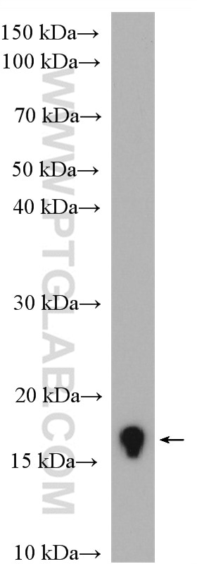
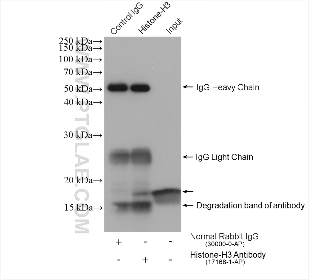
 with MCF-7 cells lysate 2120 ug.")
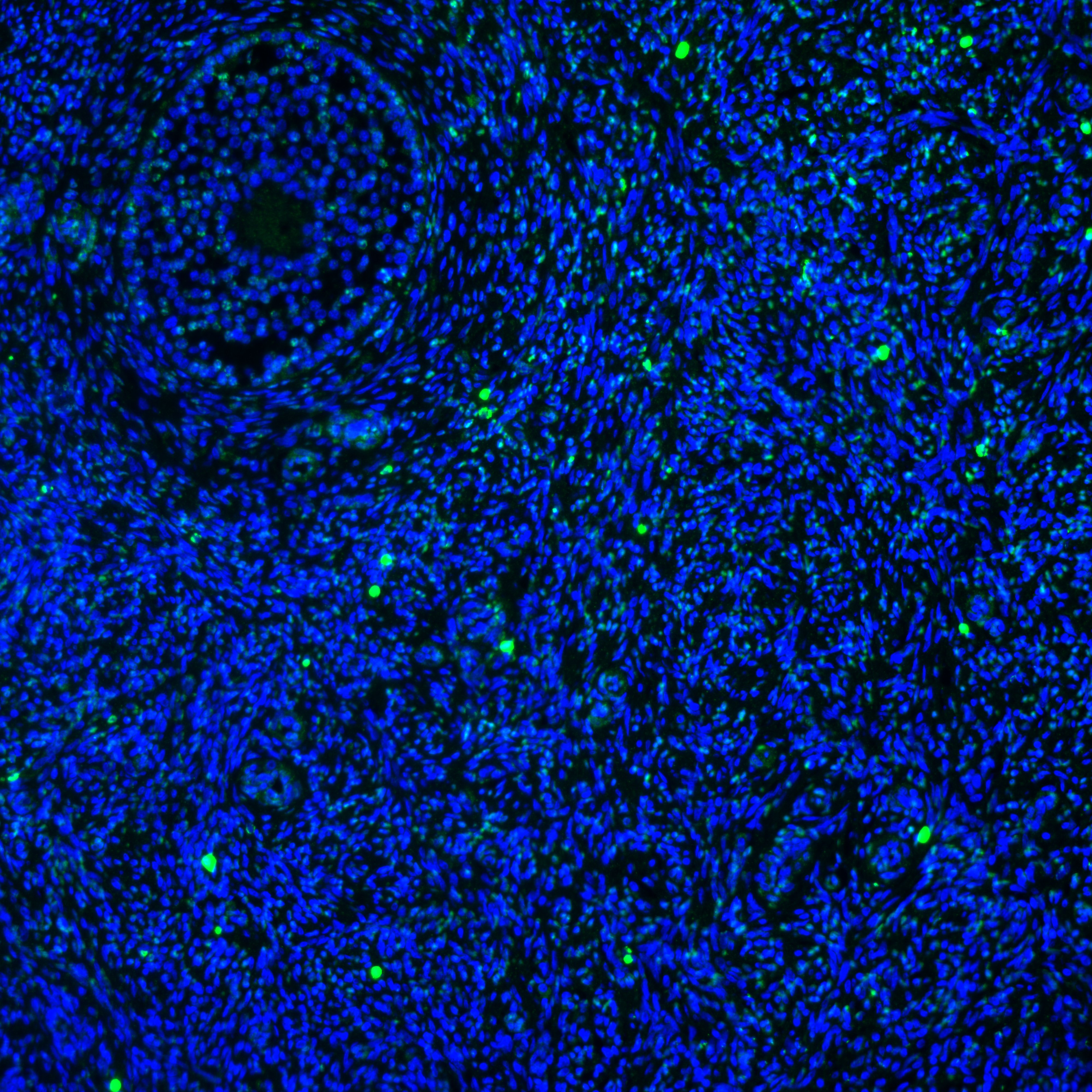
 at dilution of 1:200 (under 40x lens). Heat mediated antigen retrieval with Tris-EDTA buffer (pH 9.0).")
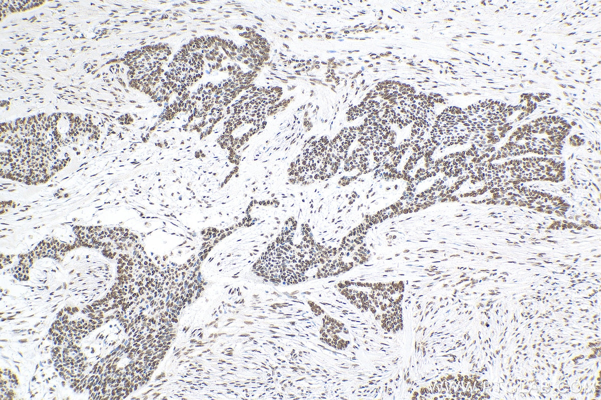
 at dilution of 1:200 (under 10x lens). Heat mediated antigen retrieval with Tris-EDTA buffer (pH 9.0).")
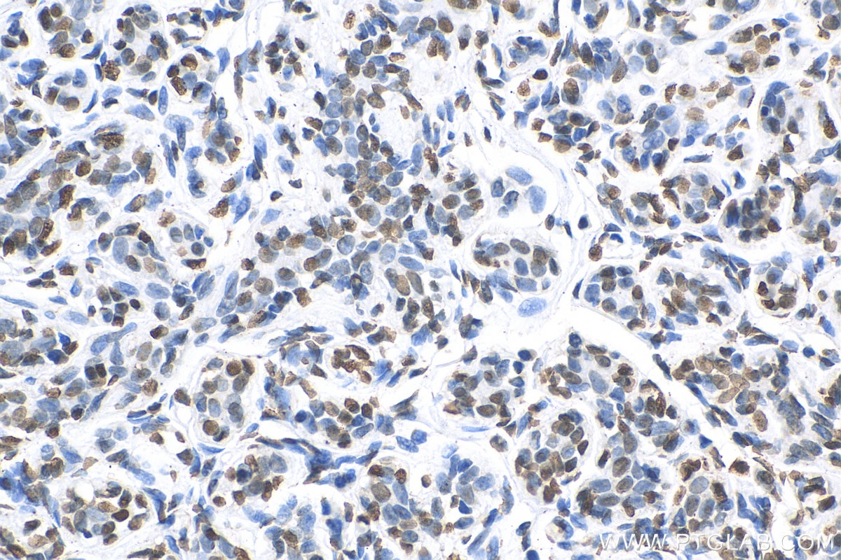
 at dilution of 1:200 (under 40x lens).")
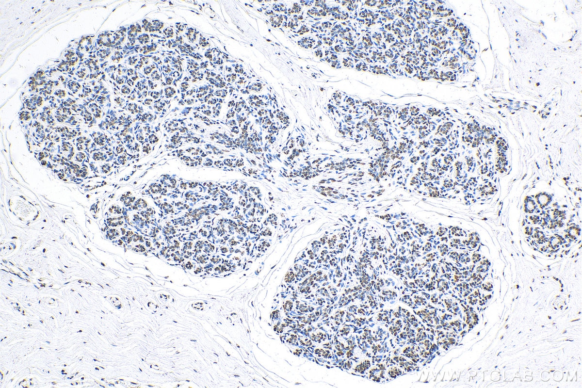
 at dilution of 1:200 (under 10x lens).")
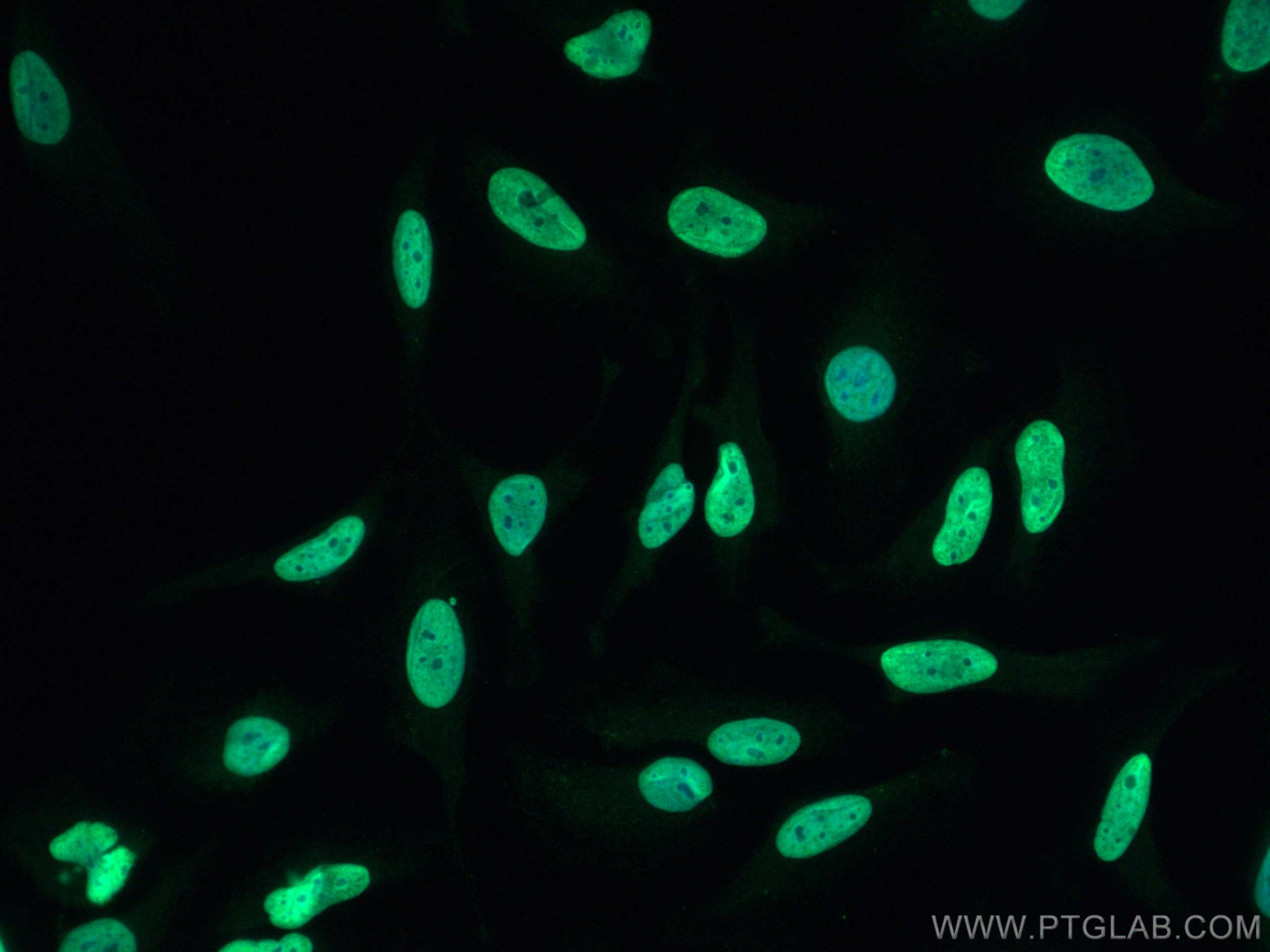
 at dilution of 1:2000 (under 10x lens). Heat mediated antigen retrieval with Tris-EDTA buffer (pH 9.0).")
 at dilution of 1:2000 (under 40x lens). Heat mediated antigen retrieval with Tris-EDTA buffer (pH 9.0).")
 fixed HeLa cells using Histone-H3 antibody (17168-1-AP) at dilution of 1:200 and CoraLite®488-Conjugated AffiniPure Goat Anti-Rabbit IgG(H+L).")
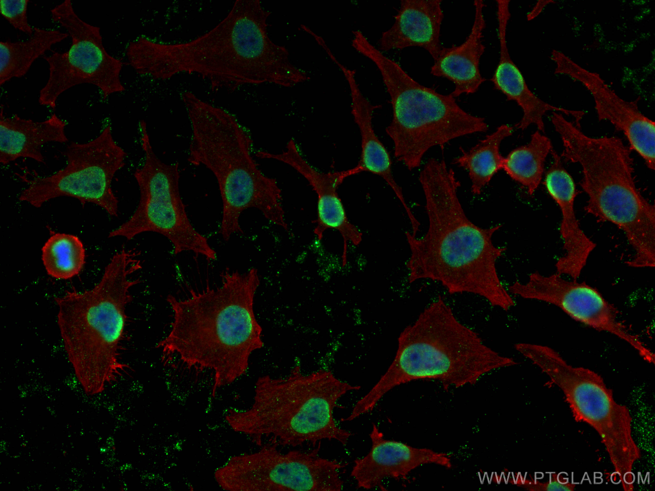
 fixed HeLa cells using Histone H3 antibody (17168-1-AP) at dilution of 1:1200 and CoraLite®488-Conjugated AffiniPure Goat Anti-Rabbit IgG(H+L), Beta Actin antibody (66009-1-Ig, Clone: 2D4H5, red).")
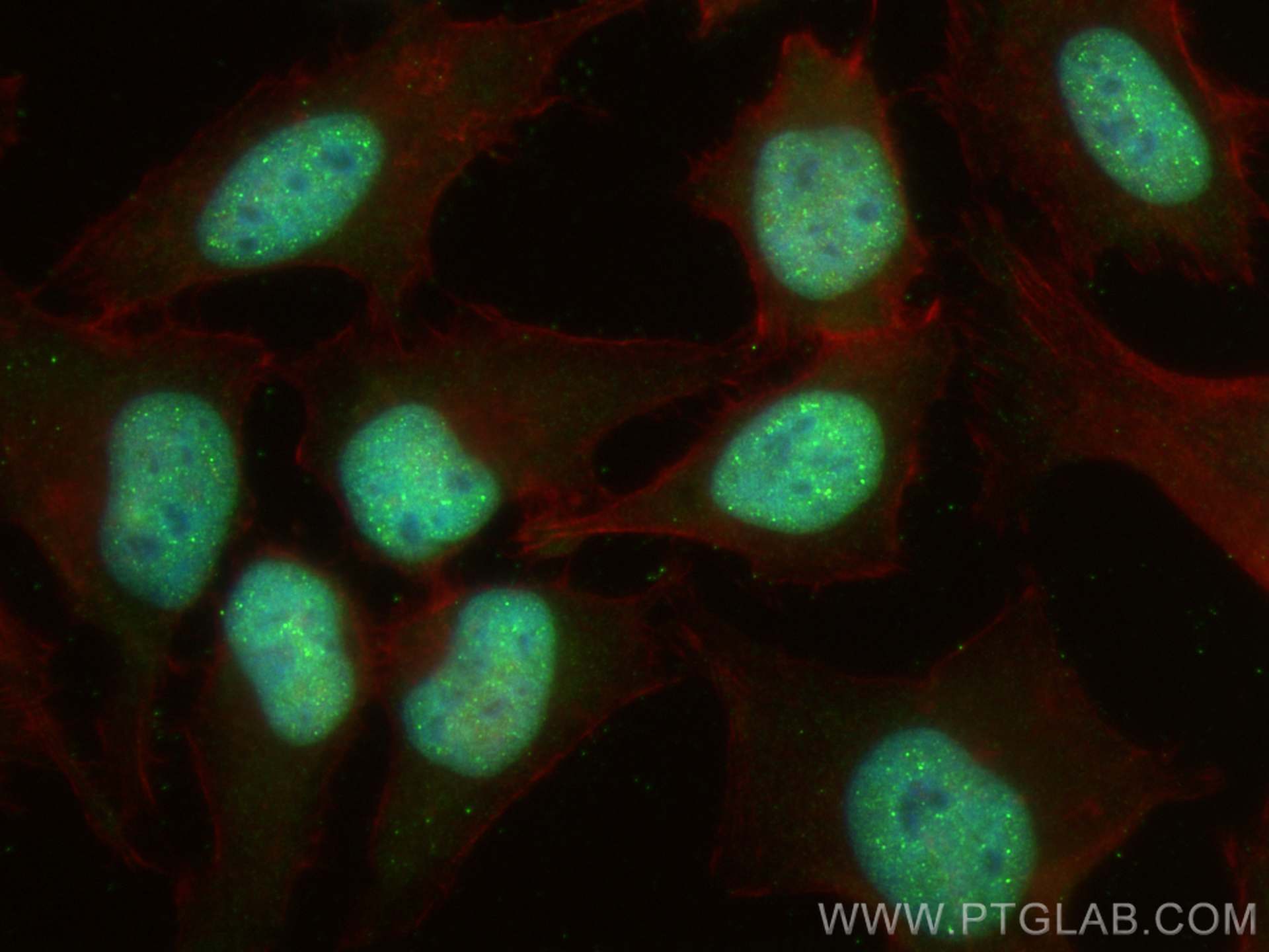
 fixed HeLa cells using Histone-H3 antibody (17168-1-AP) at dilution of 1:200 and CoraLite®488-Conjugated AffiniPure Goat Anti-Rabbit IgG(H+L), CL594-Phalloidin (red).")
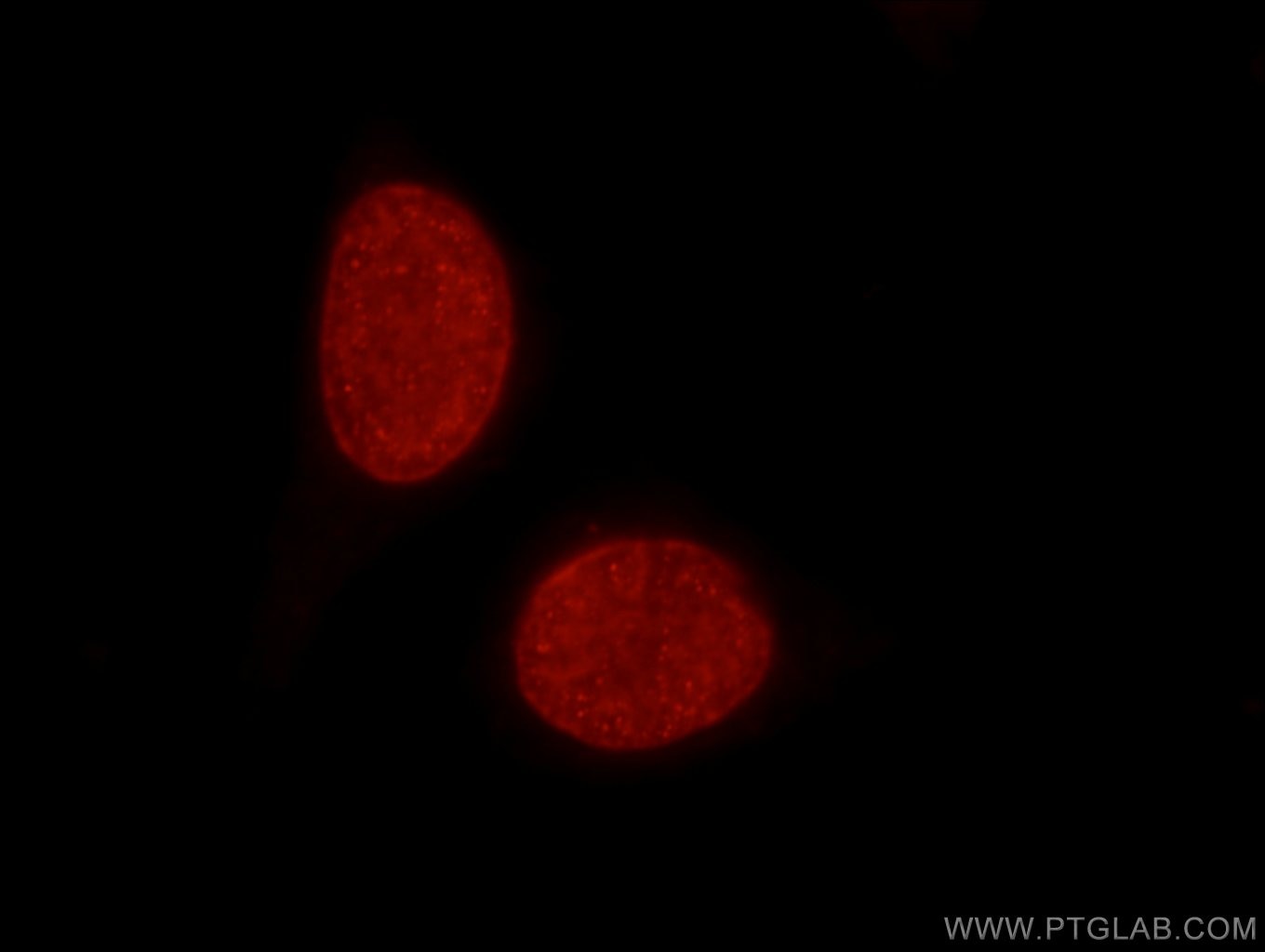
 at dilution of 1:50 and Rhodamine-Goat anti-Rabbit IgG.")
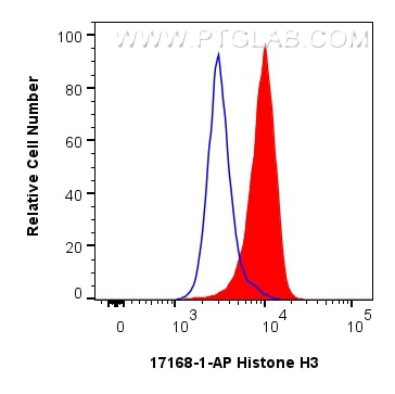
 and CoraLite®488-Conjugated Goat Anti-Rabbit IgG(H+L) (SA00013-2)(red), or 0.25 ug rabbit IgG isotype control (blue). Cells were fixed with 4% PFA and permeabilized with Flow Cytometry Perm Buffer.")
"Histone H3 Antibodies" Comparison
View side-by-side comparison of Histone H3 antibodies from other vendors to find the one that best suits your research needs.
Tested Applications
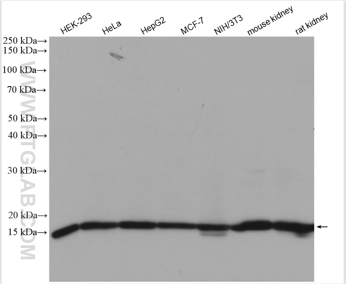
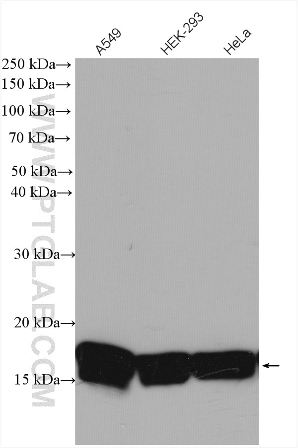
| Positive WB detected in | HEK-293 cells, A549 cells, mouse skeletal muscle tissue, mouse liver tissue, mouse brain tissue, Hela cells, HepG cells, MCF-7 cells, NIH/3T3 cells, mouse kidney tissue, rat kidney tissue |
| Positive IP detected in | MCF-7 cells |
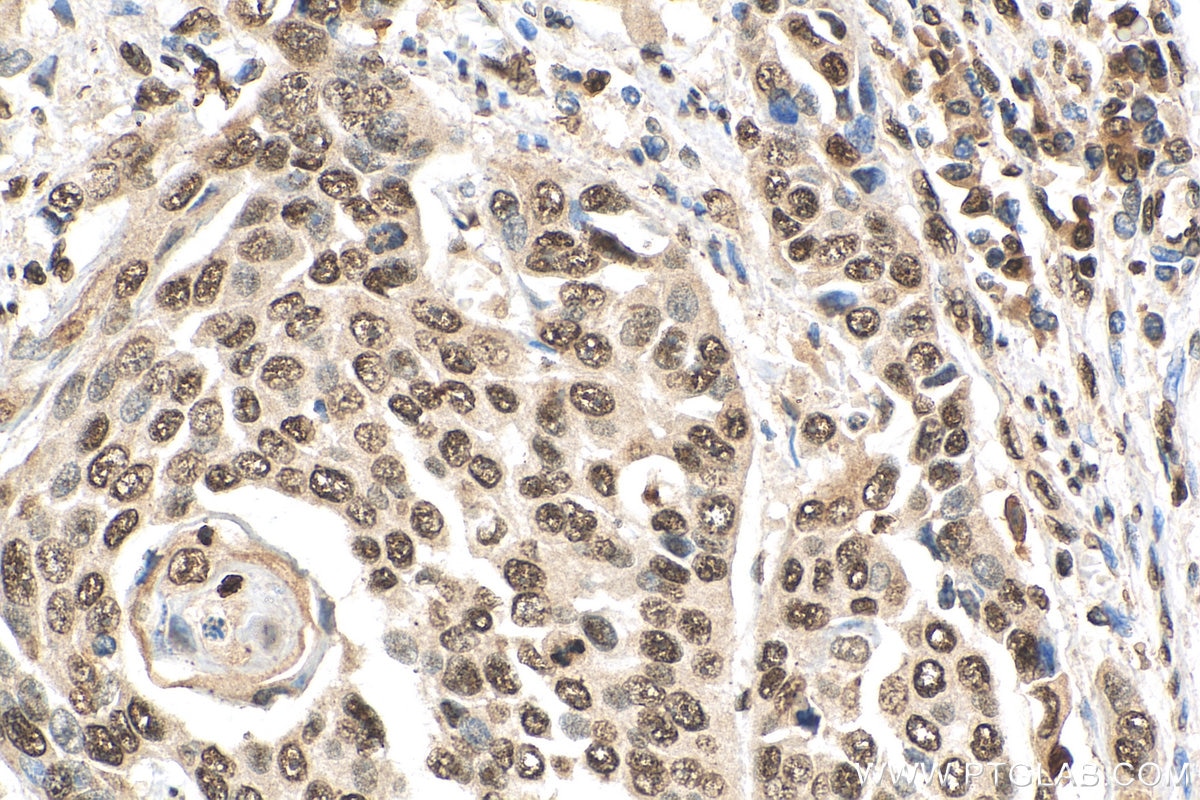
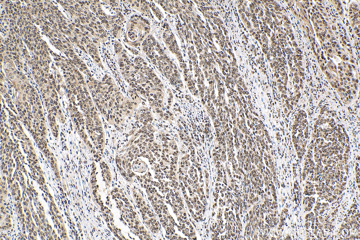
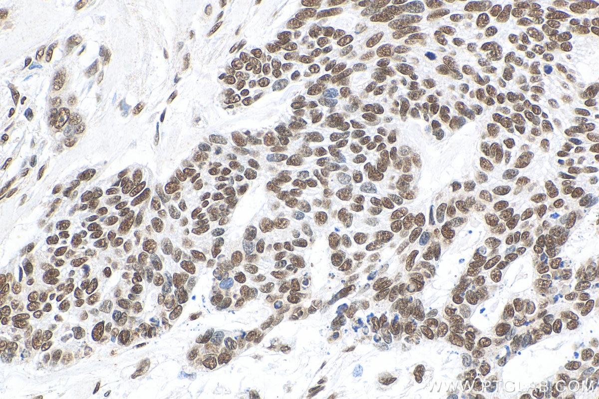
| Positive IHC detected in | human oesophagus cancer tissue, human skin cancer tissue, human breast cancer tissue Note: suggested antigen retrieval with TE buffer pH 9.0; (*) Alternatively, antigen retrieval may be performed with citrate buffer pH 6.0 |
| Positive IF/ICC detected in | HeLa cells |
| Positive FC (Intra) detected in | HeLa cells |
Recommended dilution
| Application | Dilution |
|---|---|
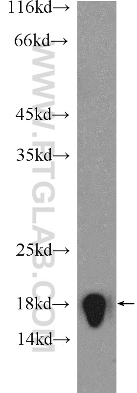
| Western Blot (WB) | WB : 1:2000-1:16000 |
| Immunoprecipitation (IP) | IP : 0.5-4.0 ug for 1.0-3.0 mg of total protein lysate |
| Immunohistochemistry (IHC) | IHC : 1:50-1:500 |
| Immunofluorescence (IF)/ICC | IF/ICC : 1:600-1:2400 |
| Flow Cytometry (FC) (INTRA) | FC (INTRA) : 0.25 ug per 10^6 cells in a 100 µl suspension |
| It is recommended that this reagent should be titrated in each testing system to obtain optimal results. | |
| Sample-dependent, Check data in validation data gallery. | |
Published Applications
| KD/KO | See 2 publications below |
| WB | See 1011 publications below |
| IHC | See 3 publications below |
| IF | See 10 publications below |
| IP | See 1 publications below |
| CoIP | See 2 publications below |
| ChIP | See 9 publications below |
Product Information
17168-1-AP targets Histone H3 in WB, IHC, IF/ICC, FC (Intra), IP, CoIP, ChIP, ELISA applications and shows reactivity with human, mouse, rat samples.
| Tested Reactivity | human, mouse, rat |
| Cited Reactivity | human, mouse, rat, monkey, chicken, zebrafish, goat, fish, arabidopsis, yellow catfish |
| Host / Isotype | Rabbit / IgG |
| Class | Polyclonal |
| Type | Antibody |
| Immunogen |
CatNo: Ag10644 Product name: Recombinant human Histone-H3 protein Source: e coli.-derived, PET28a Tag: 6*His Domain: 1-136 aa of BC015544 Sequence: MARTKQTARKSTGGKAPRKQLATKAARKSAPATGGVKKPHRYRPGTVALREIRRYQKSTELLIRKLPFQRLVREIAQDFKTDLRFQSSAVMALQEASEAYLVGLFEDTNLCAIHAKRVTIMPKDIQLARRIRGERA Predict reactive species |
| Full Name | histone cluster 2, H3a |
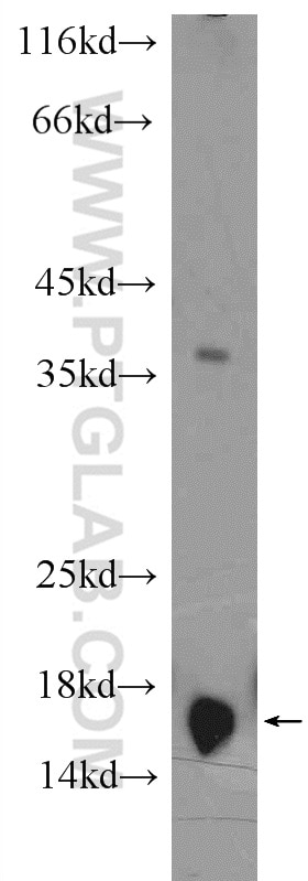
| Calculated Molecular Weight | 136 aa, 15 kDa |
| Observed Molecular Weight | 15-17 kDa |
| GenBank Accession Number | BC015544 |
| Gene Symbol | Histone H3 |
| Gene ID (NCBI) | 333932 |
| RRID | AB_2716755 |
| Conjugate | Unconjugated |
| Form | Liquid |
| Purification Method | Antigen affinity purification |
| UNIPROT ID | Q71DI3 |
| Storage Buffer | PBS with 0.02% sodium azide and 50% glycerol, pH 7.3. |
| Storage Conditions | Store at -20°C. Stable for one year after shipment. Aliquoting is unnecessary for -20oC storage. 20ul sizes contain 0.1% BSA. |
Background Information
Histone-H3, histone cluster 2, H3a is the core component of nucleosome. Nucleosomes wrap and compact DNA into chromatin, limiting DNA accessibility to the cellular machinery which requires DNA as a template. Histones thereby play a central role in transcription regulation, DNA repair, DNA replication and chromosomal stability. DNA accessibility is regulated via a complex set of post-translational modifications of histones, also called histone code, and nucleosome remodeling. Histone-H3 is expressed during S phase; then expression strongly decreases as cell division slows down during the process of differentiation.
Protocols
| Product Specific Protocols | |
|---|---|
| IF protocol for Histone H3 antibody 17168-1-AP | Download protocol |
| IHC protocol for Histone H3 antibody 17168-1-AP | Download protocol |
| IP protocol for Histone H3 antibody 17168-1-AP | Download protocol |
| WB protocol for Histone H3 antibody 17168-1-AP | Download protocol |
| Standard Protocols | |
|---|---|
| Click here to view our Standard Protocols |
Publications
| Species | Application | Title |
|---|---|---|
Signal Transduct Target Ther TRAF3 activates STING-mediated suppression of EV-A71 and target of viral evasion | ||
Mol Cancer Cell surface CD55 traffics to the nucleus leading to cisplatin resistance and stemness by inducing PRC2 and H3K27 trimethylation on chromatin in ovarian cancer | ||
Cell Targeting Epigenetic Crosstalk as a Therapeutic Strategy for EZH2-Aberrant Solid Tumors. |
Reviews
The reviews below have been submitted by verified Proteintech customers who received an incentive for providing their feedback.
FH Felipe (Verified Customer) (07-24-2025) | Histone H3 antibody is a reliable marker for detecting proliferating cells. After optimizing the dilution, I achieved good results in identifying proliferative cells in bovine ovarian tissue using an immunofluorescence assay.
 |
FH Christin (Verified Customer) (06-14-2025) | Antibody worked very vell for western blot using iPSC-derived macrophages and microglia
|
FH kis (Verified Customer) (02-21-2025) | This antibody gave u satisfactory results in our cell line for Western blot (WB) analysis.
|
FH Parijat (Verified Customer) (08-19-2024) | Worked on blot.
|
FH Wissem (Verified Customer) (06-27-2024) | Antibody works very well at detecting Histone H3 in extracts from U2OS, HCT116, HeLa, RPE1 and HEK293 cells - used at 1:10,000 to 1:15,000 incubated overnight at 4C
|
FH Roy (Verified Customer) (06-12-2024) | This antibody is great at detecting Histone H3 by WB (1/1000- 1h incubation at RT) with beautiful enrichment at the purified chromatin fraction.
|
FH Mi (Verified Customer) (06-25-2023) | It works very well in nuclear extracts from human brown adipocyte cells.
|
FH Veda (Verified Customer) (04-29-2022) | IF staining test in cryo sections of skin. It does not seem to work. Maybe it only stains the nuclear membrane.
|
FH Pradeep (Verified Customer) (09-19-2020) | Good antibody for Western blot. Selvaraju, V., Thirunavukkarasu, M., Joshi, M. et al. Deletion of newly described pro-survival molecule Pellino-1 increases oxidative stress, downregulates cIAP2/NF-κB cell survival pathway, reduces angiogenic response, and thereby aggravates tissue function in mouse ischemic models. Basic Res Cardiol 115, 45 (2020). https://doi.org/10.1007/s00395-020-0804-4
|
FH Tanusree (Verified Customer) (12-03-2019) | The antibody works great in Western blotting analysis.
|
FH Ruiting (Verified Customer) (11-11-2019) | I use the Proteintech antibodies almost every week, the antibodies never failed to me. Very gald to use their products with a great price.Ruiting
|
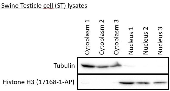
FH Robert (Verified Customer) (07-18-2019) | The Histon H3 antibody gives a signal in the three nuclear samples, but not in the cytoplasmic samples.
 |