Tested Applications
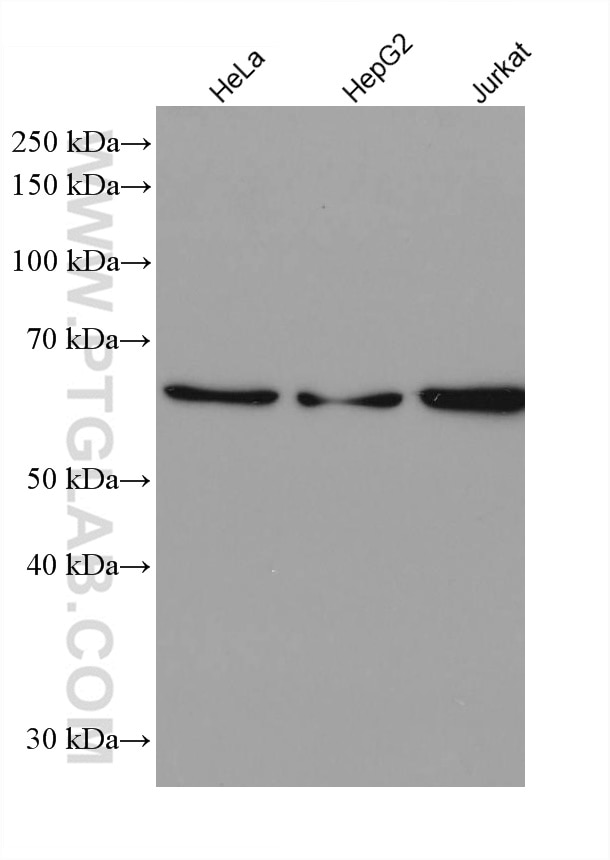
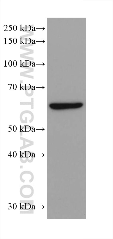
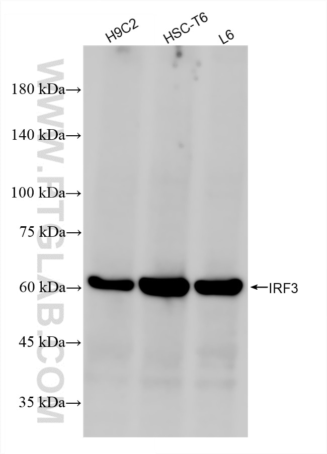
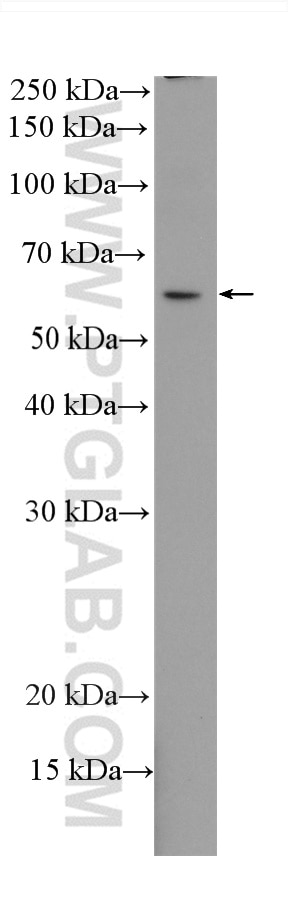
| Positive WB detected in | HeLa cells, H9C2 cells, HT-29 cells, RAW 264.7 cells, THP-1 cells, zebrafish tissue, HSC-T6 cells, L6 cells, HepG2 cells, Jurkat cells |
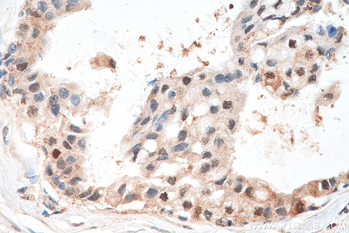
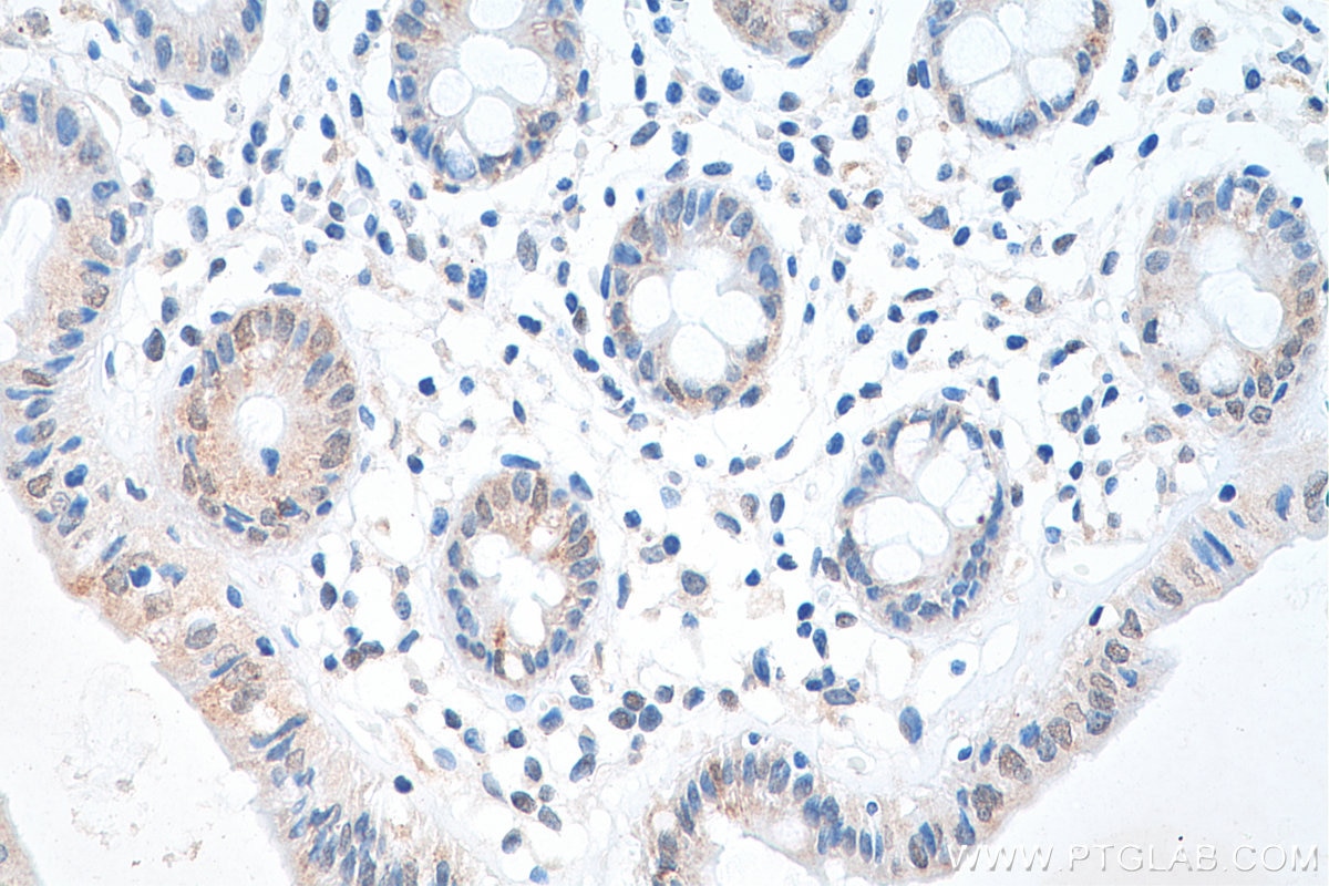
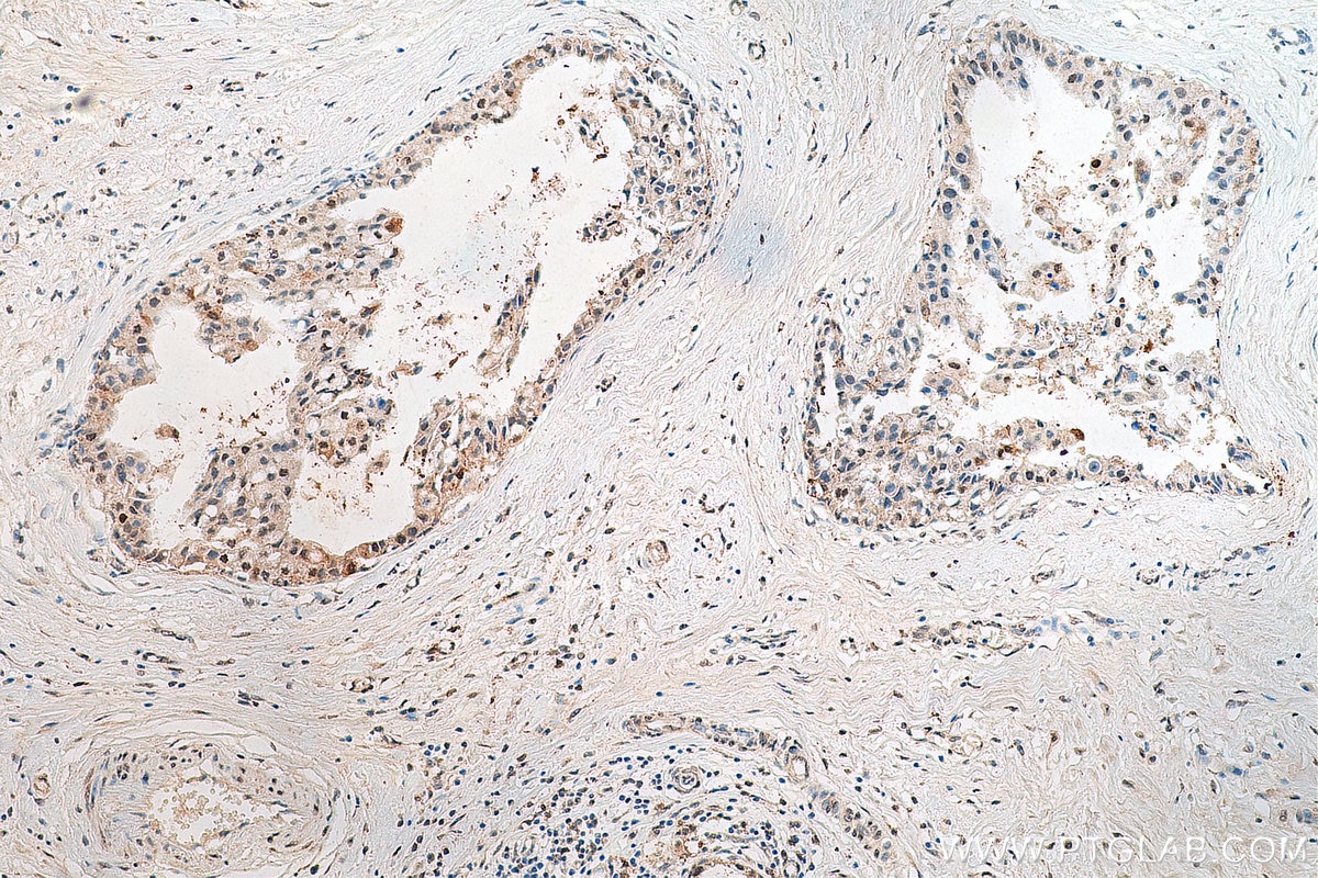
| Positive IHC detected in | human breast cancer tissue, human colon tissue Note: suggested antigen retrieval with TE buffer pH 9.0; (*) Alternatively, antigen retrieval may be performed with citrate buffer pH 6.0 |
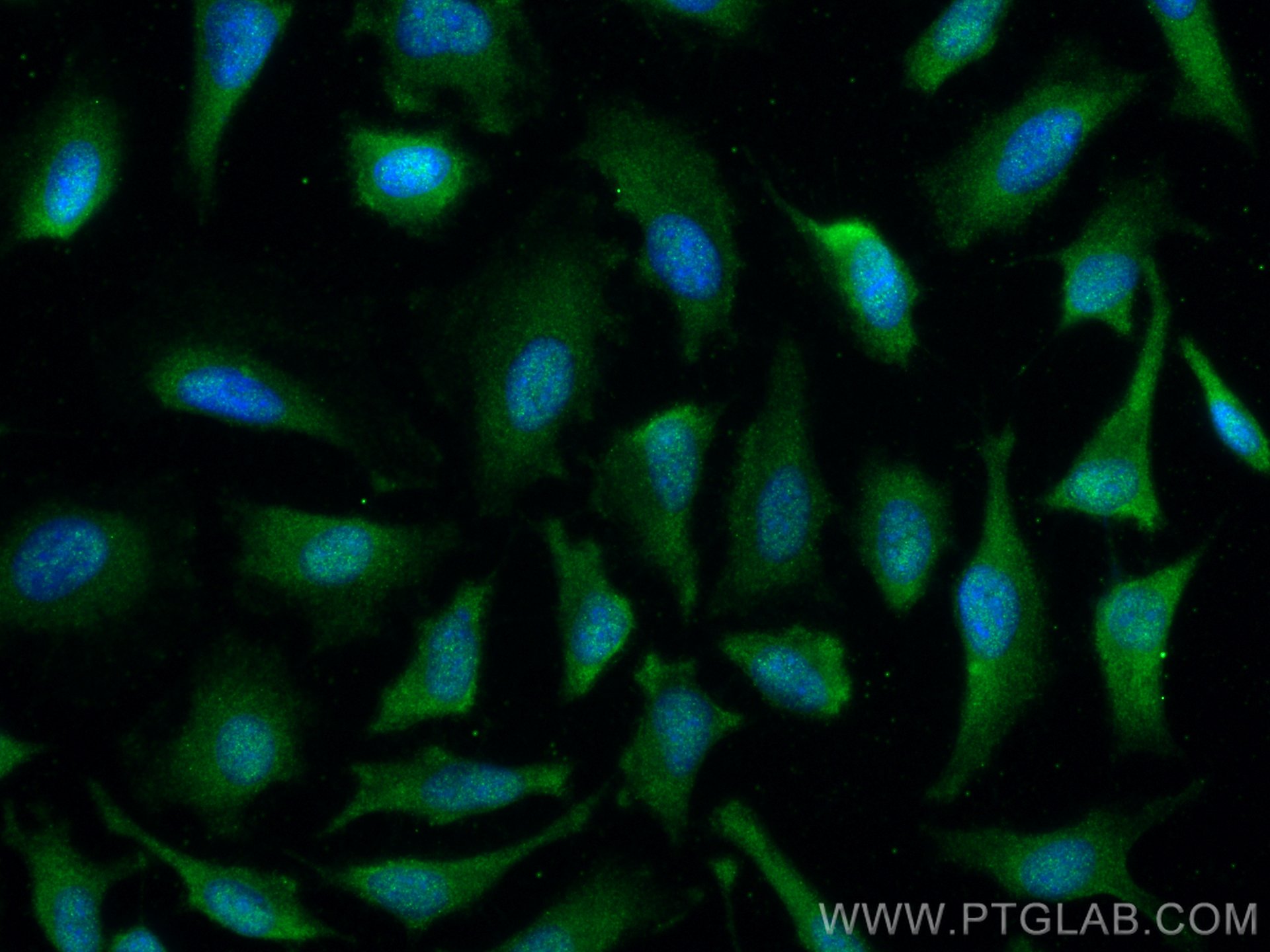
| Positive IF/ICC detected in | HeLa cells |
Recommended dilution
| Application | Dilution |
|---|---|
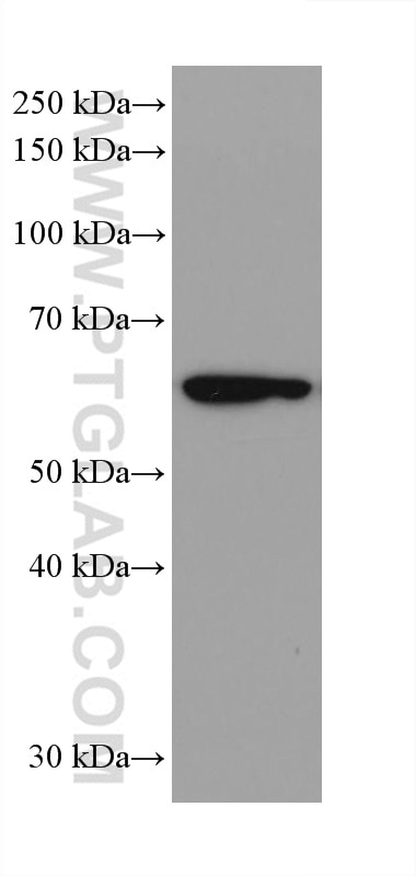
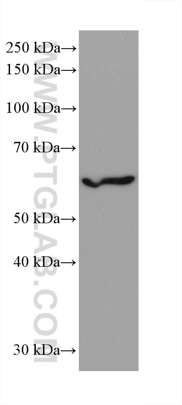
| Western Blot (WB) | WB : 1:2000-1:10000 |
| Immunohistochemistry (IHC) | IHC : 1:50-1:500 |
| Immunofluorescence (IF)/ICC | IF/ICC : 1:50-1:500 |
| It is recommended that this reagent should be titrated in each testing system to obtain optimal results. | |
| Sample-dependent, Check data in validation data gallery. | |
Published Applications
| WB | See 4 publications below |
Product Information
80519-1-RR targets IRF3 in WB, IHC, IF/ICC, ELISA applications and shows reactivity with human, mouse, rat, zebrafish samples.
| Tested Reactivity | human, mouse, rat, zebrafish |
| Cited Reactivity | human, mouse |
| Host / Isotype | Rabbit / IgG |
| Class | Recombinant |
| Type | Antibody |
| Immunogen |
Peptide Predict reactive species |
| Full Name | interferon regulatory factor 3 |
| Calculated Molecular Weight | 47 kDa |
| Observed Molecular Weight | 50-60 kDa |
| GenBank Accession Number | BC009395 |
| Gene Symbol | IRF3 |
| Gene ID (NCBI) | 3661 |
| RRID | AB_2918899 |
| Conjugate | Unconjugated |
| Form | Liquid |
| Purification Method | Protein A purification |
| UNIPROT ID | Q14653 |
| Storage Buffer | PBS with 0.02% sodium azide and 50% glycerol, pH 7.3. |
| Storage Conditions | Store at -20°C. Stable for one year after shipment. Aliquoting is unnecessary for -20oC storage. 20ul sizes contain 0.1% BSA. |
Background Information
The virul-induced expression of interferon(IFN) genes in infected cells implicate in the interplay of several constitutively expressed and virus-activated transcription factors. A family of IFN regulatory factors(IRFs) have been shown to has a role in the transcription of IFN genes as well as IFN-stimulated genes. IRF3 is a novel key transcriptional regulator of type I IFN-dependent immune responses and involves in the innate immune response against DNA and RNA viruses, by binding to the promoters of IFN. It located in the cytoplasm of uninfected cells in an inactive form, and following viral infection, double-stranded RNA (dsRNA), or toll-like receptor (TLR) signaling, could be phosphorylated by IKBKE and TBK1 kinases. This induces a conformational change, leading to its dimerization, nuclear localization and association with CREB binding protein (CREBBP) to form dsRNA-activated factor 1 (DRAF1), a complex which activates the transcription of the type I IFN and ISG genes.
Protocols
| Product Specific Protocols | |
|---|---|
| IF protocol for IRF3 antibody 80519-1-RR | Download protocol |
| IHC protocol for IRF3 antibody 80519-1-RR | Download protocol |
| WB protocol for IRF3 antibody 80519-1-RR | Download protocol |
| Standard Protocols | |
|---|---|
| Click here to view our Standard Protocols |
Publications
| Species | Application | Title |
|---|---|---|
Diabetes Application of Dental Pulp Stem Cell-Derived Intracellular Vesicles for Diabetic Wound Healing. | ||
Eur J Pharm Sci Discovery of STING antagonists Targeting cGAS-STING Pathway to Alleviate IMQ-induced Psoriasis-like Dermatitis | ||
Mater Today Bio Rational design of a V-shaped DNA-targeted photosensitizer enables endogenous DNA damage-driven cGAS-STING activation and systemic antitumor immunity. | ||
Adv Sci (Weinh) Nanoparticle-Modified Nanofibers Induce Ferroptosis and Stimulate Antitumor Immunity for Melanoma Therapy. |
Reviews
The reviews below have been submitted by verified Proteintech customers who received an incentive for providing their feedback.
FH Lisa (Verified Customer) (03-05-2026) | This antibody works ok for WB but does not have a super strong signal.
|