

Tested Applications
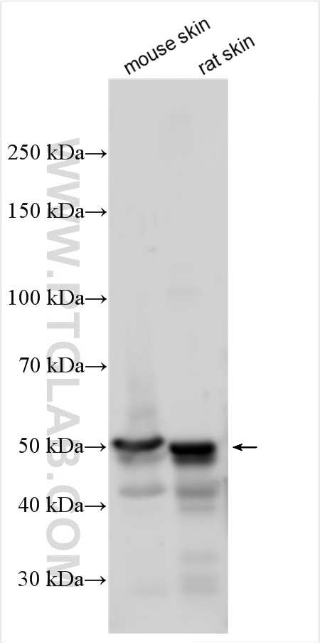
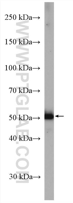
| Positive WB detected in | mouse skin tissue, A431 cells, rat skin tissue |
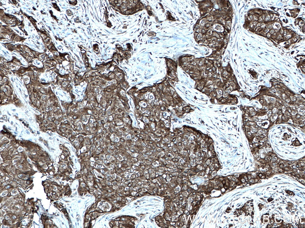
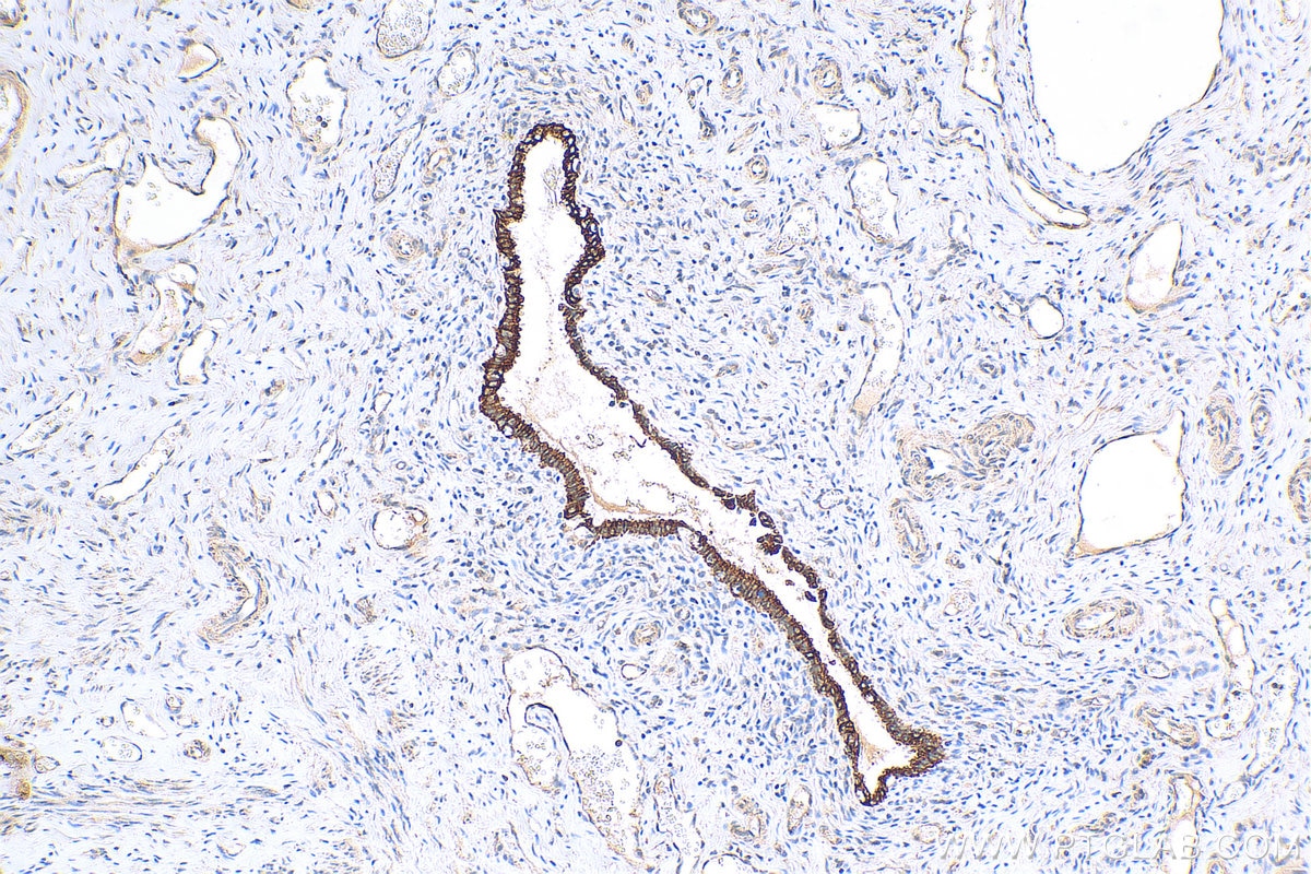
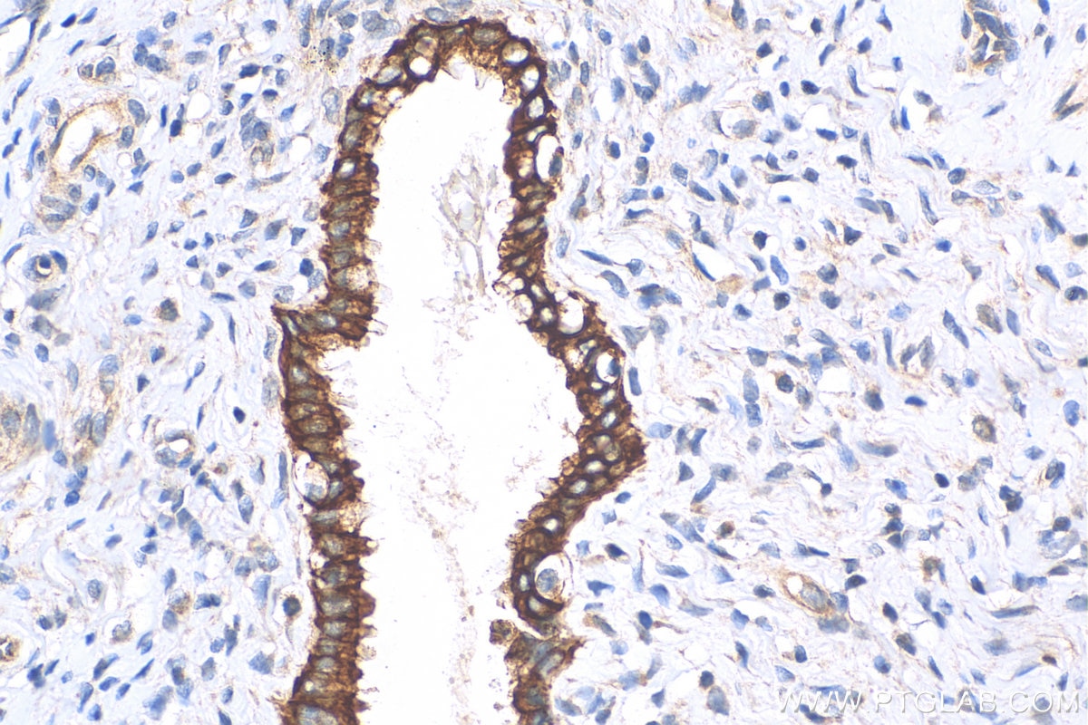
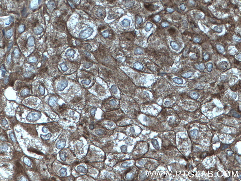
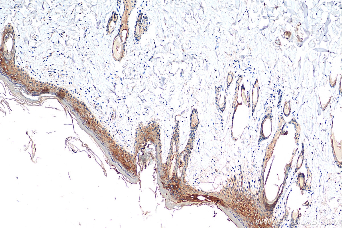
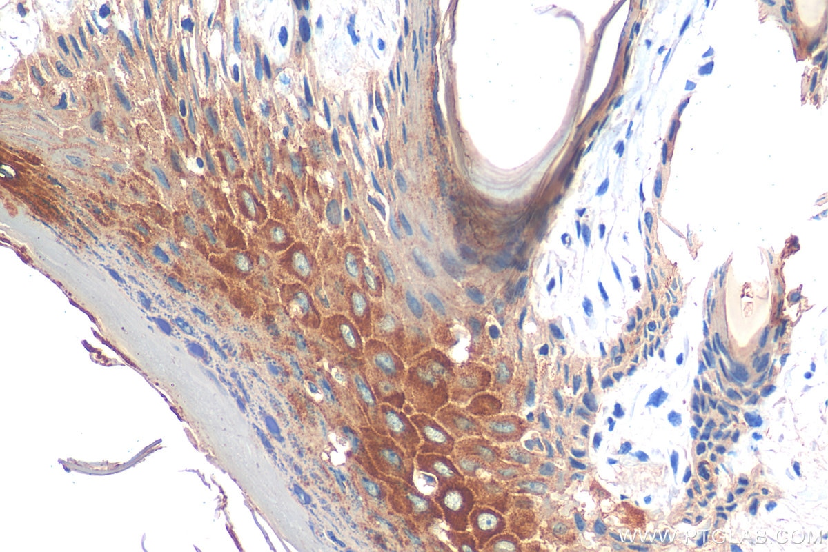
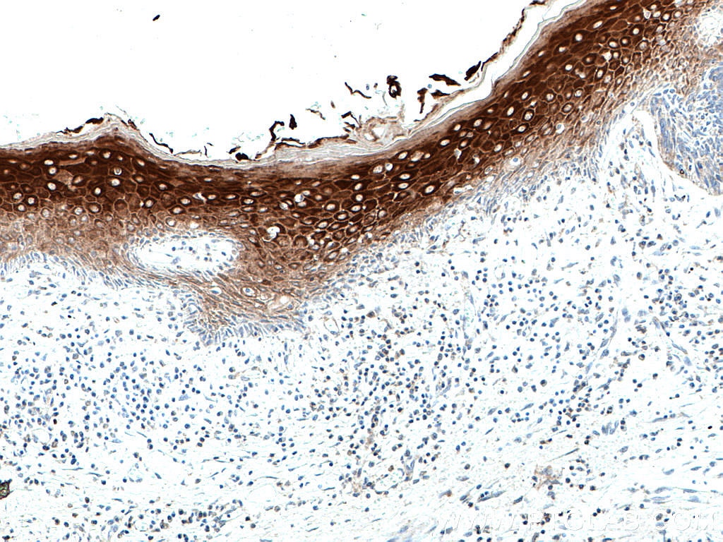
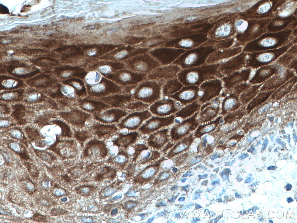
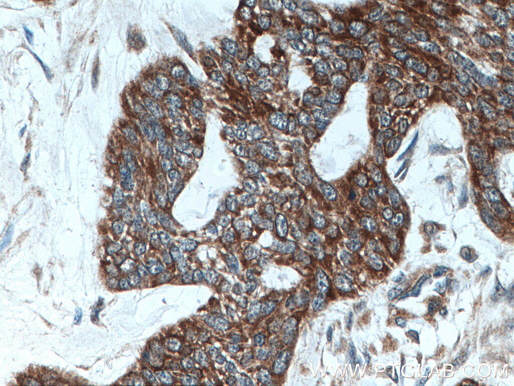
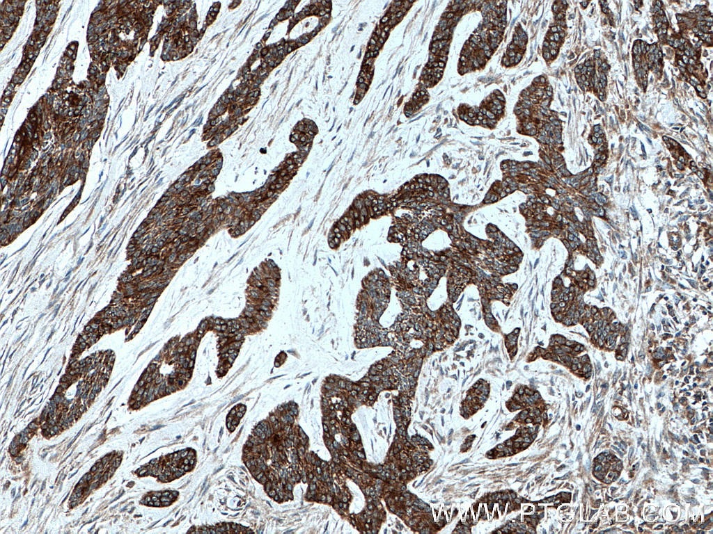
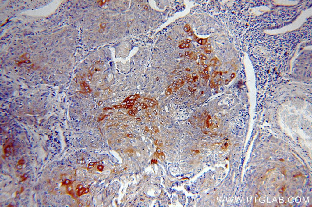
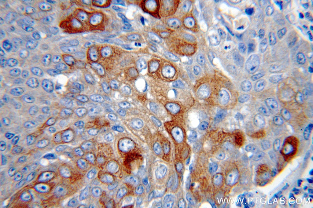
| Positive IHC detected in | human cervical cancer tissue, human breast cancer tissue, human lung cancer tissue, human skin cancer tissue, rat skin tissue Note: suggested antigen retrieval with TE buffer pH 9.0; (*) Alternatively, antigen retrieval may be performed with citrate buffer pH 6.0 |
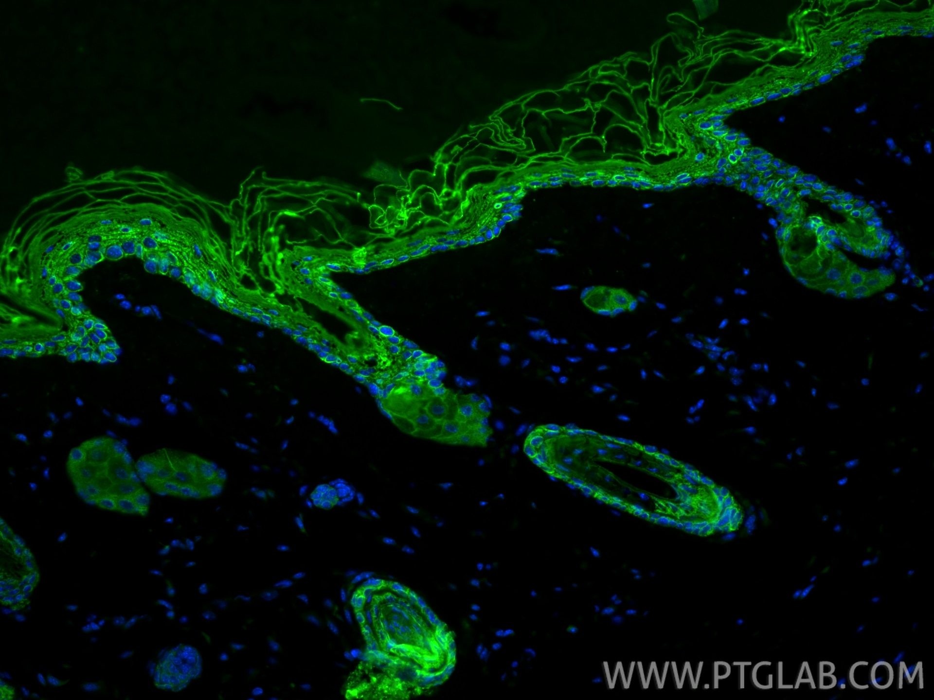
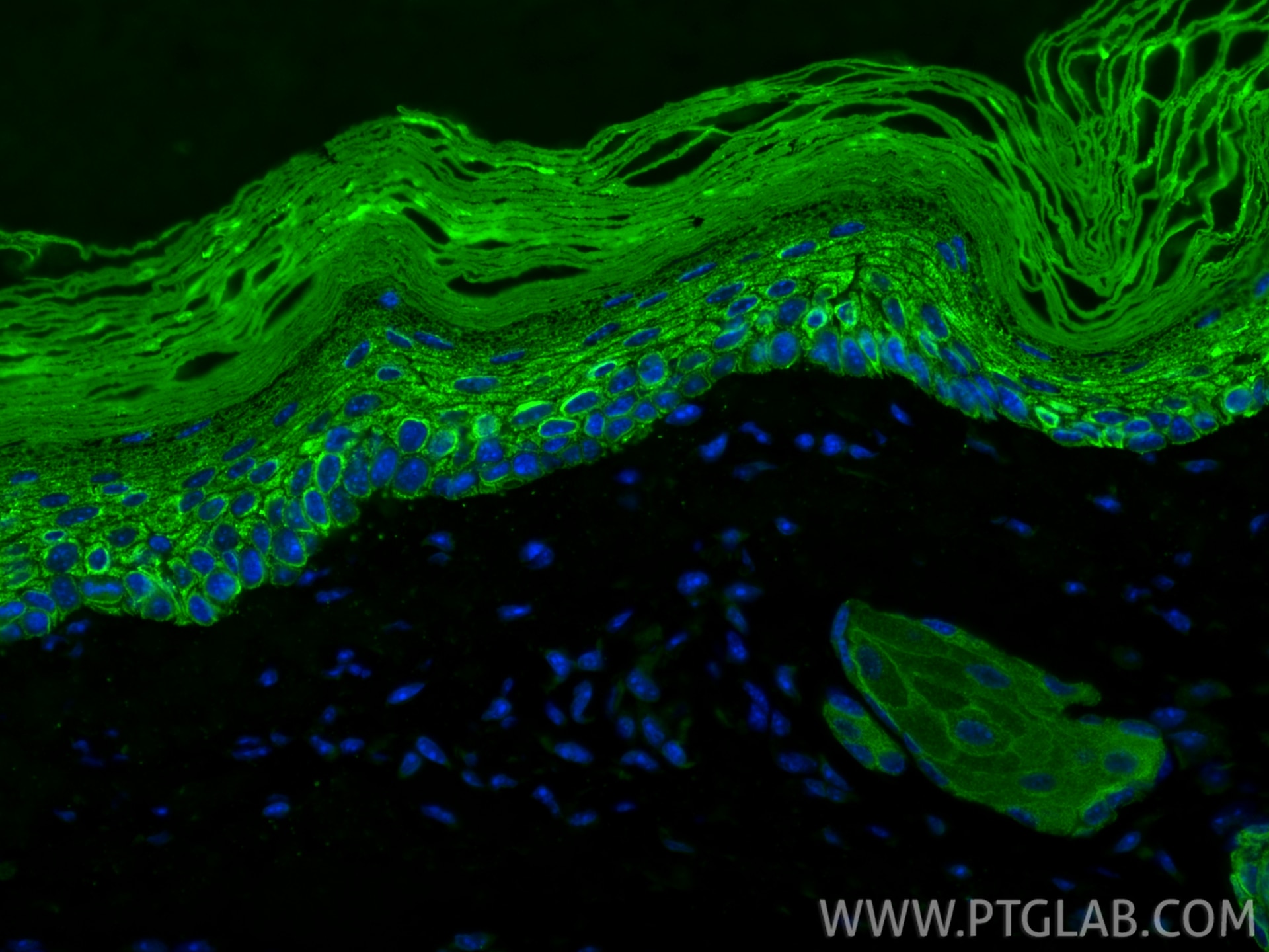
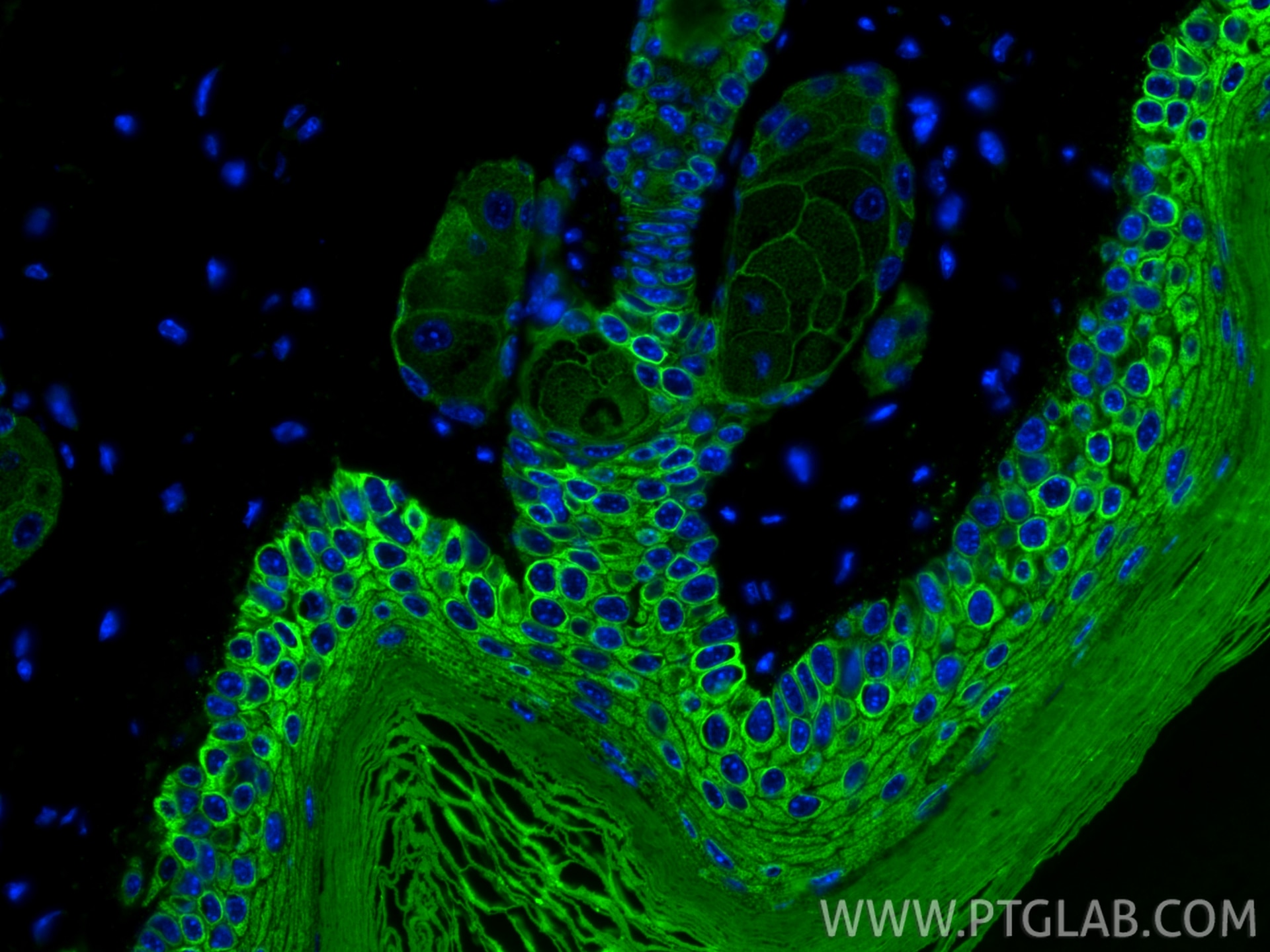
| Positive IF-P detected in | mouse skin tissue |
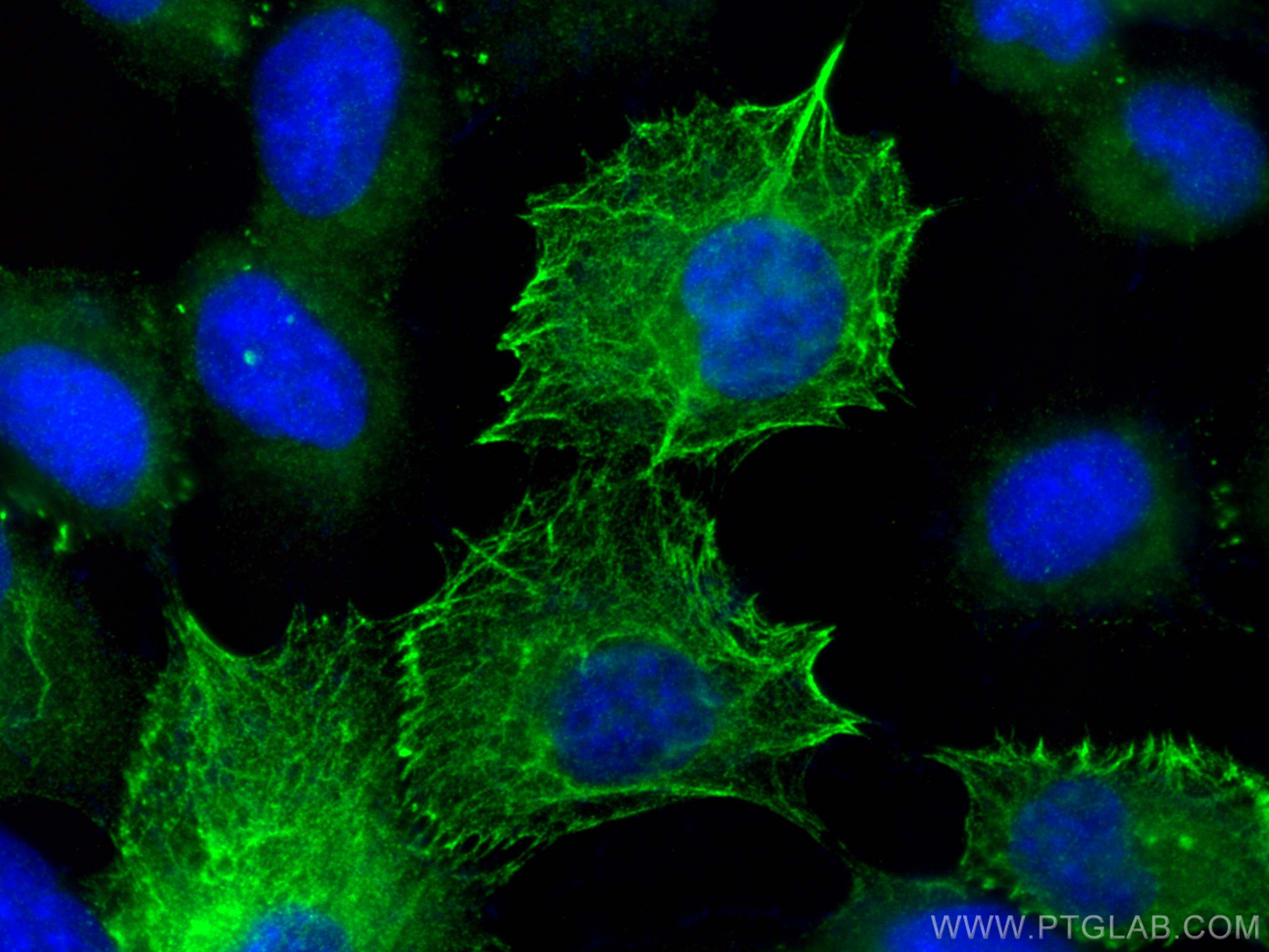
| Positive IF/ICC detected in | A431 cells |
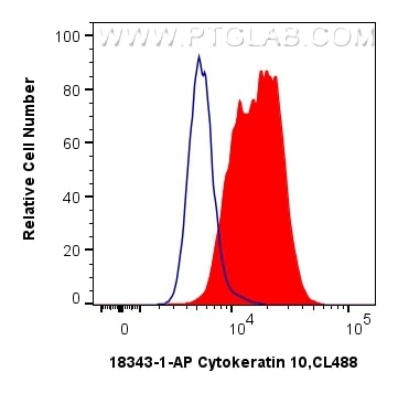
| Positive FC (Intra) detected in | A431 cells |
Recommended dilution
| Application | Dilution |
|---|---|
| Western Blot (WB) | WB : 1:5000-1:20000 |
| Immunohistochemistry (IHC) | IHC : 1:500-1:2000 |
| Immunofluorescence (IF)-P | IF-P : 1:50-1:500 |
| Immunofluorescence (IF)/ICC | IF/ICC : 1:50-1:500 |
| Flow Cytometry (FC) (INTRA) | FC (INTRA) : 0.40 ug per 10^6 cells in a 100 µl suspension |
| It is recommended that this reagent should be titrated in each testing system to obtain optimal results. | |
| Sample-dependent, Check data in validation data gallery. | |
Product Information
18343-1-AP targets Cytokeratin 10 in WB, IHC, IF/ICC, IF-P, FC (Intra), ELISA applications and shows reactivity with human, mouse, rat samples.
| Tested Reactivity | human, mouse, rat |
| Cited Reactivity | human, mouse, rat |
| Host / Isotype | Rabbit / IgG |
| Class | Polyclonal |
| Type | Antibody |
| Immunogen |
CatNo: Ag13136 Product name: Recombinant human KRT10 protein Source: e coli.-derived, PGEX-4T Tag: GST Domain: 235-584 aa of BC034697 Sequence: RLKYENEVALRQSVEADINGLRRVLDELTLTKADLEMQIESLTEELAYLKKNHEEEMKDLRNVSTGDVNVEMNAAPGVDLTQLLNNMRSQYEQLAEQNRKDAEAWFNEKSKELTTEIDNNIEQISSYKSEITELRRNVQALEIELQSQLALKQSLEASLAETEGRYCVQLSQIQAQISALEEQLQQIRAETECQNTEYQQLLDIKIRLENEIQTYRSLLEGEGSSGGGGRGGGSFGGGYGGGSSGGGSSGGGYGGGHGGSSGGGYGGGSSGGGSSGGGYGGGSSSGGHGGSSSGGYGGGSSGGGGGGYGGGSSGGGSSSGGGYGGGSSSGGHKSSSSGSVGESSSKGPRY Predict reactive species |
| Full Name | keratin 10 |
| Calculated Molecular Weight | 584 aa, 59 kDa |
| Observed Molecular Weight | 50-59 kDa |
| GenBank Accession Number | BC034697 |
| Gene Symbol | Cytokeratin 10 |
| Gene ID (NCBI) | 3858 |
| RRID | AB_10863654 |
| Conjugate | Unconjugated |
| Form | Liquid |
| Purification Method | Antigen affinity purification |
| UNIPROT ID | P13645 |
| Storage Buffer | PBS with 0.02% sodium azide and 50% glycerol, pH 7.3. |
| Storage Conditions | Store at -20°C. Stable for one year after shipment. Aliquoting is unnecessary for -20oC storage. 20ul sizes contain 0.1% BSA. |
Background Information
Keratins are a large family of proteins that form the intermediate filament cytoskeleton of epithelial cells, which are classified into two major sequence types. Type I keratins are a group of acidic intermediate filament proteins, including K9-K23, and the hair keratins Ha1-Ha8. Type II keratins are the basic or neutral courterparts to the acidic type I keratins, including K1-K8, and the hair keratins, Hb1-Hb6. As a type I keratin, keratin 10 is a suprabasal marker of differentiation in stratified squamous epithelia.
Protocols
| Product Specific Protocols | |
|---|---|
| FC protocol for Cytokeratin 10 antibody 18343-1-AP | Download protocol |
| IF protocol for Cytokeratin 10 antibody 18343-1-AP | Download protocol |
| IHC protocol for Cytokeratin 10 antibody 18343-1-AP | Download protocol |
| WB protocol for Cytokeratin 10 antibody 18343-1-AP | Download protocol |
| Standard Protocols | |
|---|---|
| Click here to view our Standard Protocols |
Publications
What published studies show
The Cytokeratin 10 Polyclonal antibody (Cat# 18343-1-AP) has accumulated 39 citations across journals including Nature Aging, Bioactive Materials, Microbiome, Cell Death & Differentiation, Journal of Controlled Release, International Journal of Biological Macromolecules, Acta Biomaterialia, mBio, Scientific Reports, iScience, and Experimental Dermatology. Associated research fields include dermatology (psoriasis, skin aging, wound healing), epithelial cell biology, tissue engineering, and biomaterials.
| Species | Application | Title |
|---|---|---|
Nat Aging Skin chronological aging drives age-related bone loss via secretion of cystatin-A | ||
Bioact Mater Engineering biomimetic tarsal microtissue via structurally zoned hydrogel scaffold for integrated eyelid reconstruction. | ||
Bioact Mater Biomimetic gender-specific human skin model based on gonads/epidermis-on-a-chip | ||
Microbiome Multi-omics characterization of the skin microbiota reveals the anti-aging roles of Stenotrophomonas maltophilia. | ||
Cell Death Differ The parkin-γ-tubulin axis regulates epidermal homeostasis and is associated with the susceptibility to psoriasis. | ||
J Control Release Improving ocular bioavailability of hydrophilic drugs through dynamic covalent complexation |
Reviews
What customers say
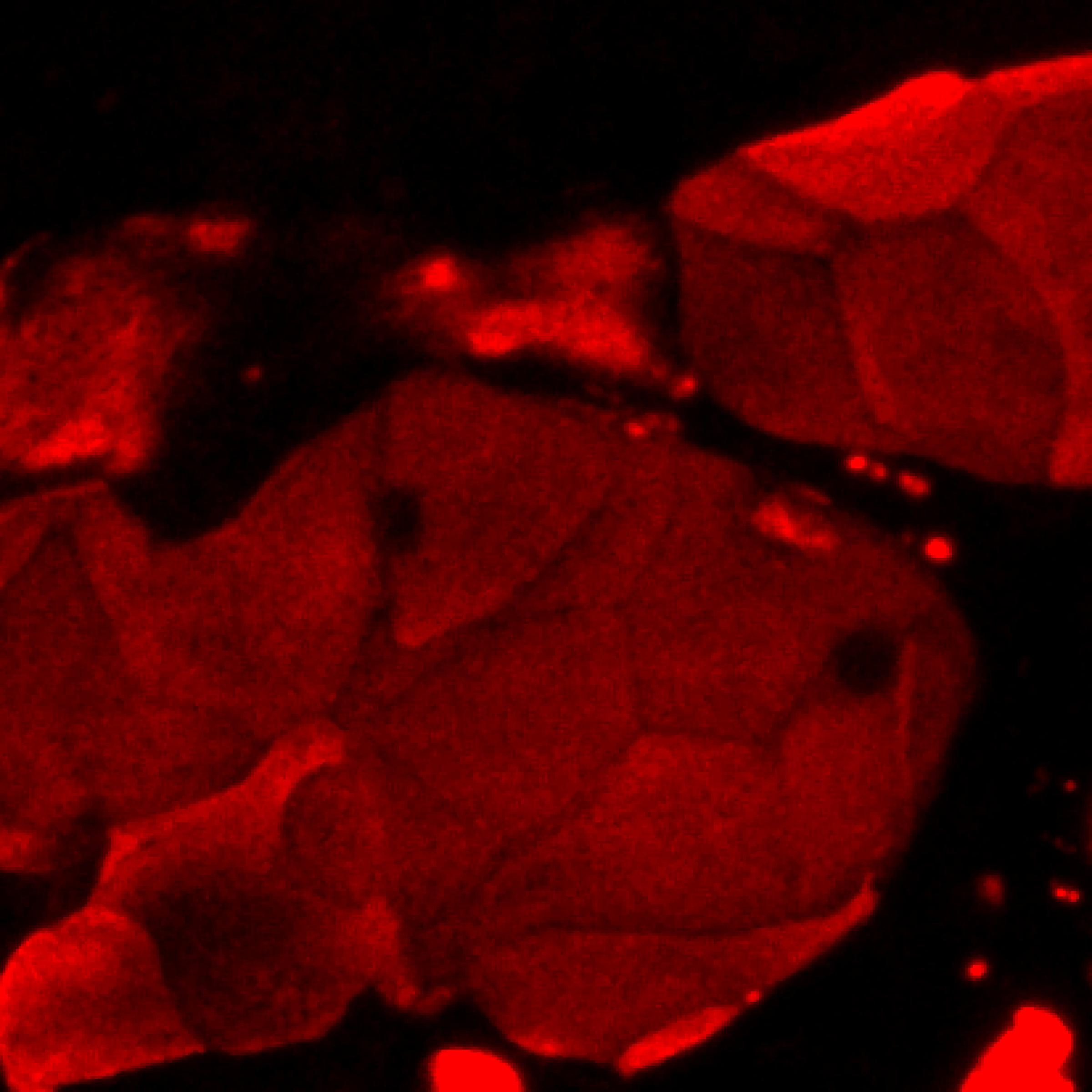
The single review for 18343-1-AP is a 5-star rating from Joshua Morgan (July 2019). He used the antibody at a 1:500 dilution on human keratinocytes differentiated at an air-liquid interface. The cells were fixed in 4% paraformaldehyde and stained at 4°C overnight for immunofluorescence. He reported bright staining with minimal background, indicating strong performance and low non-specific signal under his conditions.
The reviews below have been submitted by verified Proteintech customers who received an incentive for providing their feedback.
Joshua (Verified Customer) (07-27-2019) | Cells differentiated at air-liquid interface. Cells fixed in 4% paraformaldehyde and stained at 4C overnight. Bright staining with minimal background.
 |




















