Tested Applications
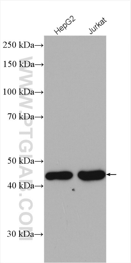
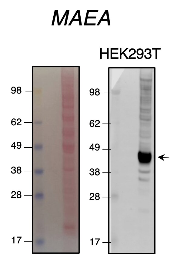
| Positive WB detected in | HepG2 cells, Jurkat cells |
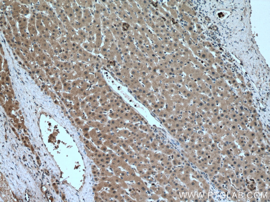
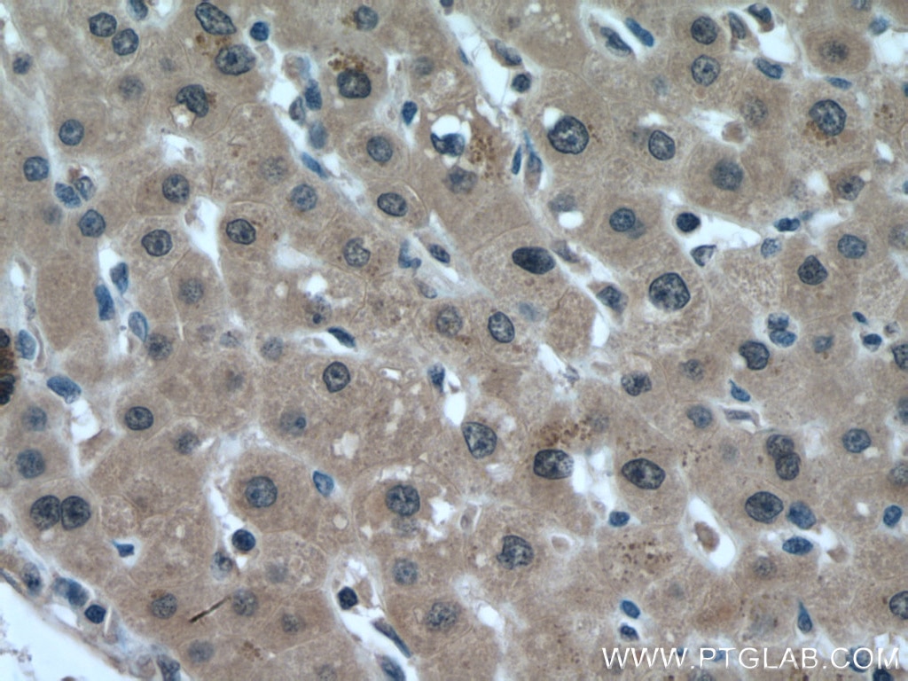
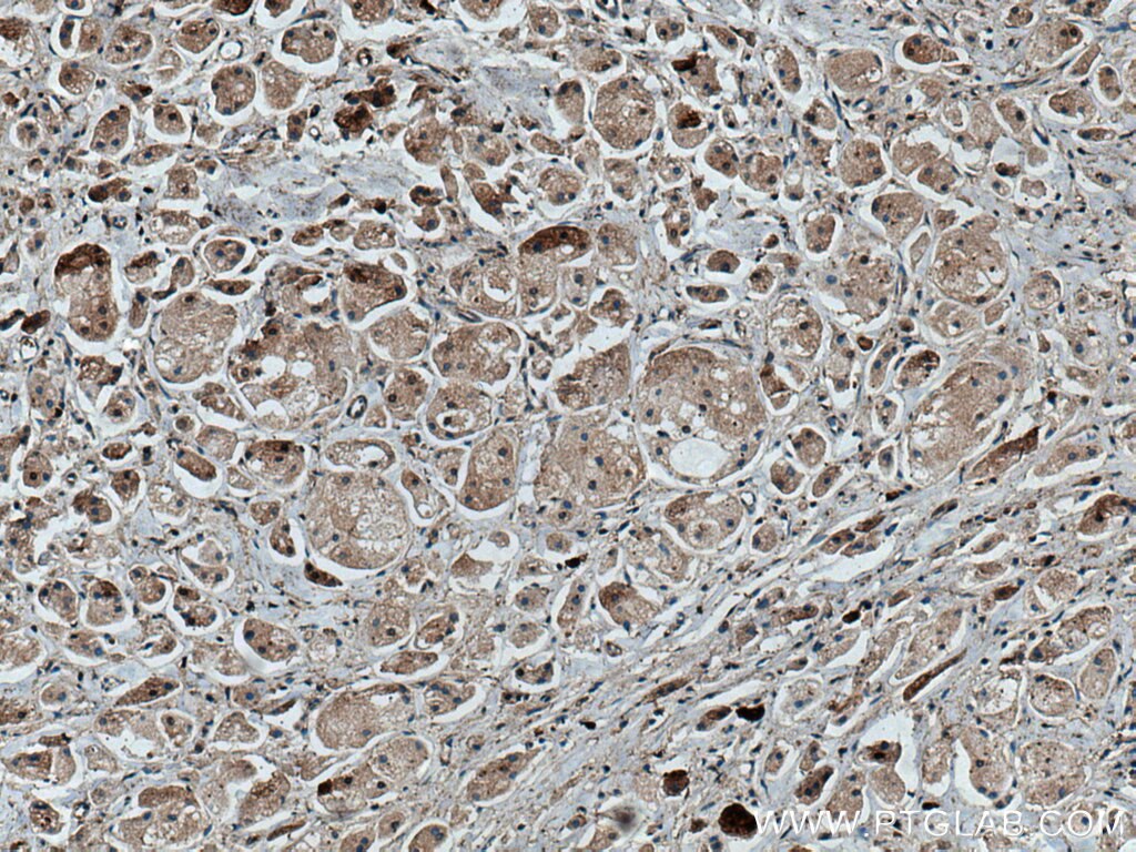
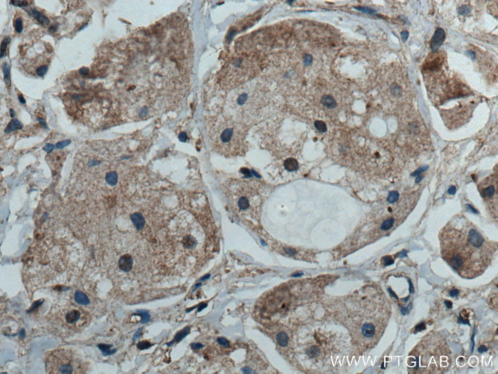
| Positive IHC detected in | human liver cancer tissue, human breast cancer tissue Note: suggested antigen retrieval with TE buffer pH 9.0; (*) Alternatively, antigen retrieval may be performed with citrate buffer pH 6.0 |
Recommended dilution
| Application | Dilution |
|---|---|
| Western Blot (WB) | WB : 1:1000-1:4000 |
| Immunohistochemistry (IHC) | IHC : 1:50-1:500 |
| It is recommended that this reagent should be titrated in each testing system to obtain optimal results. | |
| Sample-dependent, Check data in validation data gallery. | |
Published Applications
| KD/KO | See 3 publications below |
| WB | See 8 publications below |
| IHC | See 2 publications below |
| IP | See 2 publications below |
Product Information
28363-1-AP targets MAEA in WB, IHC, IP, ELISA applications and shows reactivity with Human samples.
| Tested Reactivity | Human |
| Cited Reactivity | human, mouse |
| Host / Isotype | Rabbit / IgG |
| Class | Polyclonal |
| Type | Antibody |
| Immunogen |
CatNo: Ag27150 Product name: Recombinant human MAEA protein Source: e coli.-derived, PGEX-4T Tag: GST Domain: 26-239 aa of BC001225 Sequence: YETLNKRFRAAQKNIDRETSHVTMVVAELEKTLSGCPAVDSVVSLLDGVVEKLSVLKRKAVESIQAEDESAKLCKRRIEHLKEHSSDQPAAASVWKRKRMDRMMVEHLLRCGYYNTAVKLARQSGIEDLVNIEMFLTAKEVEESLERRETATCLAWCHDNKSRLRKMKSCLEFSLRIQEFIELIRQNKRLDAVRHARKHFSQAEGSQLDEVRQA Predict reactive species |
| Full Name | macrophage erythroblast attacher |
| Calculated Molecular Weight | 45 kDa |
| Observed Molecular Weight | 45 kDa |
| GenBank Accession Number | BC001225 |
| Gene Symbol | MAEA |
| Gene ID (NCBI) | 10296 |
| RRID | AB_2881122 |
| Conjugate | Unconjugated |
| Form | Liquid |
| Purification Method | Antigen affinity purification |
| UNIPROT ID | Q7L5Y9 |
| Storage Buffer | PBS with 0.02% sodium azide and 50% glycerol, pH 7.3. |
| Storage Conditions | Store at -20°C. Stable for one year after shipment. Aliquoting is unnecessary for -20oC storage. 20ul sizes contain 0.1% BSA. |
Background Information
MAEA(macrophage erythroblast attacher) is a core component of the CTLH E3 ubiquitin-protein ligase complex. MAEA is required for normal cell proliferation and plays a role in erythroblast enucleation during erythrocyte maturation and in the development of mature macrophages. N-terminus MAEA and full-length MAEA are mostly expressed in nuclear and can be detected at low level in the cytoplasm of some cells, while C-terminus MAEA can be detected in the cytoplasm and nucleoli within the nucleus. MAEA mediates the attachment of erythroid cell to mature macrophages which can inhibit erythroid cell apoptosis. The expression of MAEA may be associated with the type 2 diabetes mellitus.(PubMed: 28955747, 24143168, 29911972, 29911972, 30674470, 9763581).
Protocols
| Product Specific Protocols | |
|---|---|
| IHC protocol for MAEA antibody 28363-1-AP | Download protocol |
| WB protocol for MAEA antibody 28363-1-AP | Download protocol |
| Standard Protocols | |
|---|---|
| Click here to view our Standard Protocols |
Publications
| Species | Application | Title |
|---|---|---|
Cancers (Basel) Oncopeptide MBOP Encoded by LINC01234 Promotes Colorectal Cancer through MAPK Signaling Pathway. | ||
bioRxiv Proteome-wide C-degron activity profiling connects conditional regulation of the CTLH E3 ligase complex to ribosome biogenesis.
| ||
Mol Cell mTORC1-CTLH E3 ligase regulates the degradation of HMG-CoA synthase 1 through the Pro/N-degron pathway | ||
Oncogene E3 ligase MAEA-mediated ubiquitination and degradation of PHD3 promotes glioblastoma progression
| ||
Int J Biol Sci The E3 ubiquitin ligase MAEA promotes macrophage phagocytosis and inhibits gastrointestinal cancer progression by mediating PARP1 ubiquitination and degradation | ||
J Transl Med LAD1 promotes malignant progression by diminishing ubiquitin-dependent degradation of vimentin in gastric cancer |
Reviews
The reviews below have been submitted by verified Proteintech customers who received an incentive for providing their feedback.
FH S (Verified Customer) (12-31-2021) | Quite a good quality antibody. KO validated.
 |
FH H (Verified Customer) (07-09-2020) | This is very good antibody. MAEA band is very thick and clear.
 |