Tested Applications
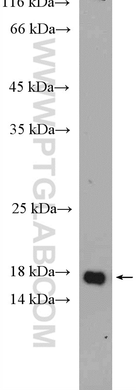
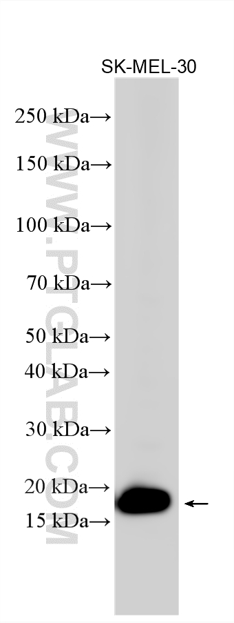
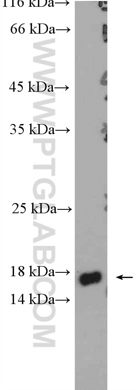
| Positive WB detected in | SK-MEL-30 cells, mouse eye tissue |
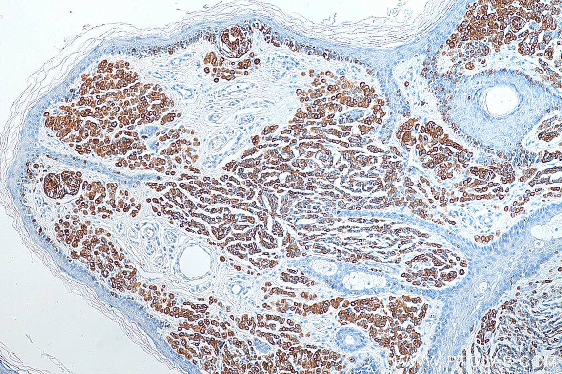
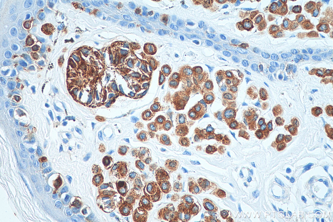
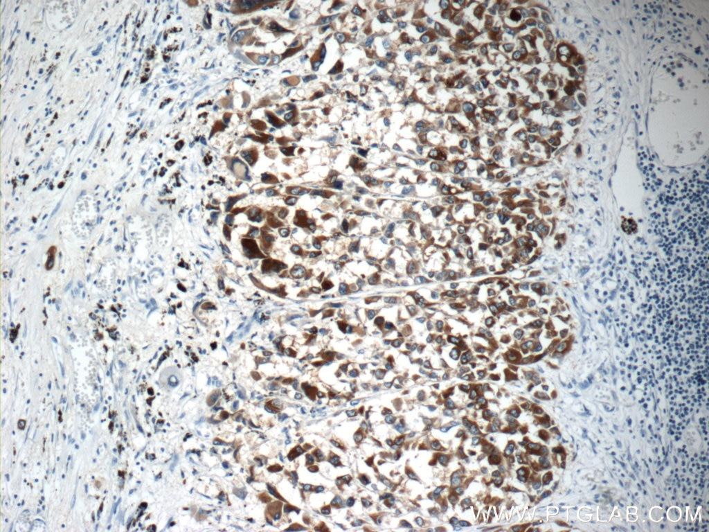
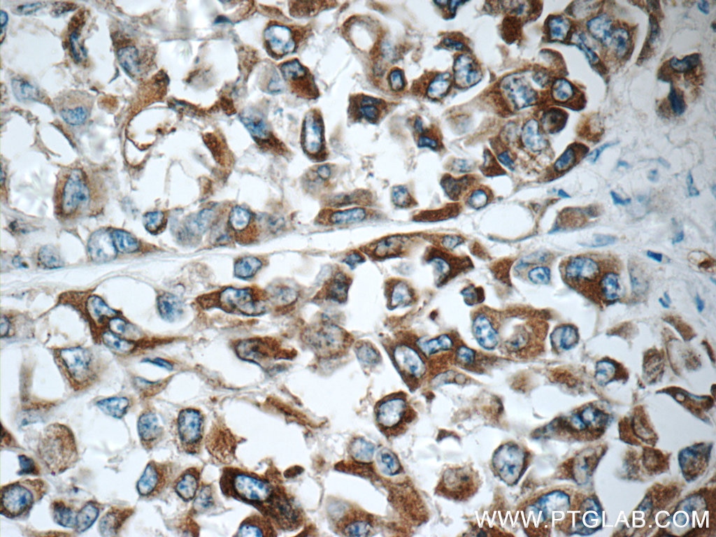
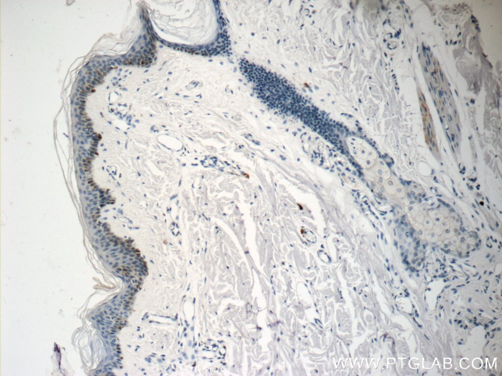
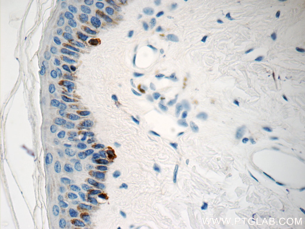
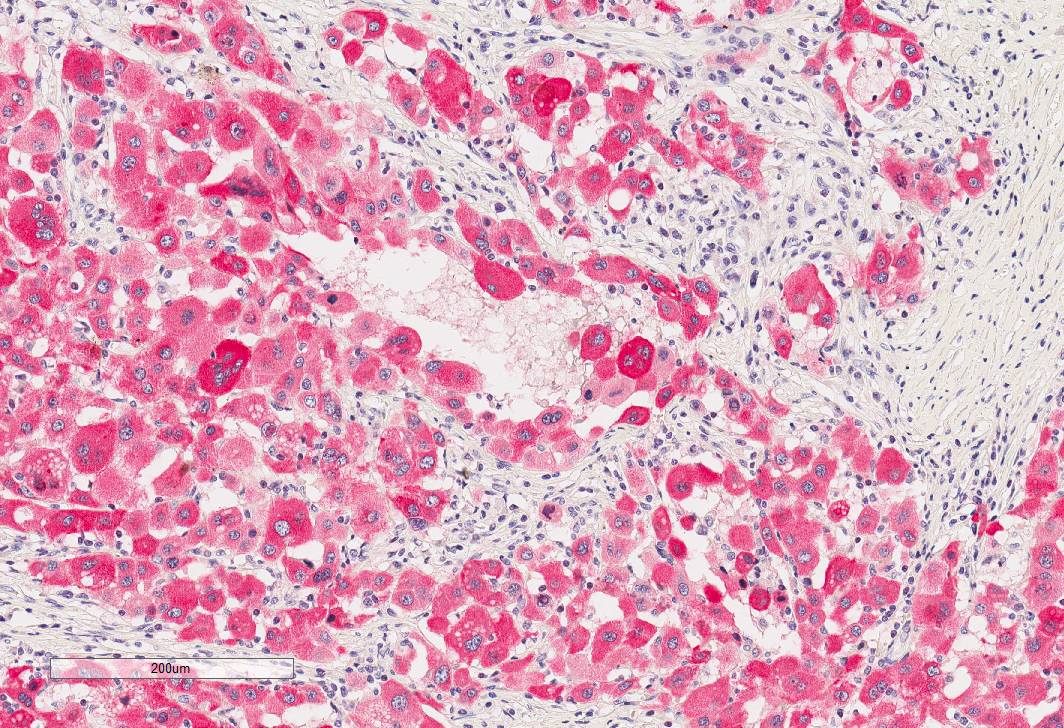
| Positive IHC detected in | human malignant melanoma tissue, human skin tissue Note: suggested antigen retrieval with TE buffer pH 9.0; (*) Alternatively, antigen retrieval may be performed with citrate buffer pH 6.0 |
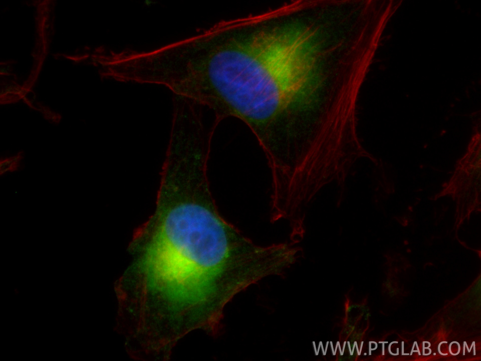
| Positive IF/ICC detected in | SK-MEL-28 cells |
Recommended dilution
| Application | Dilution |
|---|---|
| Western Blot (WB) | WB : 1:500-1:1000 |
| Immunohistochemistry (IHC) | IHC : 1:500-1:2000 |
| Immunofluorescence (IF)/ICC | IF/ICC : 1:200-1:800 |
| It is recommended that this reagent should be titrated in each testing system to obtain optimal results. | |
| Sample-dependent, Check data in validation data gallery. | |
Published Applications
| WB | See 1 publications below |
| IHC | See 2 publications below |
| IF | See 2 publications below |
Product Information
18472-1-AP targets Melan-A in WB, IHC, IF/ICC, ELISA applications and shows reactivity with human, mouse samples.
| Tested Reactivity | human, mouse |
| Cited Reactivity | human, mouse |
| Host / Isotype | Rabbit / IgG |
| Class | Polyclonal |
| Type | Antibody |
| Immunogen |
CatNo: Ag13346 Product name: Recombinant human MLANA protein Source: e coli.-derived, PGEX-4T Tag: GST Domain: 1-118 aa of BC014423 Sequence: MPREDAHFIYGYPKKGHGHSYTTAEEAAGIGILTVILGVLLLIGCWYCRRRNGYRALMDKSLHVGTQCALTRRCPQEGFDHRDSKVSLQEKNCEPVVPNAPPAYEKLSAEQSPPPYSP Predict reactive species |
| Full Name | melan-A |
| Calculated Molecular Weight | 13 kDa |
| Observed Molecular Weight | 13-20 kDa |
| GenBank Accession Number | BC014423 |
| Gene Symbol | MelanA |
| Gene ID (NCBI) | 2315 |
| RRID | AB_2878545 |
| Conjugate | Unconjugated |
| Form | Liquid |
| Purification Method | Antigen affinity purification |
| UNIPROT ID | Q16655 |
| Storage Buffer | PBS with 0.02% sodium azide and 50% glycerol, pH 7.3. |
| Storage Conditions | Store at -20°C. Stable for one year after shipment. Aliquoting is unnecessary for -20oC storage. 20ul sizes contain 0.1% BSA. |
Background Information
Melan-A is a palmitoylated integral membrane protein of 118 amino acids with a short amino-terminal luminal domain and a longer carboxy-terminal cytoplasmic domain . The protein does not possess any detectable enzymatic activity and has not been linked to any of the numerous genetic defects that affect skin pigmentation. Melan-A is new immunohistochemical markers that can be used in the diagnosis of melanocytic lesions. (PMID: 15703212, PMID: 17445277)
Protocols
| Product Specific Protocols | |
|---|---|
| IF protocol for Melan-A antibody 18472-1-AP | Download protocol |
| IHC protocol for Melan-A antibody 18472-1-AP | Download protocol |
| WB protocol for Melan-A antibody 18472-1-AP | Download protocol |
| Standard Protocols | |
|---|---|
| Click here to view our Standard Protocols |
Publications
| Species | Application | Title |
|---|---|---|
Life Sci Differentiation-inducing factor-1 reduces lipopolysaccharide-induced vascular cell adhesion molecule-1 by suppressing mTORC1-S6K signaling in vascular endothelial cells | ||
Biochim Biophys Acta Mol Cell Res MitoQ for vitiligo by mitigating PARP1 translocation aberrations: Network pharmacology and experimental validation | ||
Cell Death Discov Unique lipid composition maintained by extracellular blockade leads to prooncogenicity | ||
Mol Ther Nucleic Acids IL-12 and PD-1 peptide combination gene therapy for the treatment of melanoma |
Reviews
The reviews below have been submitted by verified Proteintech customers who received an incentive for providing their feedback.
FH Federica (Verified Customer) (12-08-2023) | Leica Bond Rxm Red Chromogenic Kit from Leica Antigen Retrival ER1 (ph6) for 30 min
 |