Tested Applications
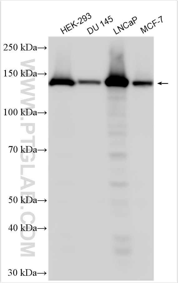
| Positive WB detected in | HEK-293 cells, mouse small intestine tissue, DU 145 cells, LNCaP cells, MCF-7 cells |
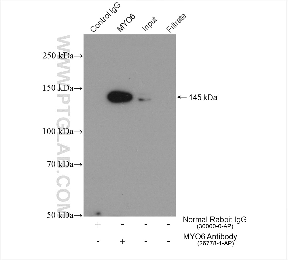
| Positive IP detected in | PC-3 cells |
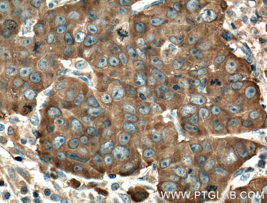
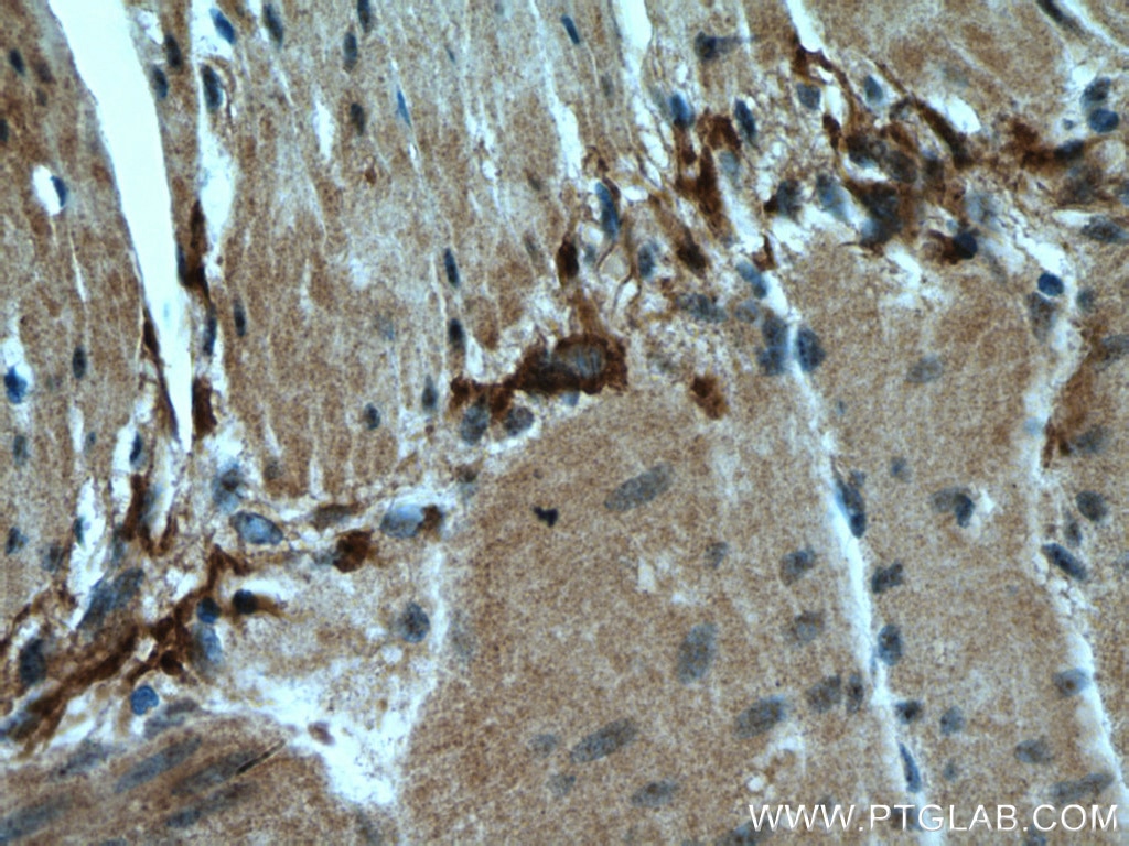
| Positive IHC detected in | human prostate cancer tissue, human small intestine tissue Note: suggested antigen retrieval with TE buffer pH 9.0; (*) Alternatively, antigen retrieval may be performed with citrate buffer pH 6.0 |
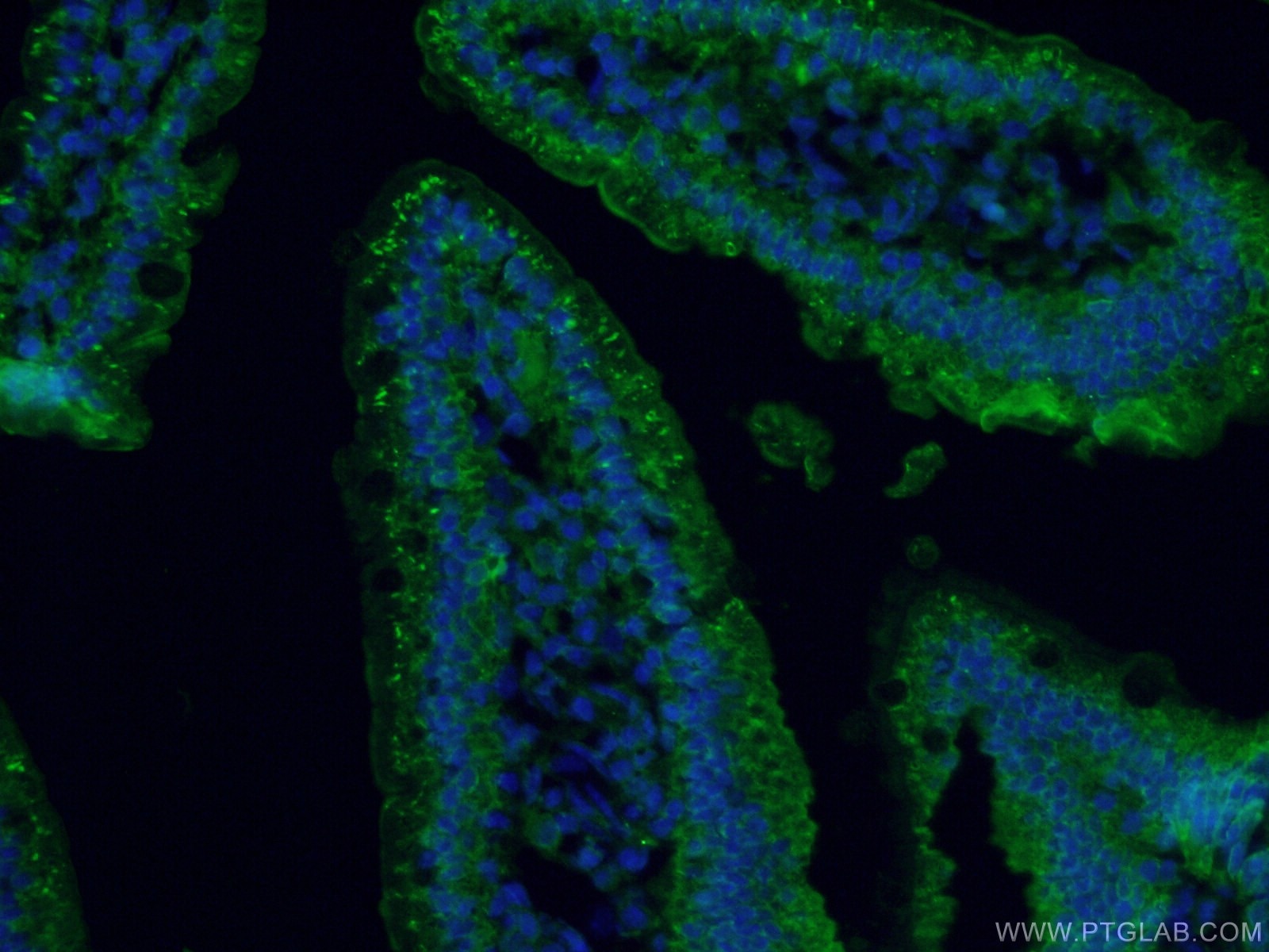
| Positive IF-P detected in | mouse small intestine tissue |
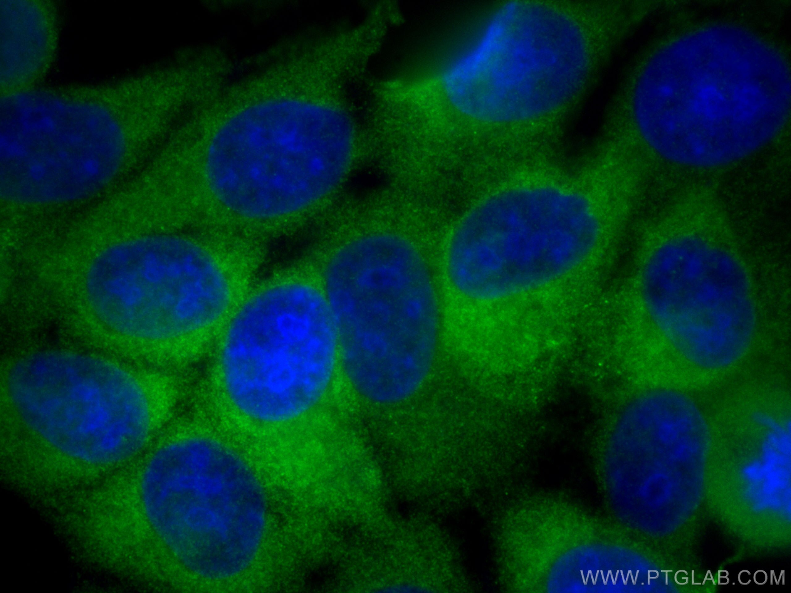
| Positive IF/ICC detected in | MCF-7 cells |
Recommended dilution
| Application | Dilution |
|---|---|
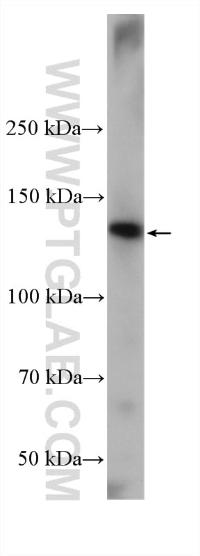
| Western Blot (WB) | WB : 1:5000-1:50000 |
| Immunoprecipitation (IP) | IP : 0.5-4.0 ug for 1.0-3.0 mg of total protein lysate |
| Immunohistochemistry (IHC) | IHC : 1:50-1:500 |
| Immunofluorescence (IF)-P | IF-P : 1:50-1:500 |
| Immunofluorescence (IF)/ICC | IF/ICC : 1:200-1:800 |
| It is recommended that this reagent should be titrated in each testing system to obtain optimal results. | |
| Sample-dependent, Check data in validation data gallery. | |
Published Applications
| KD/KO | See 1 publications below |
| WB | See 6 publications below |
| IHC | See 3 publications below |
| IF | See 5 publications below |
Product Information
26778-1-AP targets MYO6 in WB, IHC, IF/ICC, IF-P, IP, ELISA applications and shows reactivity with human, mouse samples.
| Tested Reactivity | human, mouse |
| Cited Reactivity | human, mouse, rat |
| Host / Isotype | Rabbit / IgG |
| Class | Polyclonal |
| Type | Antibody |
| Immunogen |
CatNo: Ag24906 Product name: Recombinant human MYO6 protein Source: e coli.-derived, PGEX-4T Tag: GST Domain: 1-145 aa of BC146764 Sequence: MEDGKPVWAPHPTDGFQMGNIVDIGPDSLTIEPLNQKGKTFLALINQVFPAEEDSKKDVEDNCSLMYLNEATLLHNIKVRYSKDRIYTYVANILIAVNPYFDIPKIYSSEAIKSYQGKSLGTRPPHVFAIADKAFRDMKVLKMSQ Predict reactive species |
| Full Name | myosin VI |
| Calculated Molecular Weight | 1285 aa, 149 kDa |
| Observed Molecular Weight | 145-150 kDa |
| GenBank Accession Number | BC146764 |
| Gene Symbol | MYO6 |
| Gene ID (NCBI) | 4646 |
| RRID | AB_2880631 |
| Conjugate | Unconjugated |
| Form | Liquid |
| Purification Method | Antigen affinity purification |
| UNIPROT ID | Q9UM54 |
| Storage Buffer | PBS with 0.02% sodium azide and 50% glycerol, pH 7.3. |
| Storage Conditions | Store at -20°C. Stable for one year after shipment. Aliquoting is unnecessary for -20oC storage. 20ul sizes contain 0.1% BSA. |
Background Information
MYO6, an actin-based motor protein, is the only myosin known to move toward the minus end of actin filaments. MYO6 is highly expressed in the inner and outer hair cells of the ear, retina, and polarized epithelial cells such as kidney proximal tubule cells and intestinal enterocytes. And it participates in a wide range of biological processes within cells, including clathrin-mediated endocytosis, vesicular membrane traffic, polarized secretion, and autophagy (PMID: 23620821; PMID: 28591580). Previous studies showed that MYO6 is upregulated in various types of cancer, and it has been widely reported to contribute to tumor cell migration and metastasis. Some articles indicate that MYO6 is associated with prostate cancer, lung cancer, human colorectal cancer and gastric cancer (PMID: 29022908).
Protocols
| Product Specific Protocols | |
|---|---|
| IF protocol for MYO6 antibody 26778-1-AP | Download protocol |
| IHC protocol for MYO6 antibody 26778-1-AP | Download protocol |
| IP protocol for MYO6 antibody 26778-1-AP | Download protocol |
| WB protocol for MYO6 antibody 26778-1-AP | Download protocol |
| Standard Protocols | |
|---|---|
| Click here to view our Standard Protocols |
Publications
| Species | Application | Title |
|---|---|---|
Nat Commun Structure of Myosin VI/Tom1 complex reveals a cargo recognition mode of Myosin VI for tethering. | ||
Front Cardiovasc Med Herb pair of Astragali Radix-Descurainiae Semen attenuate heart failure through the myosin VI-Tom1 complex mediated autophagy | ||
Int J Mol Sci The Suppression of the Epithelial to Mesenchymal Transition in Prostate Cancer through the Targeting of MYO6 Using MiR-145-5p | ||
Cancer Manag Res The Actin Motor Protein Myosin 6 Contributes to Cell Migration and Expression of GIPC1 and Septins in Breast Cancer Cells | ||
Cell Signal Elevated expression of myosin VI contributes to breast cancer progression via MAPK/ERK signaling pathway
|
Reviews
The reviews below have been submitted by verified Proteintech customers who received an incentive for providing their feedback.
FH Wojciech (Verified Customer) (04-20-2023) | We used this antibody to assess protein levels in human T cells. We got a good signal with 1:1000 dilution with observed molecular weight at ~150kDa.
|
FH Sara (Verified Customer) (10-25-2020) | B cells (mouse) in RIPA buffer. 10 µg of protein per lane were loaded on a 10% PAGE-SDS gel. Transfer is not optimised for high MW proteins, so I could get a better blot. Two bands are detected, one of 150 kDa and one > 200 kDa.
 |