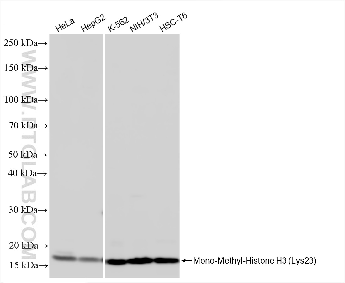
Various lysates were subjected to SDS PAGE followed by western blot with 86861-1-RR (Mono-Methyl-Histone H3 (Lys23) antibody) at dilution of 1:5000 incubated at room temperature for 1.5 hours.
Various lysates were subjected to SDS PAGE followed by western blot with 86861-1-RR (Mono-Methyl-Histone H3 (Lys23) antibody) at dilution of 1:5000 incubated at room temperature for 1.5 hours.
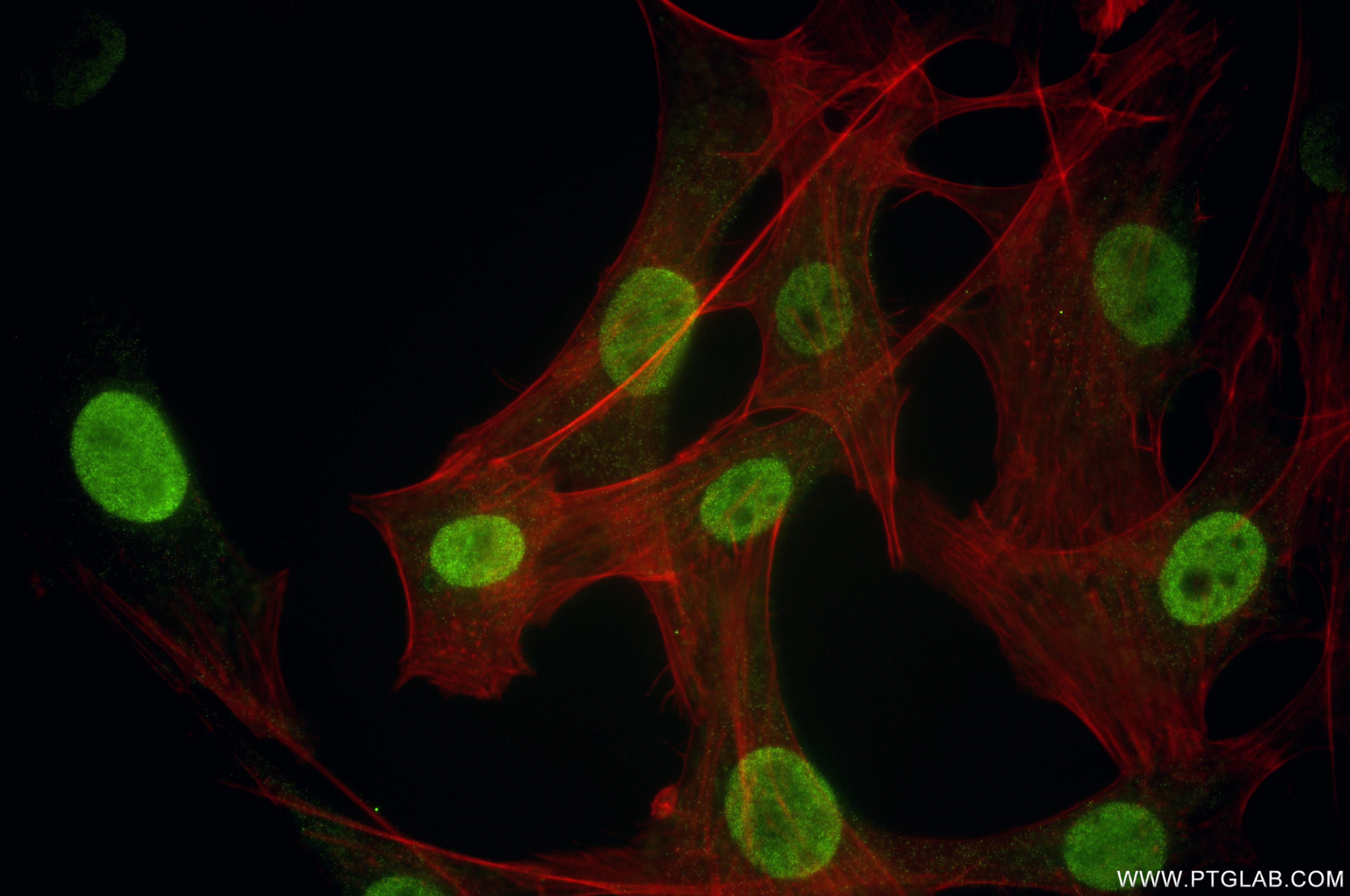
IF Staining of NIH/3T3 using 86861-1-RR
Immunofluorescent analysis of (4% PFA) fixed NIH/3T3 cells using Mono-Methyl-Histone H3 (Lys23) antibody (86861-1-RR, Clone: 250368B2 ) at dilution of 1:1000 and CoraLite®488-Conjugated Goat Anti-Rabbit IgG(H+L) (SA00013-2), CL594-Phalloidin (red).
Immunofluorescent analysis of (4% PFA) fixed NIH/3T3 cells using Mono-Methyl-Histone H3 (Lys23) antibody (86861-1-RR, Clone: 250368B2 ) at dilution of 1:1000 and CoraLite®488-Conjugated Goat Anti-Rabbit IgG(H+L) (SA00013-2), CL594-Phalloidin (red).
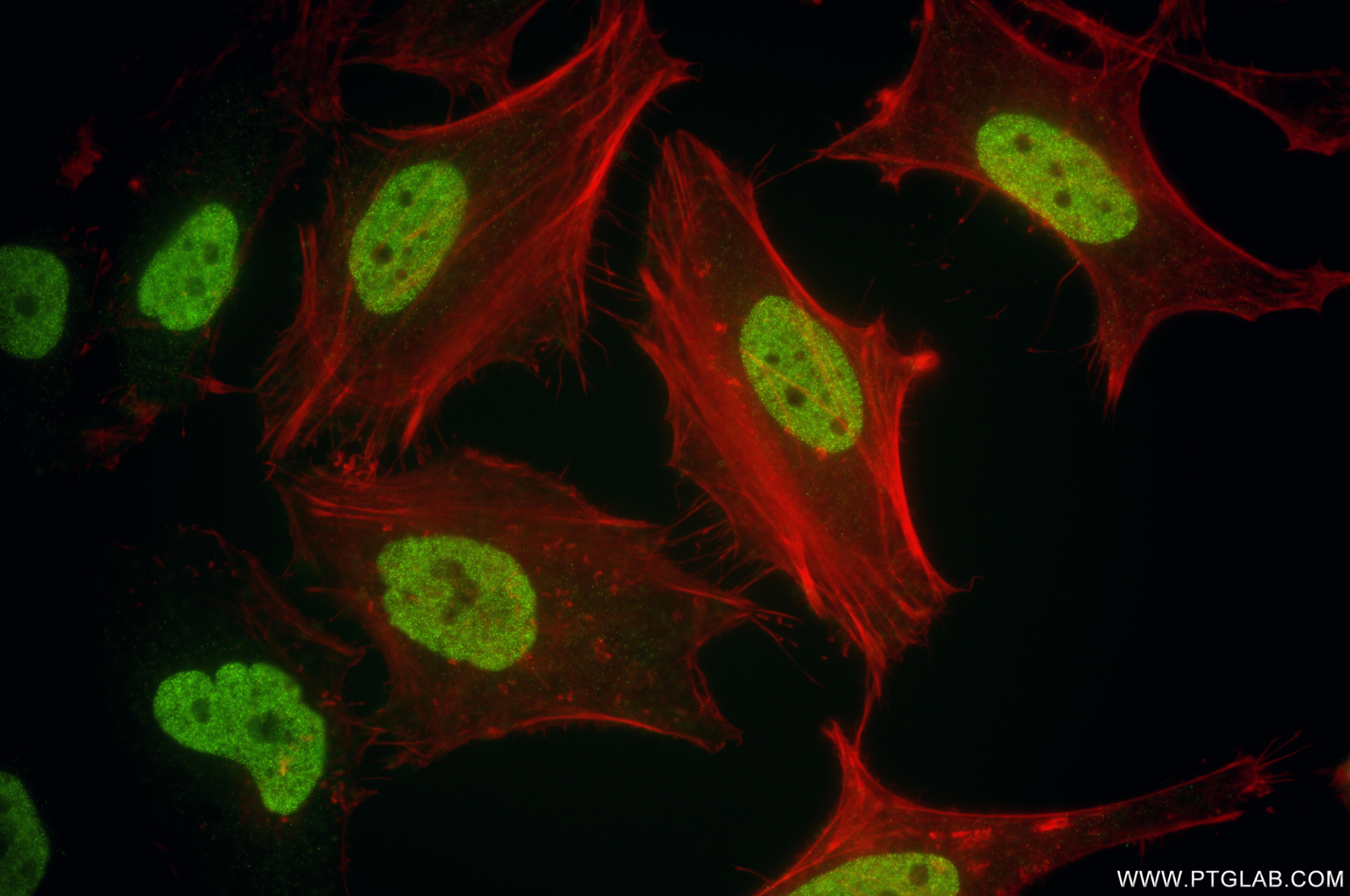
IF Staining of HeLa using 86861-1-RR
Immunofluorescent analysis of (4% PFA) fixed HeLa cells using Mono-Methyl-Histone H3 (Lys23) antibody (86861-1-RR, Clone: 250368B2 ) at dilution of 1:1000 and CoraLite®488-Conjugated Goat Anti-Rabbit IgG(H+L) (SA00013-2), CL594-Phalloidin (red).
Immunofluorescent analysis of (4% PFA) fixed HeLa cells using Mono-Methyl-Histone H3 (Lys23) antibody (86861-1-RR, Clone: 250368B2 ) at dilution of 1:1000 and CoraLite®488-Conjugated Goat Anti-Rabbit IgG(H+L) (SA00013-2), CL594-Phalloidin (red).
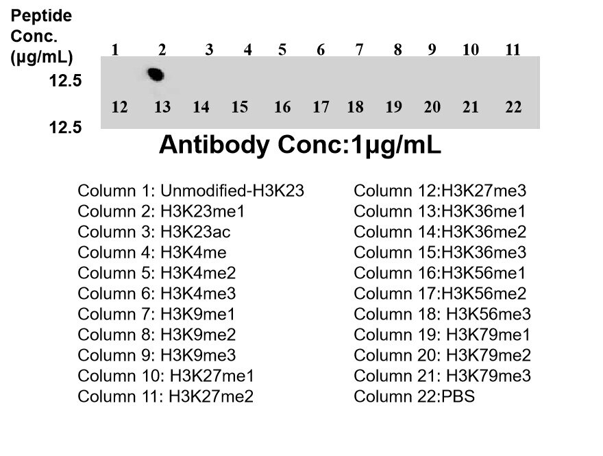
Dot Blot experiment of / using 86861-1-RR
Dot blot analysis was used to confirm the specificity of 86861-1-RR Mono-Methyl-Histone H3 (Lys23) antibody. peptides were spotted onto NC and probed with antibody at 1 µg/ml.The amount of peptide (μg/mL) spotted is indicated next to each row.
Dot blot analysis was used to confirm the specificity of 86861-1-RR Mono-Methyl-Histone H3 (Lys23) antibody. peptides were spotted onto NC and probed with antibody at 1 µg/ml.The amount of peptide (μg/mL) spotted is indicated next to each row.
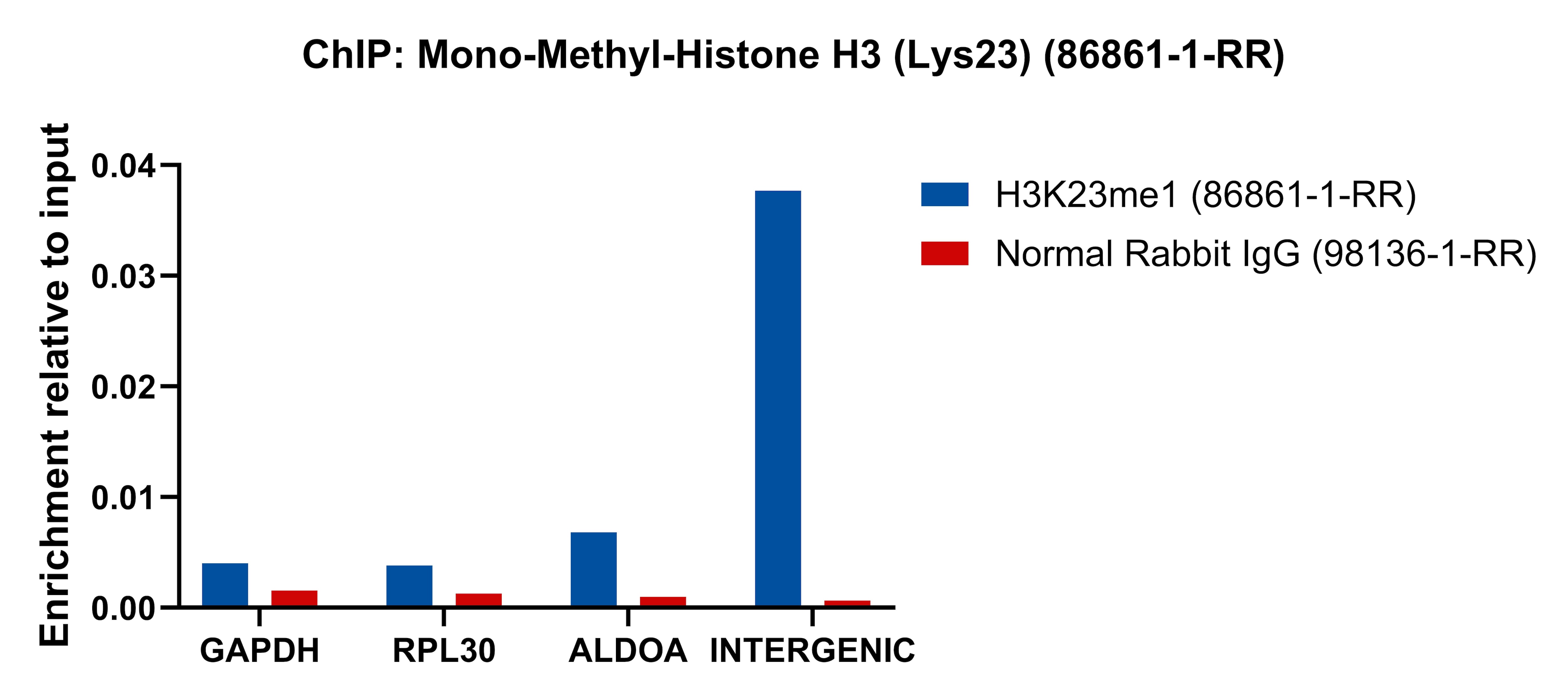
ChIP experiment of HeLa using 86861-1-RR
Chromatin was prepared from HeLa cells. Cells were fixed with formaldehyde for 10 minutes. The ChIP was performed with 15 µg of cross-linked chromatin, 5 µg of Mono-Methyl-Histone H3 (Lys23) (86861-1-RR) or 5 ug of Normal Rabbit IgG (98136-1-RR), and 20 µl of Protein A Magarose Beads. The immunoprecipitated DNA was quantified by real-time PCR.
Chromatin was prepared from HeLa cells. Cells were fixed with formaldehyde for 10 minutes. The ChIP was performed with 15 µg of cross-linked chromatin, 5 µg of Mono-Methyl-Histone H3 (Lys23) (86861-1-RR) or 5 ug of Normal Rabbit IgG (98136-1-RR), and 20 µl of Protein A Magarose Beads. The immunoprecipitated DNA was quantified by real-time PCR.
The Proteintech guarantee covers Proteintech antibodies in any species and any application, including those not listed on the datasheet. If the antibody doesn’t perform, you can receive a hassle-free refund or credit note.
Various lysates were subjected to SDS PAGE followed by western blot with 86861-1-RR (Mono-Methyl-Histone H3 (Lys23) antibody) at dilution of 1:5000 incubated at room temperature for 1.5 hours.
IF/ICC Figures
IF Staining of NIH/3T3 using 86861-1-RR
Immunofluorescent analysis of (4% PFA) fixed NIH/3T3 cells using Mono-Methyl-Histone H3 (Lys23) antibody (86861-1-RR, Clone: 250368B2 ) at dilution of 1:1000 and CoraLite®488-Conjugated Goat Anti-Rabbit IgG(H+L) (SA00013-2), CL594-Phalloidin (red).
IF Staining of HeLa using 86861-1-RR
Immunofluorescent analysis of (4% PFA) fixed HeLa cells using Mono-Methyl-Histone H3 (Lys23) antibody (86861-1-RR, Clone: 250368B2 ) at dilution of 1:1000 and CoraLite®488-Conjugated Goat Anti-Rabbit IgG(H+L) (SA00013-2), CL594-Phalloidin (red).
DOT BLOT Figures
Dot Blot experiment of / using 86861-1-RR
Dot blot analysis was used to confirm the specificity of 86861-1-RR Mono-Methyl-Histone H3 (Lys23) antibody. peptides were spotted onto NC and probed with antibody at 1 µg/ml.The amount of peptide (μg/mL) spotted is indicated next to each row.
CHIP-QPCR Figures
ChIP experiment of HeLa using 86861-1-RR
Chromatin was prepared from HeLa cells. Cells were fixed with formaldehyde for 10 minutes. The ChIP was performed with 15 µg of cross-linked chromatin, 5 µg of Mono-Methyl-Histone H3 (Lys23) (86861-1-RR) or 5 ug of Normal Rabbit IgG (98136-1-RR), and 20 µl of Protein A Magarose Beads. The immunoprecipitated DNA was quantified by real-time PCR.
The species listed in Tested Reactivity are in-house verified and applicable species. For unlisted species, please refer to the homology analysis of the immunogen sequence and related species. For rabbit polyclonal antibodies, homology >70% is recommended. For mouse monoclonal antibodies and rabbit recombinant antibodies, homology >90% is recommended. Generally, the higher the homology, the greater the applicability. However, there will be certain differences in protein expression in different species, tissues or cells. Therefore, the homology analysis results are for reference only and do not serve as a guarantee.
At Proteintech, we pride ourselves on our antibody quality, customer service and transparency. As such, we are comparing our antibodies with other vendors, enabling easy identification and comparisons of key data to help you choose the suitable antibody for your needs.
We have selected the top cited antibodies from these vendors for you to compare.

 antibody) at dilution of 1:5000 incubated at room temperature for 1.5 hours.")
 fixed NIH/3T3 cells using Mono-Methyl-Histone H3 (Lys23) antibody (86861-1-RR, Clone: 250368B2 ) at dilution of 1:1000 and CoraLite®488-Conjugated Goat Anti-Rabbit IgG(H+L) (SA00013-2), CL594-Phalloidin (red).")
 fixed HeLa cells using Mono-Methyl-Histone H3 (Lys23) antibody (86861-1-RR, Clone: 250368B2 ) at dilution of 1:1000 and CoraLite®488-Conjugated Goat Anti-Rabbit IgG(H+L) (SA00013-2), CL594-Phalloidin (red).")
 antibody. peptides were spotted onto NC and probed with antibody at 1 µg/ml.The amount of peptide (μg/mL) spotted is indicated next to each row.")
 (86861-1-RR) or 5 ug of Normal Rabbit IgG (98136-1-RR), and 20 µl of Protein A Magarose Beads. The immunoprecipitated DNA was quantified by real-time PCR.")