Tested Applications
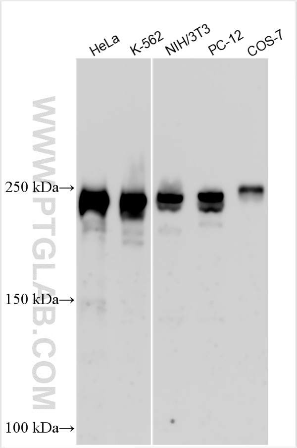
| Positive WB detected in | HeLa cells, K-562 cells, NIH/3T3 cells, PC-12 cells, COS-7 cells |
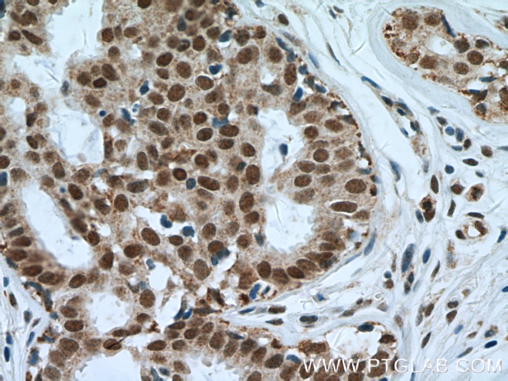
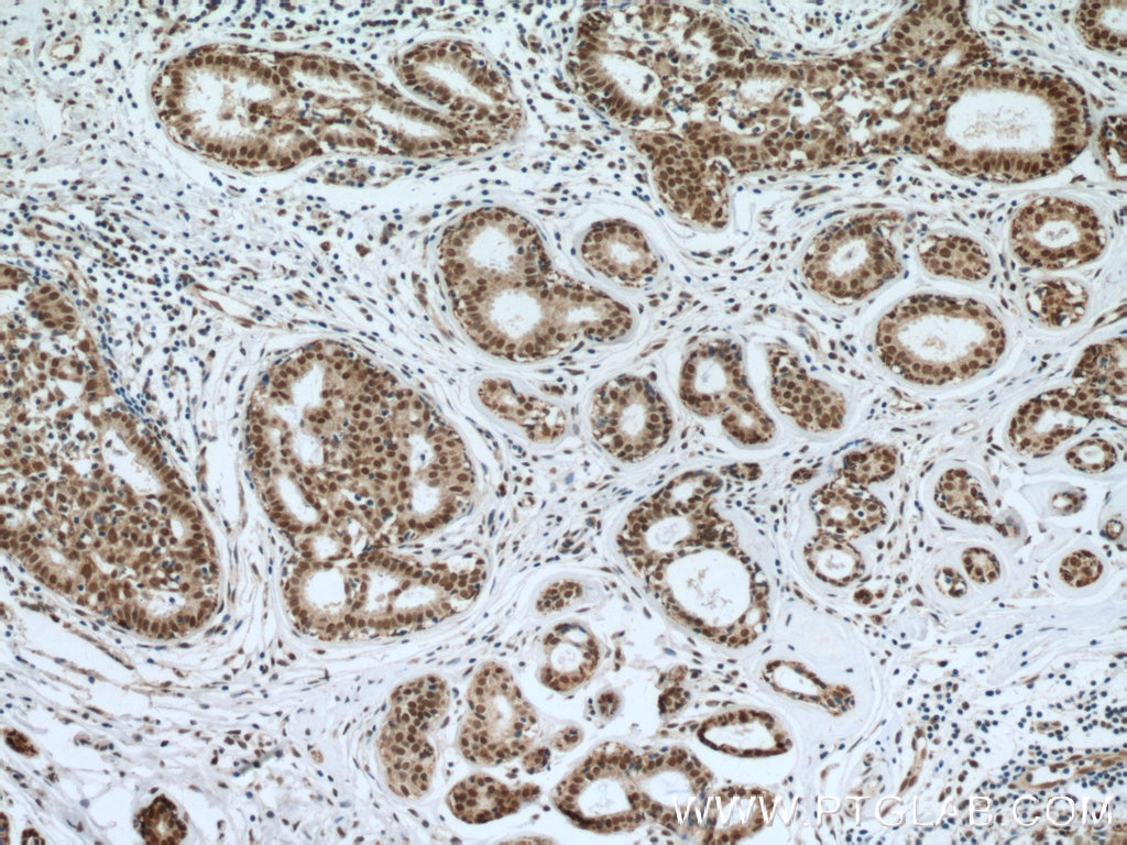
| Positive IHC detected in | human breast cancer tissue Note: suggested antigen retrieval with TE buffer pH 9.0; (*) Alternatively, antigen retrieval may be performed with citrate buffer pH 6.0 |
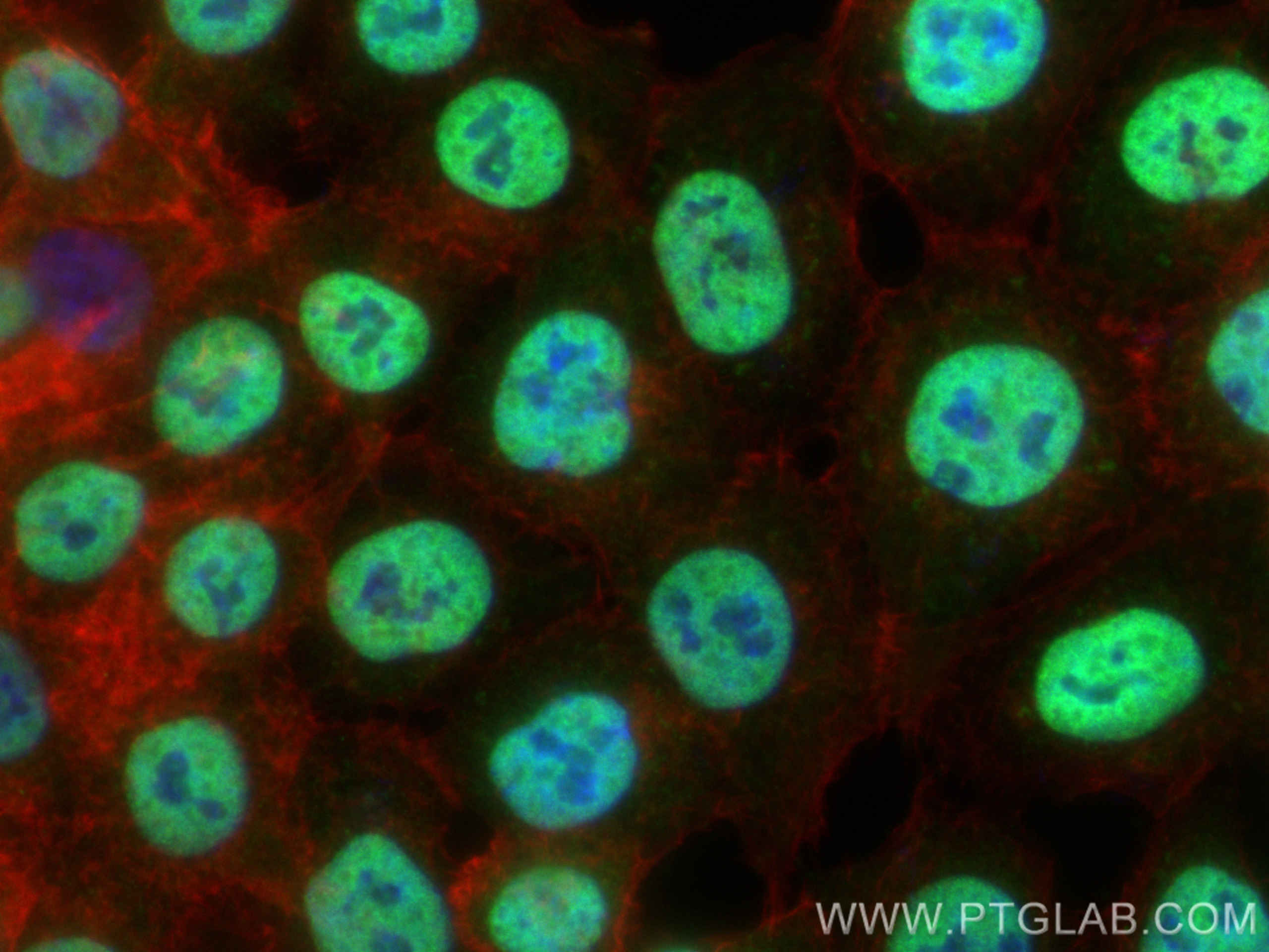
| Positive IF/ICC detected in | HCT 116 cells |
Recommended dilution
| Application | Dilution |
|---|---|
| Western Blot (WB) | WB : 1:500-1:2000 |
| Immunohistochemistry (IHC) | IHC : 1:50-1:500 |
| Immunofluorescence (IF)/ICC | IF/ICC : 1:200-1:800 |
| It is recommended that this reagent should be titrated in each testing system to obtain optimal results. | |
| Sample-dependent, Check data in validation data gallery. | |
Published Applications
| WB | See 1 publications below |
| IF | See 1 publications below |
Product Information
16607-1-AP targets NuMA in WB, IHC, IF/ICC, ELISA applications and shows reactivity with human, mouse, rat, monkey samples.
| Tested Reactivity | human, mouse, rat, monkey |
| Cited Reactivity | human, pig |
| Host / Isotype | Rabbit / IgG |
| Class | Polyclonal |
| Type | Antibody |
| Immunogen |
CatNo: Ag9815 Product name: Recombinant human NUMA1 protein Source: e coli.-derived, PET28a Tag: 6*His Domain: 845-1193 aa of BC013023 Sequence: VESLESLYFTPIPARSQAPLESSLDSLGDVFLDSGRKTRSARRRTTQIINITMTKKLDVEEPDSANSSFYSTRSAPASQASLRATSSTQSLARLGSPDYGNSALLSLPGYRPTTRSSARRSQAGVSSGAPPGRNSFYMGTCQDEPEQLDDWNRIAELQQRNRVCPPHLKTCYPLESRPSLSLGTITDEEMKTGDPQETLRRASMQPIQIAEGTGITTRQQRKRVSLEPHQGPGTPESKKATSCFPRPMTPRDRHEGRKQSTTEAQKKAAPASTKQADRRQSMAFSILNTPKKLGNSLLRRGASKKALSKASPNTRSGTRRSPRIATTTASAATAAAIGATPRAKGKAKH Predict reactive species |
| Full Name | nuclear mitotic apparatus protein 1 |
| Calculated Molecular Weight | 238 kDa |
| Observed Molecular Weight | 238 kDa |
| GenBank Accession Number | BC013023 |
| Gene Symbol | NuMA |
| Gene ID (NCBI) | 4926 |
| RRID | AB_2154616 |
| Conjugate | Unconjugated |
| Form | Liquid |
| Purification Method | Antigen affinity purification |
| UNIPROT ID | Q14980 |
| Storage Buffer | PBS with 0.02% sodium azide and 50% glycerol, pH 7.3. |
| Storage Conditions | Store at -20°C. Stable for one year after shipment. Aliquoting is unnecessary for -20oC storage. 20ul sizes contain 0.1% BSA. |
Background Information
NUMA1, also named as Nuclear matrix protein-22, is a 2115 amino acid protein, which forms multiarm oligomers by association of C-terminal tail domains. NUMA1 resides in the nuclear matrix during interphase. NUMA1 is a highly abundant component of the nuclear matrix where it may serve a non-mitotic structural role and occupies the majority if the nuclear volume. NUMA1 is required for maintenance and establishment of the mitotic spindle poles during symmetric cell divisions, functioning as a tether linking bulk microtubules of the spindle to centrosomes.
Protocols
| Product Specific Protocols | |
|---|---|
| IF protocol for NuMA antibody 16607-1-AP | Download protocol |
| IHC protocol for NuMA antibody 16607-1-AP | Download protocol |
| WB protocol for NuMA antibody 16607-1-AP | Download protocol |
| Standard Protocols | |
|---|---|
| Click here to view our Standard Protocols |
Publications
| Species | Application | Title |
|---|---|---|
Cell Death Dis CBX2 phase-separation contributes to homologous recombination repair and drug resistance in ovarian cancer. | ||
J Cell Sci RNA localization to the mitotic spindle is essential for early development and is regulated by kinesin-1 and dynein |
Reviews
The reviews below have been submitted by verified Proteintech customers who received an incentive for providing their feedback.
FH Sara (Verified Customer) (12-10-2025) | Performed well in Western Blot and Immunofluorescence, particularly Proximity Ligation Assays in combination with other primary antibodies.
|