Tested Applications
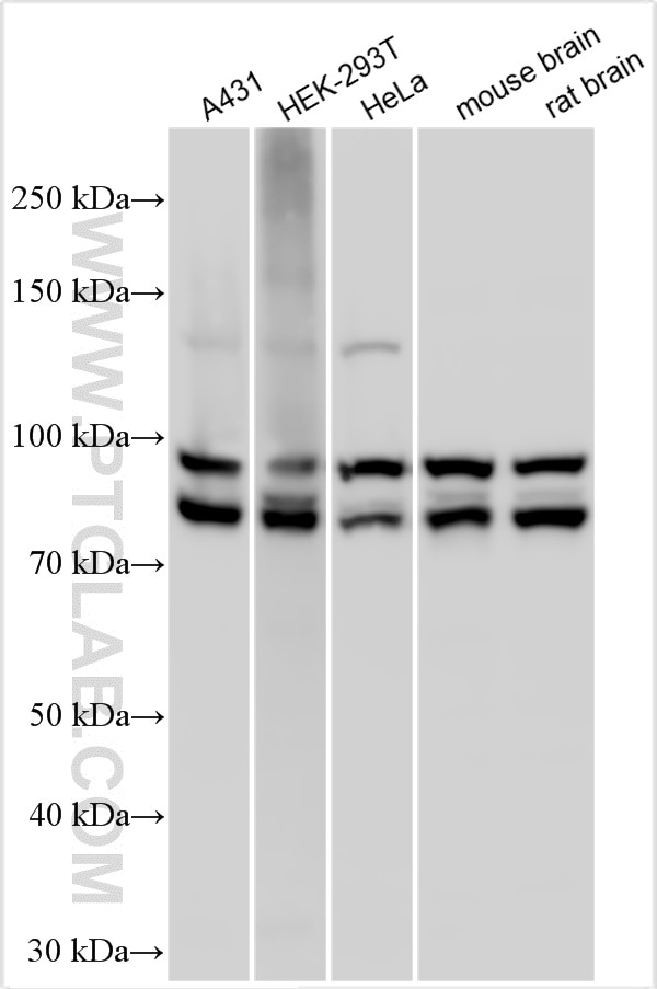
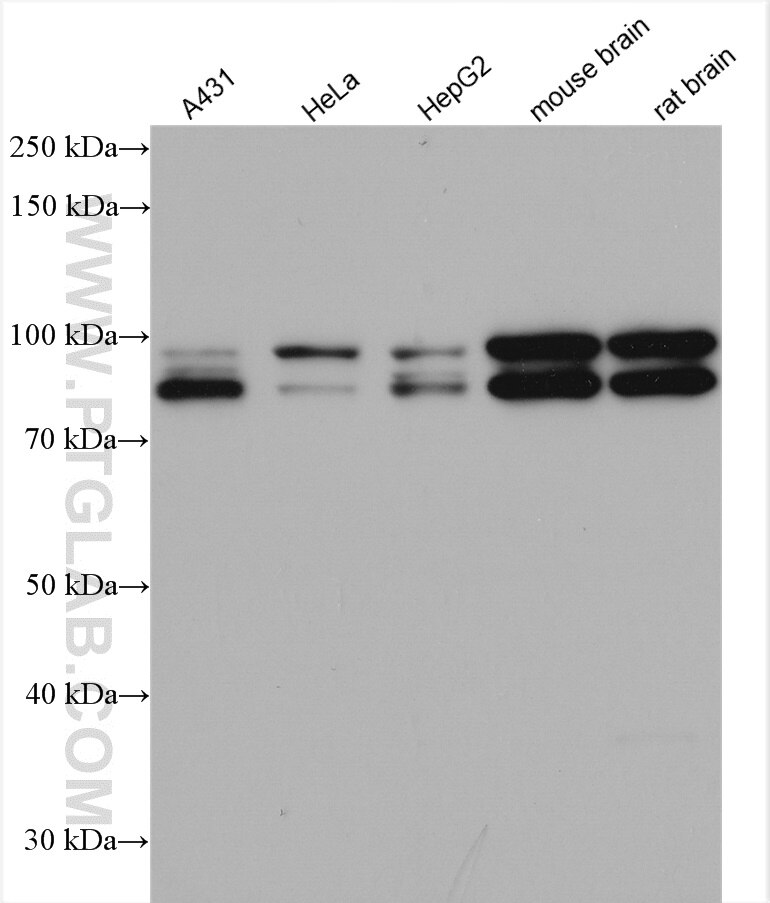
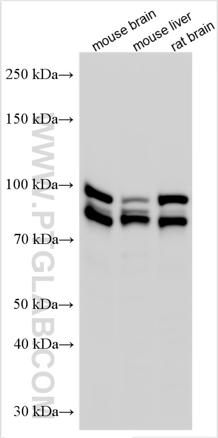
| Positive WB detected in | A431 cells, HEK-293 cells, mouse brain tissue, HeLa cells, HepG2 cells, rat brain tissue, HEK-293T cells, L02 cells, mouse liver tissue |
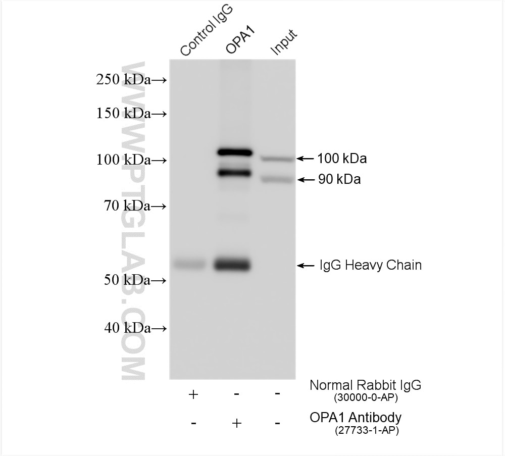
| Positive IP detected in | mouse brain tissue |
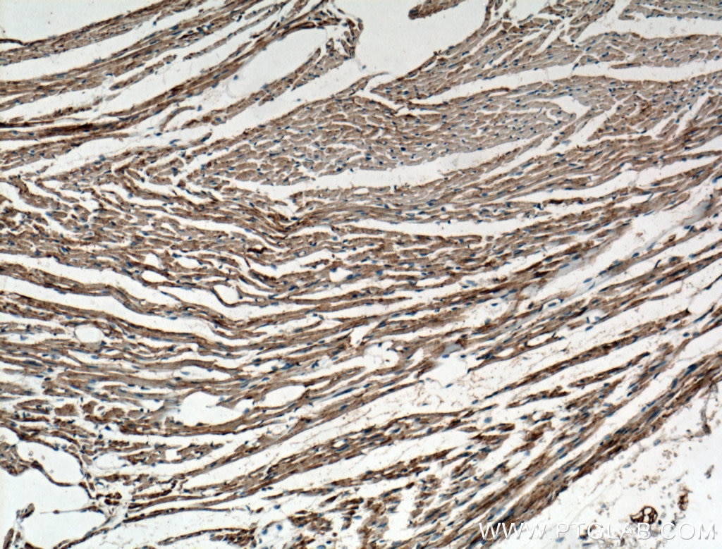
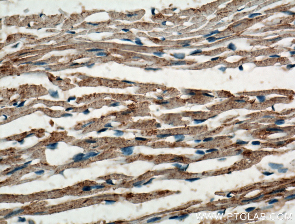
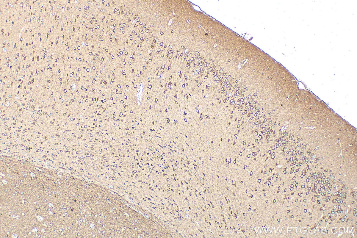
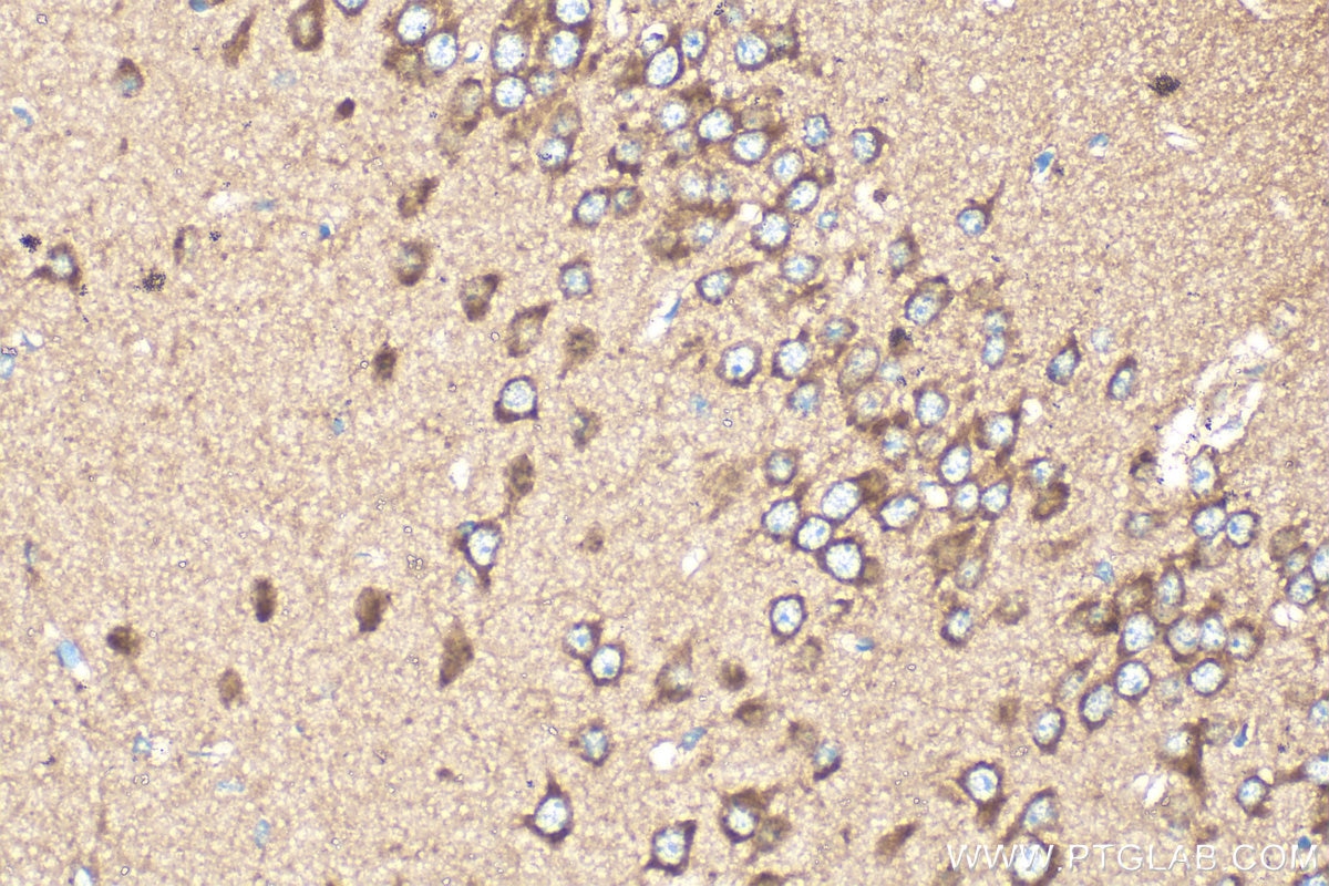
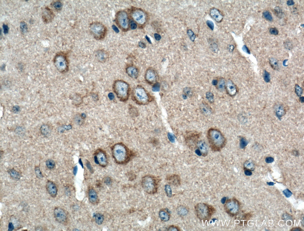
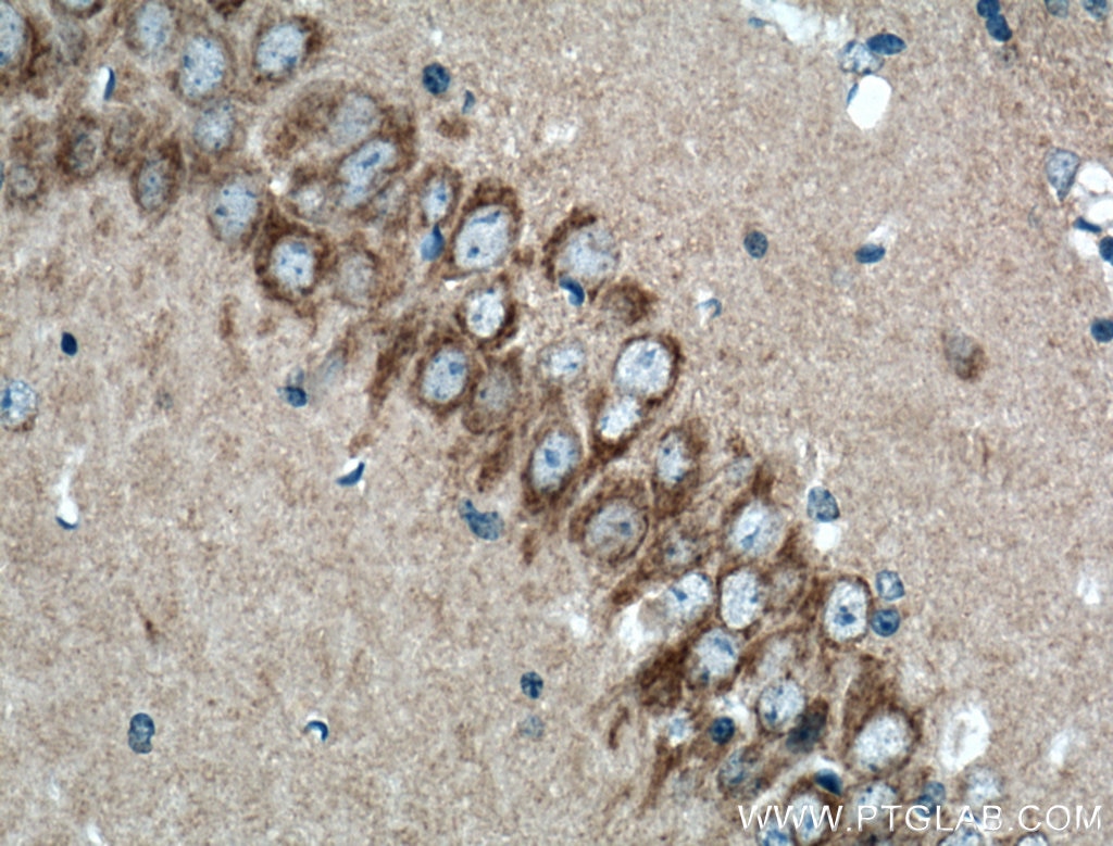
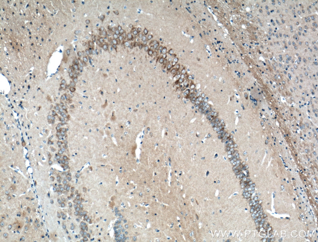
| Positive IHC detected in | mouse brain tissue, mouse heart tissue Note: suggested antigen retrieval with TE buffer pH 9.0; (*) Alternatively, antigen retrieval may be performed with citrate buffer pH 6.0 |
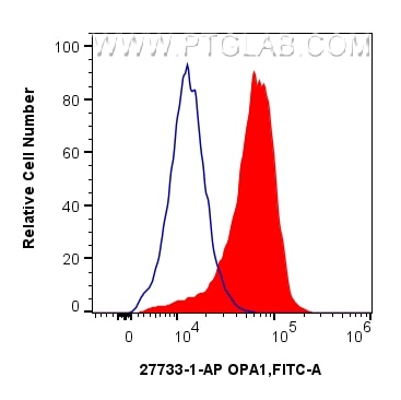
| Positive FC (Intra) detected in | HeLa cells |
Recommended dilution
| Application | Dilution |
|---|---|
| Western Blot (WB) | WB : 1:5000-1:50000 |
| Immunoprecipitation (IP) | IP : 0.5-4.0 ug for 1.0-3.0 mg of total protein lysate |
| Immunohistochemistry (IHC) | IHC : 1:500-1:2000 |
| Flow Cytometry (FC) (INTRA) | FC (INTRA) : 0.25 ug per 10^6 cells in a 100 µl suspension |
| It is recommended that this reagent should be titrated in each testing system to obtain optimal results. | |
| Sample-dependent, Check data in validation data gallery. | |
Published Applications
| KD/KO | See 5 publications below |
| WB | See 247 publications below |
| IHC | See 13 publications below |
| IF | See 17 publications below |
| IP | See 1 publications below |
| CoIP | See 3 publications below |
Product Information
27733-1-AP targets OPA1 in WB, IHC, IF, FC (Intra), IP, CoIP, ELISA applications and shows reactivity with human, mouse, rat samples.
| Tested Reactivity | human, mouse, rat |
| Cited Reactivity | human, mouse, rat, pig, chicken, zebrafish, bovine, hamster, goat, duck |
| Host / Isotype | Rabbit / IgG |
| Class | Polyclonal |
| Type | Antibody |
| Immunogen |
CatNo: Ag26887 Product name: Recombinant human OPA1 protein Source: e coli.-derived, PET30a Tag: 6*His Domain: 607-960 aa of BC075805 Sequence: SLSQVTPKHWEEILQQSLWERVSTHVIENIYLPAAQTMNSGTFNTTVDIKLKQWTDKQLPNKAVEVAWETLQEEFSRFMTEPKGKEHDDIFDKLKEAVKEESIKRHKWNDFAEDSLRVIQHNALEDRSISDKQQWDAAIYFMEEALQARLKDTENAIENMVGPDWKKRWLYWKNRTQEQCVHNETKNELEKMLKCNEEHPAYLASDEITTVRKNLESRGVEVDPSLIKDTWHQVYRRHFLKTALNHCNLCRRGFYYYQRHFVDSELECNDVVLFWRIQRMLAITANTLRQQLTNTEVRRLEKNVKEVLEDFAEDGEKKIKLLTGKRVQLAEDLKKVREIQEKLDAFIEALHQEK Predict reactive species |
| Full Name | optic atrophy 1 (autosomal dominant) |
| Calculated Molecular Weight | 960 aa, 112 kDa |
| Observed Molecular Weight | 80-100 kDa |
| GenBank Accession Number | BC075805 |
| Gene Symbol | OPA1 |
| Gene ID (NCBI) | 4976 |
| RRID | AB_2810292 |
| Conjugate | Unconjugated |
| Form | Liquid |
| Purification Method | Antigen affinity purification |
| UNIPROT ID | O60313 |
| Storage Buffer | PBS with 0.02% sodium azide and 50% glycerol, pH 7.3. |
| Storage Conditions | Store at -20°C. Stable for one year after shipment. Aliquoting is unnecessary for -20oC storage. 20ul sizes contain 0.1% BSA. |
Background Information
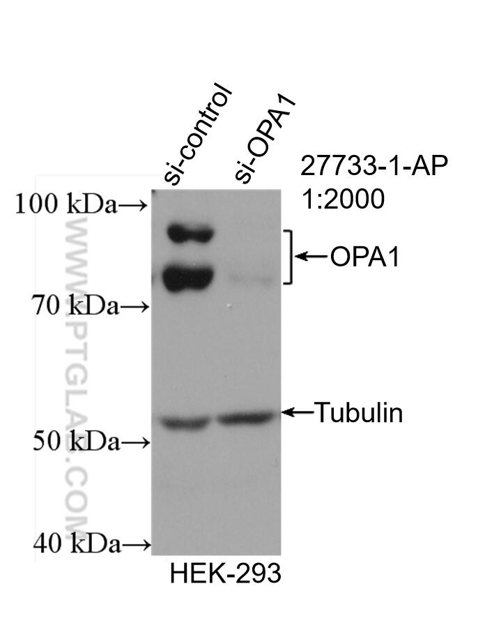
OPA1 is a nuclear-encoded mitochondrial protein with similarity to dynamin-related GTPases. OPA1 localizes to the inner mitochondrial membrane and helps regulate mitochondrial stability and energy output. This protein also sequesters cytochrome c. OPA1 is associated with the inner membrane and protects cells from apoptosis by regulating inner membrane dynamics. Mutation of OPA1 causes the disease dominant optic atrophy, a degeneration of the retinal ganglion cells. OPA1 undergoes complex posttranscriptional regulation and posttranslational proteolysis. OPA1 is regulated by proteolytic cleavage, which degrades long OPA1 isoforms into short isoforms. The gene OPA1 can be cleaved into some chains with MW 100 kDa and 80-90 kDa.
Protocols
| Product Specific Protocols | |
|---|---|
| WB protocol for OPA1 antibody 27733-1-AP | Download protocol |
| FC protocol for OPA1 antibody 27733-1-AP | Download protocol |
| IHC protocol for OPA1 antibody 27733-1-AP | Download protocol |
| IP protocol for OPA1 antibody 27733-1-AP | Download protocol |
| Standard Protocols | |
|---|---|
| Click here to view our Standard Protocols |
Publications
| Species | Application | Title |
|---|---|---|
Cell Res Mitochondria-localized cGAS suppresses ferroptosis to promote cancer progression | ||
ACS Cent Sci Macrophage Inactivation by Small Molecule Wedelolactone via Targeting sEH for the Treatment of LPS-Induced Acute Lung Injury | ||
Mol Cell Serine synthesis sustains macrophage IL-1β production via NAD+-dependent protein acetylation | ||
Mol Cell β-hydroxybutyrate facilitates mitochondrial-derived vesicle biogenesis and improves mitochondrial functions | ||
Mol Cell Filamentous GLS1 promotes ROS-induced apoptosis upon glutamine deprivation via insufficient asparagine synthesis.
| ||
Bone Res Mechanism of Piezo1 regulating chondrocyte mitochondrial function and promoting fracture healing through β-catenin/LARS2 signaling pathway |
Reviews
The reviews below have been submitted by verified Proteintech customers who received an incentive for providing their feedback.
FH Chun (Verified Customer) (12-22-2025) | This is an excellent antibody for Immunoblotting.
|
FH Dhanwini (Verified Customer) (09-24-2025) | GOOD
|
FH Robert (Verified Customer) (09-24-2025) | Didn't get two bands in my application, but otherwise turned out as expected
|
FH Sneha (Verified Customer) (09-24-2025) | GOOD
|
FH Michael (Verified Customer) (09-23-2025) | Excellent
|
FH Henry (Verified Customer) (09-23-2025) | Ease of use
|
FH Kahimbi (Verified Customer) (01-31-2025) | It was used as a primary antibody for a western blot and the bands were clearly visible. The antibody worked well.
|
FH P (Verified Customer) (09-23-2024) | excellent!
 |
FH Mi (Verified Customer) (02-21-2023) | Works well in human adipocyte cells, we got clean bands at the expected size.
|