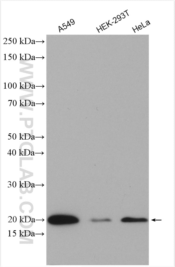
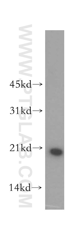
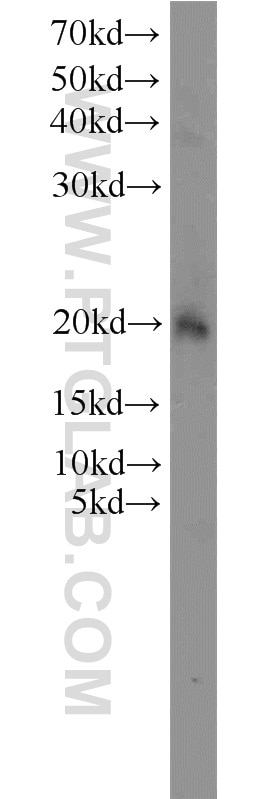
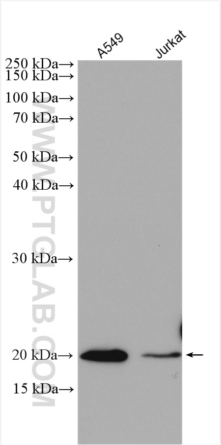
Tested Applications
| Positive WB detected in | A549 cells, human kidney tissue, mouse thymus tissue, Jurkat cells, HeLa cells, HEK-293T cells |
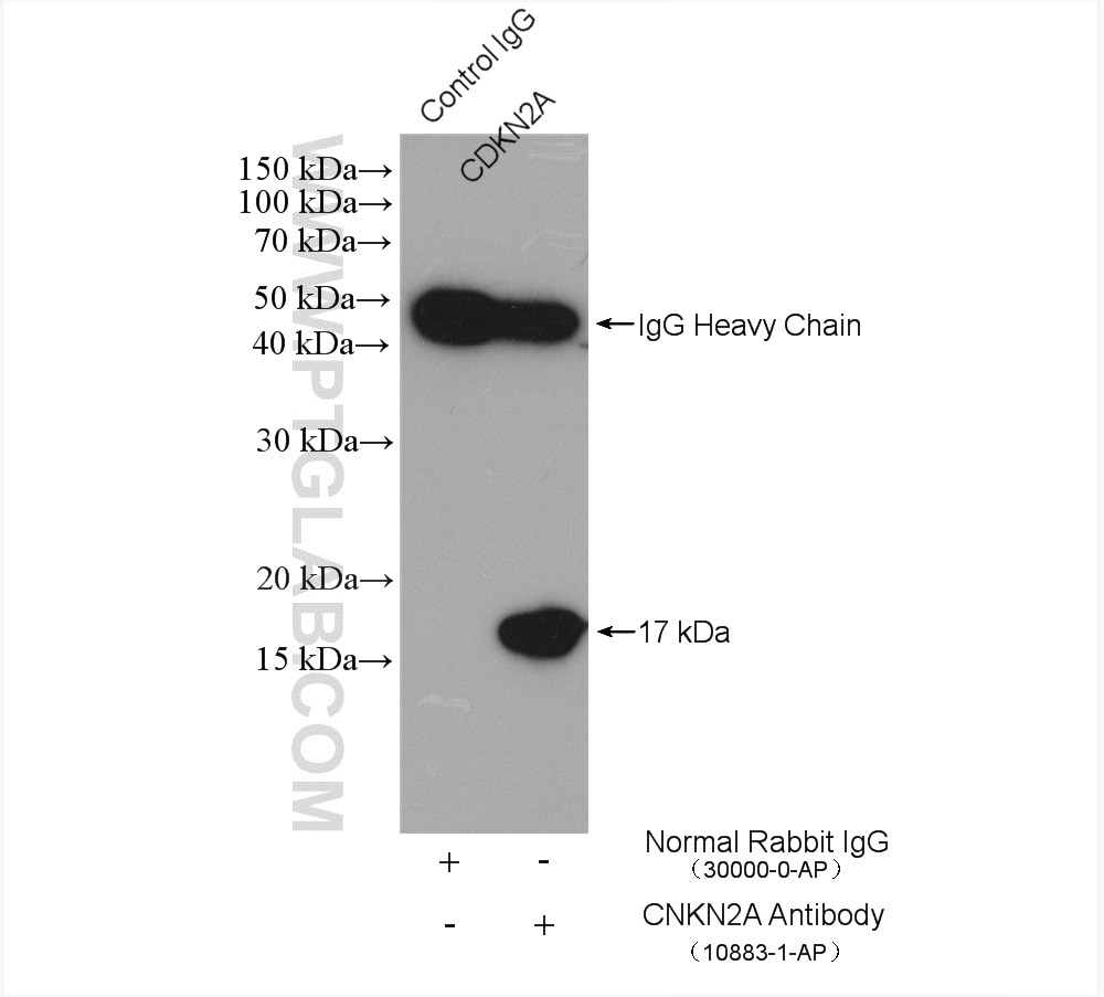
| Positive IP detected in | HeLa cells |
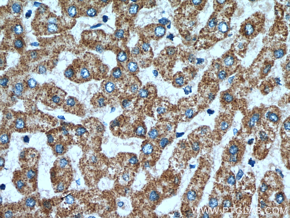
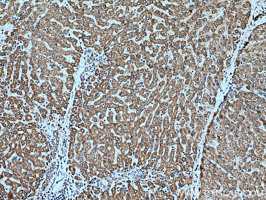
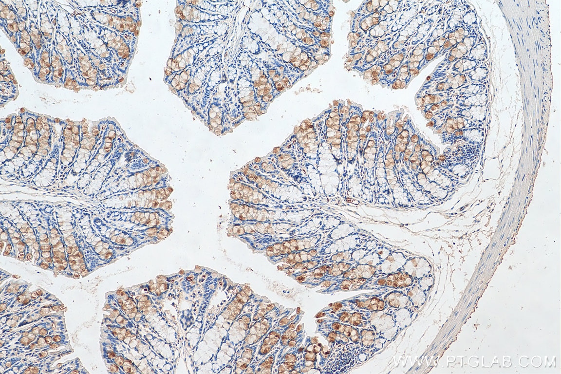
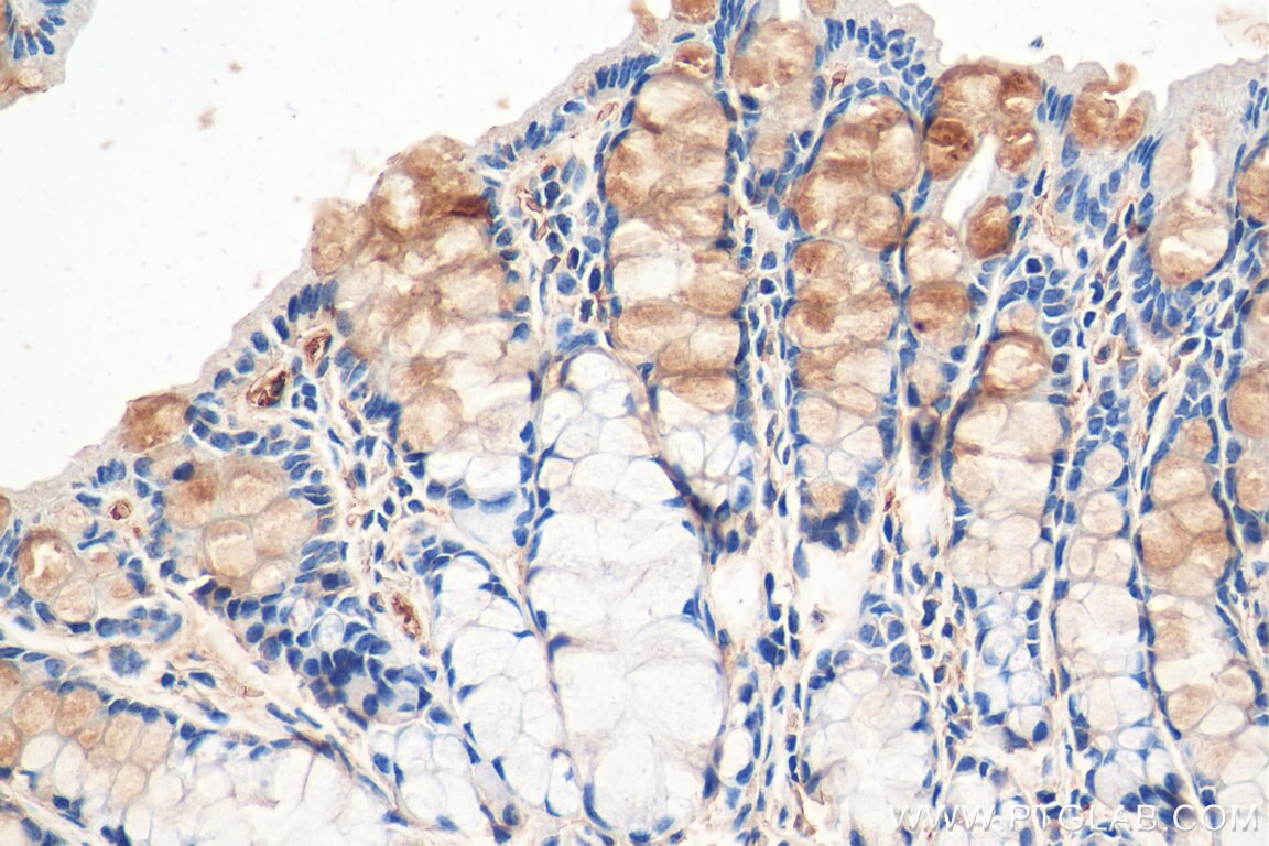
| Positive IHC detected in | human liver tissue, mouse colon tissue Note: suggested antigen retrieval with TE buffer pH 9.0; (*) Alternatively, antigen retrieval may be performed with citrate buffer pH 6.0 |
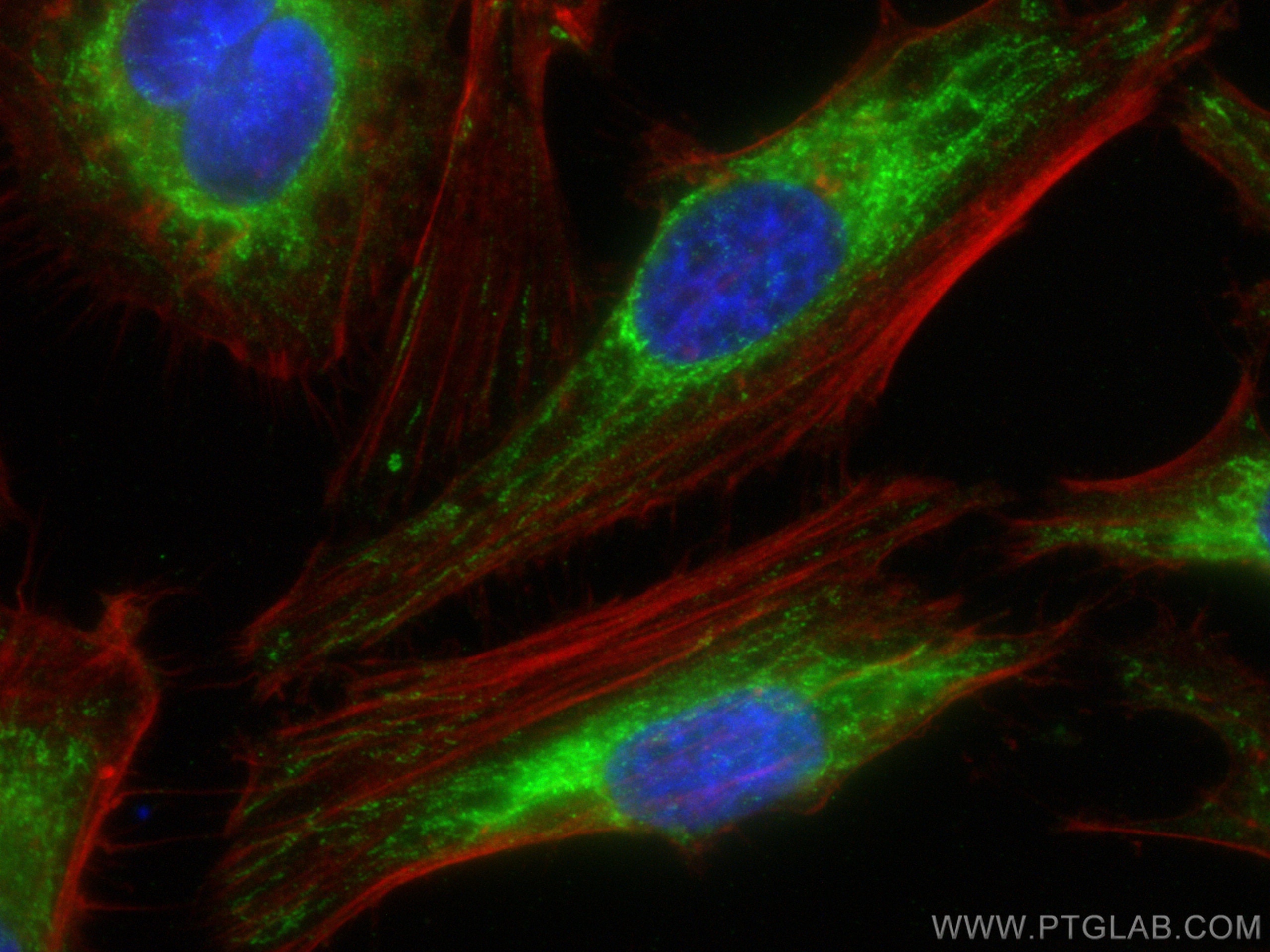
| Positive IF/ICC detected in | HeLa cells |
Recommended dilution
| Application | Dilution |
|---|---|
| Western Blot (WB) | WB : 1:500-1:2000 |
| Immunoprecipitation (IP) | IP : 0.5-4.0 ug for 1.0-3.0 mg of total protein lysate |
| Immunohistochemistry (IHC) | IHC : 1:50-1:500 |
| Immunofluorescence (IF)/ICC | IF/ICC : 1:50-1:500 |
| It is recommended that this reagent should be titrated in each testing system to obtain optimal results. | |
| Sample-dependent, Check data in validation data gallery. | |
Published Applications
| KD/KO | See 1 publications below |
| WB | See 5 publications below |
| IHC | See 3 publications below |
| IP | See 1 publications below |
Product Information
15638-1-AP targets OPA3 in WB, IHC, IF/ICC, IP, ELISA applications and shows reactivity with human, mouse, rat samples.
| Tested Reactivity | human, mouse, rat |
| Cited Reactivity | human, mouse, rat, bovine |
| Host / Isotype | Rabbit / IgG |
| Class | Polyclonal |
| Type | Antibody |
| Immunogen |
CatNo: Ag8103 Product name: Recombinant human OPA3 protein Source: e coli.-derived, PGEX-4T Tag: GST Domain: 24-179 aa of BC005059 Sequence: ANRIKEAARRSEFFKTYICLPPAQLYHWVEMRTKMRIMGFRGTVIKPLNEEAAAELGAELLGEATIFIVGGGCLVLEYWRHQAQQRHKEEEQRAAWNALRDEVGHLALALEALQAQVQAAPPQGALEELRTELQEVRAQLCNPGRSASHAVPASKK Predict reactive species |
| Full Name | optic atrophy 3 (autosomal recessive, with chorea and spastic paraplegia) |
| Calculated Molecular Weight | 179 aa, 20 kDa |
| Observed Molecular Weight | 20 kDa |
| GenBank Accession Number | BC005059 |
| Gene Symbol | OPA3 |
| Gene ID (NCBI) | 80207 |
| RRID | AB_2158168 |
| Conjugate | Unconjugated |
| Form | Liquid |
| Purification Method | Antigen affinity purification |
| UNIPROT ID | Q9H6K4 |
| Storage Buffer | PBS with 0.02% sodium azide and 50% glycerol, pH 7.3. |
| Storage Conditions | Store at -20°C. Stable for one year after shipment. Aliquoting is unnecessary for -20oC storage. 20ul sizes contain 0.1% BSA. |
Background Information
The OPA3 cDNA encodes a deduced 179-amino acid protein. Northern blot analysis demonstrated a primary transcript of approximately 5.0 kb that was ubiquitously expressed, most prominently in skeletal muscle and kidney. Mutations in this gene have been shown to result in 3-methylglutaconic aciduria type III and autosomal dominant optic atrophy and cataract.
Protocols
| Product Specific Protocols | |
|---|---|
| IF protocol for OPA3 antibody 15638-1-AP | Download protocol |
| IHC protocol for OPA3 antibody 15638-1-AP | Download protocol |
| IP protocol for OPA3 antibody 15638-1-AP | Download protocol |
| WB protocol for OPA3 antibody 15638-1-AP | Download protocol |
| Standard Protocols | |
|---|---|
| Click here to view our Standard Protocols |
Publications
| Species | Application | Title |
|---|---|---|
Hum Mol Genet Disrupted mitochondrial function in the Opa3L122P mouse model for Costeff Syndrome impairs skeletal integrity. | ||
Cell Signal Hydrogen sulfide alleviates mitochondrial damage and ferroptosis by regulating OPA3-NFS1 axis in doxorubicin-induced cardiotoxicity | ||
Invest Ophthalmol Vis Sci Mitochondrial localization and ocular expression of mutant Opa3 in a mouse model of 3-methylglutaconicaciduria type III. | ||
Genomics A nonsense mutation in the optic atrophy 3 gene (OPA3) causes dilated cardiomyopathy in Red Holstein cattle. | ||
Bioengineered MYB proto-oncogene like 2 promotes hepatocellular carcinoma growth and glycolysis via binding to the Optic atrophy 3 promoter and activating its expression.
| ||
Cancers (Basel) Oncogenic K-ras Induces Mitochondrial OPA3 Expression to Promote Energy Metabolism in Pancreatic Cancer Cells. |