Tested Applications
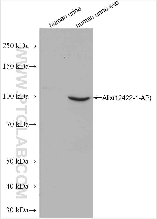
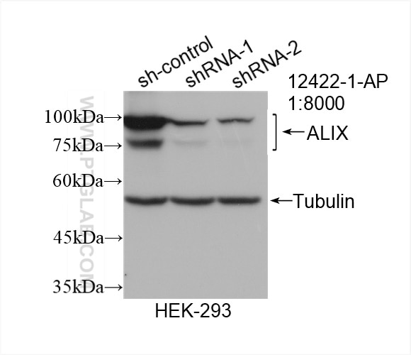
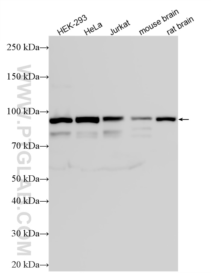
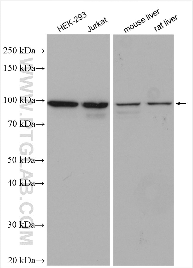
| Positive WB detected in | HEK-293 cells, human urine exosomes tissue, Jurkat cells, mouse liver tissue, rat liver tissue, HeLa cells, mouse brain tissue, rat brain tissue |
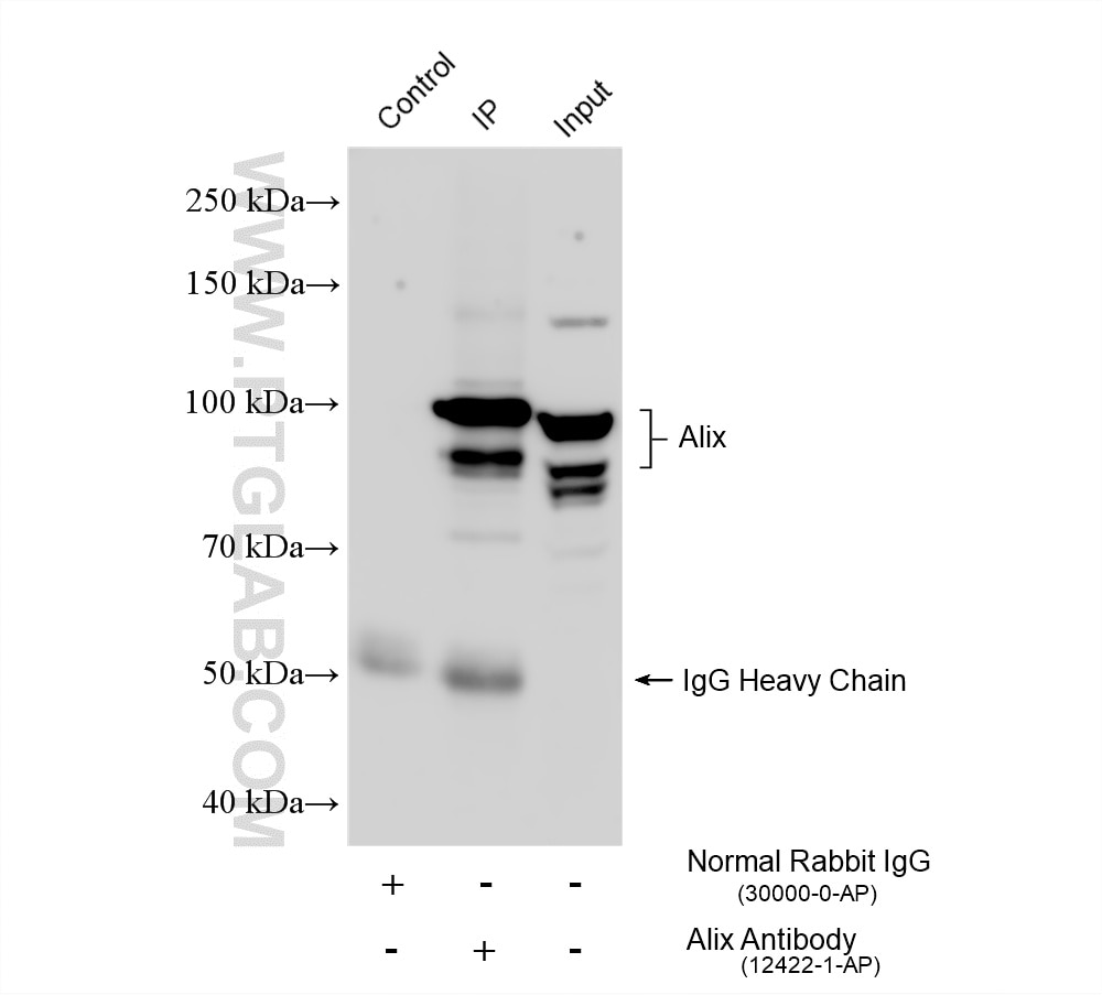
| Positive IP detected in | HeLa cells |
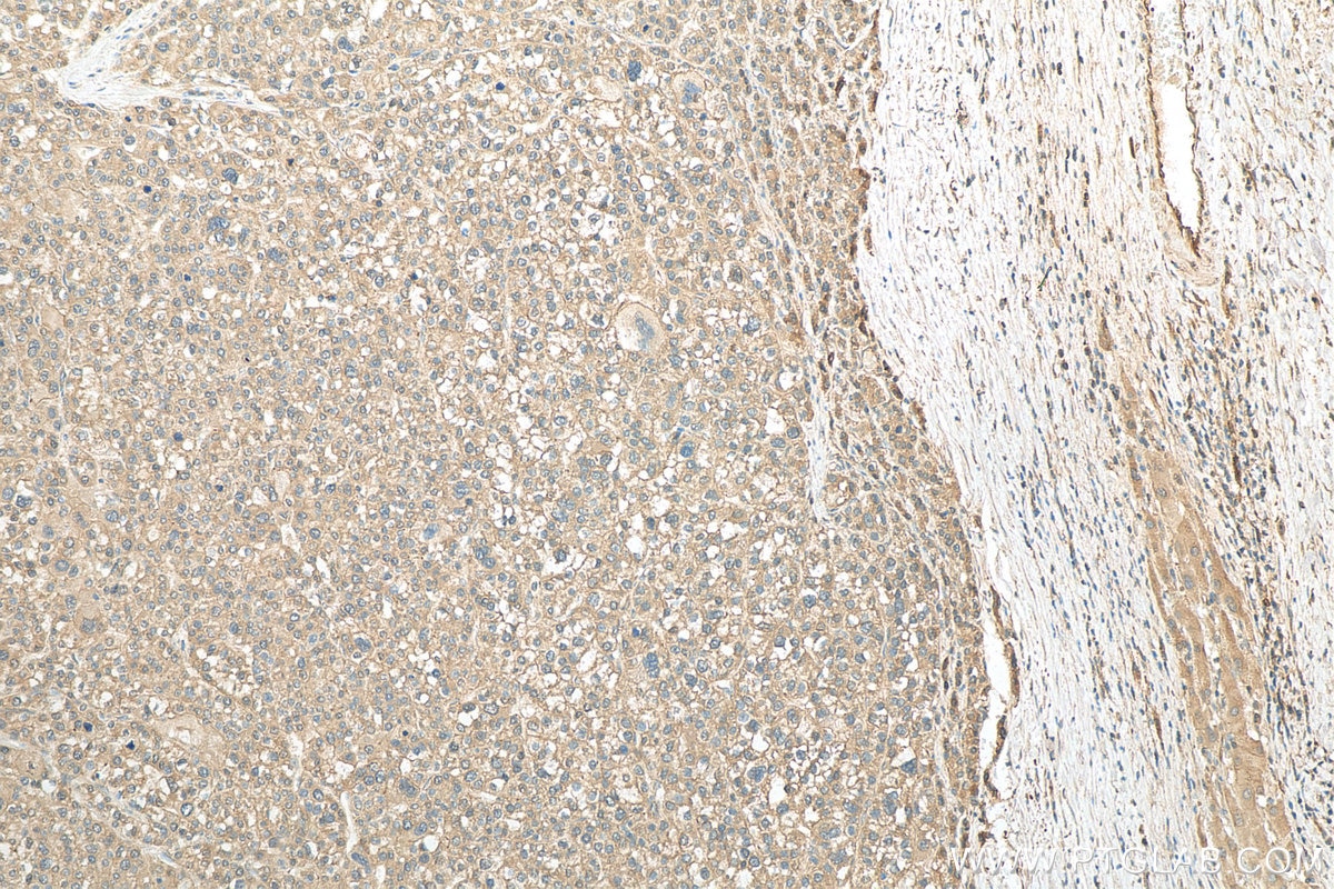
| Positive IHC detected in | human liver cancer tissue, human colon tissue Note: suggested antigen retrieval with TE buffer pH 9.0; (*) Alternatively, antigen retrieval may be performed with citrate buffer pH 6.0 |
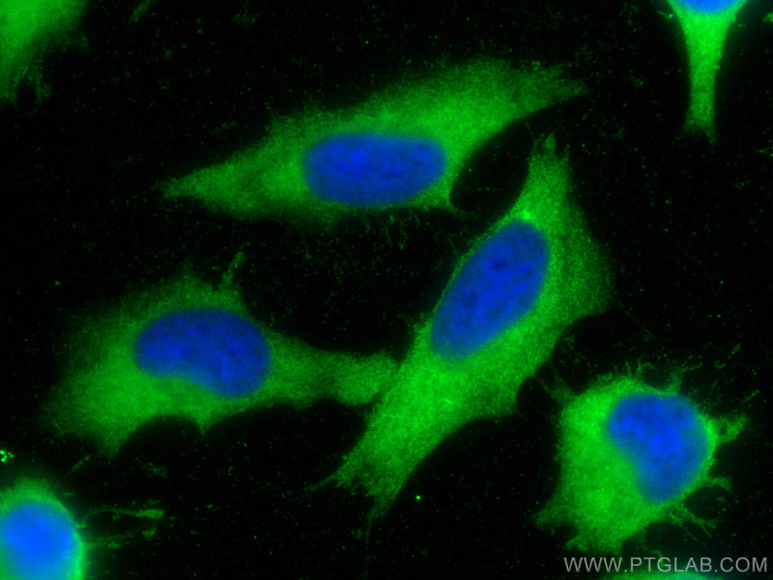
| Positive IF/ICC detected in | HeLa cells |
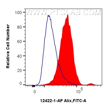
| Positive FC (Intra) detected in | HeLa cells |
Recommended dilution
| Application | Dilution |
|---|---|
| Western Blot (WB) | WB : 1:5000-1:60000 |
| Immunoprecipitation (IP) | IP : 0.5-4.0 ug for 1.0-3.0 mg of total protein lysate |
| Immunohistochemistry (IHC) | IHC : 1:50-1:500 |
| Immunofluorescence (IF)/ICC | IF/ICC : 1:200-1:800 |
| Flow Cytometry (FC) (INTRA) | FC (INTRA) : 0.25 ug per 10^6 cells in a 100 µl suspension |
| It is recommended that this reagent should be titrated in each testing system to obtain optimal results. | |
| Sample-dependent, Check data in validation data gallery. | |
Published Applications
| KD/KO | See 5 publications below |
| WB | See 338 publications below |
| IHC | See 2 publications below |
| IF | See 16 publications below |
| IP | See 3 publications below |
| CoIP | See 1 publications below |
Product Information
12422-1-AP targets Alix in WB, IHC, IF/ICC, IP, CoIP, ELISA applications and shows reactivity with human, mouse, rat samples.
| Tested Reactivity | human, mouse, rat |
| Cited Reactivity | human, mouse, rat, pig, rabbit, canine, monkey, hamster |
| Host / Isotype | Rabbit / IgG |
| Class | Polyclonal |
| Type | Antibody |
| Immunogen |
CatNo: Ag3074 Product name: Recombinant human ALIX; AIP1 protein Source: e coli.-derived, PGEX-4T Tag: GST Domain: 515-868 aa of BC020066 Sequence: SHRDTIVLLCKPEPELNAAIPSANPAKTMQGSEVVNVLKSLLSNLDEVKKEREGLENDLKSVNFDMTSKFLTALAQDGVINEEALSVTELDRVYGGLTTKVQESLKKQEGLLKNIQVSHQEFSKMKQSNNEANLREEVLKNLATAYDNFVELVANLKEGTKFYNELTEILVRFQNKCSDIVFARKTERDELLKDLQQSIAREPSAPSIPTPAYQSSPAGGHAPTPPTPAPRTMPPTKPQPPARPPPPVLPANRAPSATAPSPVGAGTAAPAPSQTPGSAPPPQAQGPPYPTYPGYPGYCQMPMPMGYNPYAYGQYNMPYPPVYHQSPGQAPYPGPQQPSYPFPQPPQQSYYPQQ Predict reactive species |
| Full Name | programmed cell death 6 interacting protein |
| Calculated Molecular Weight | 868 aa, 96 kDa |
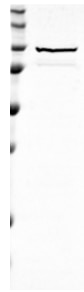
| Observed Molecular Weight | 75-100 kDa |
| GenBank Accession Number | BC020066 |
| Gene Symbol | Alix |
| Gene ID (NCBI) | 10015 |
| RRID | AB_2162467 |
| Conjugate | Unconjugated |
| Form | Liquid |
| Purification Method | Antigen affinity purification |
| UNIPROT ID | Q8WUM4 |
| Storage Buffer | PBS with 0.02% sodium azide and 50% glycerol, pH 7.3. |
| Storage Conditions | Store at -20°C. Stable for one year after shipment. Aliquoting is unnecessary for -20oC storage. 20ul sizes contain 0.1% BSA. |
Background Information
ALG-2-interacting protein 1 (ALIX), also known as AIP1 or Hp95, is encoded by PDCD6IP gene and is involved in cell death through mechanisms involving its binding partner ALG-2 (apoptosis-linked gene-2). ALG-2 is a 22-kDa protein containing five serially repetitive EF-hand structures and is defined as a regulator of calcium-induced apoptosis following endoplasmic reticulum (ER) stress. ALIX interacts with ALG-2 through its C-terminal proline-rich region and participates in formation of multivesicular bodies. Recent finding suggest that ALIX is a critical component of caspase 9 activation and apoptosis triggered by calcium. The alix antibody recognizes an additional band of 75-80 kDa which has also been observed in cells and exosomes.
Protocols
| Product Specific Protocols | |
|---|---|
| FC protocol for Alix antibody 12422-1-AP | Download protocol |
| IF protocol for Alix antibody 12422-1-AP | Download protocol |
| IHC protocol for Alix antibody 12422-1-AP | Download protocol |
| IP protocol for Alix antibody 12422-1-AP | Download protocol |
| WB protocol for Alix antibody 12422-1-AP | Download protocol |
| Standard Protocols | |
|---|---|
| Click here to view our Standard Protocols |
Publications
| Species | Application | Title |
|---|---|---|
Cell Metab Nicotinamide metabolism face-off between macrophages and fibroblasts manipulates the microenvironment in gastric cancer | ||
Nat Immunol Exosomes mediate the cell-to-cell transmission of IFN-α-induced antiviral activity. | ||
Cell Res Intercellular transfer of activated STING triggered by RAB22A-mediated non-canonical autophagy promotes antitumor immunity | ||
Nat Cell Biol Endosomal membrane tension regulates ESCRT-III-dependent intra-lumenal vesicle formation.
| ||
ACS Nano Mesenchymal Stem Cell-Derived Extracellular Vesicles Attenuate Mitochondrial Damage and Inflammation by Stabilizing Mitochondrial DNA. |
Reviews
The reviews below have been submitted by verified Proteintech customers who received an incentive for providing their feedback.
FH Sai Sindhura (Verified Customer) (01-08-2026) | Alix antibody works good
|
FH Sonam (Verified Customer) (12-09-2025) | Highly recommend. Even works at higher dilution ( 1:3000). Proteintech antibodies are good.
|
FH Kamal (Verified Customer) (02-15-2024) | Strong bands appeared between 70 and 100 kDa.
|
FH Guorong (Verified Customer) (03-22-2022) | Excellent performance with a specific band of expected size
 |
FH Susmita (Verified Customer) (02-11-2022) | all antibodies from Proteintech works great for me
|
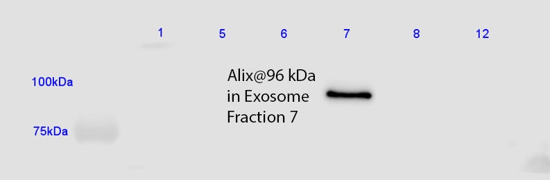
FH Jorge (Verified Customer) (05-15-2019) | Gave a very strong signal using the lowest recommended antibody dilution for Western Blot, diluted it to 1:2000 and worked just as well with a low protein detection substrate. Detected exosomes in fraction 7 of a sucrose gradient as expected.
 |