

Tested Applications
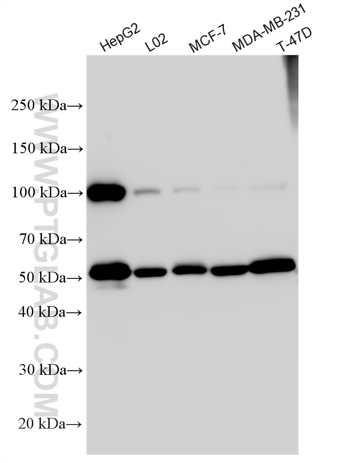
| Positive WB detected in | HepG2 cells, L02 cells, MCF-7 cells, MDA-MB-231 cells, T-47D cells |
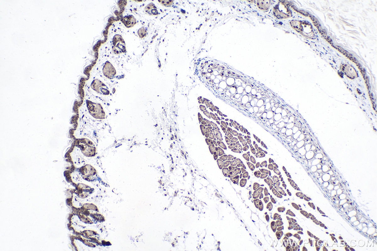
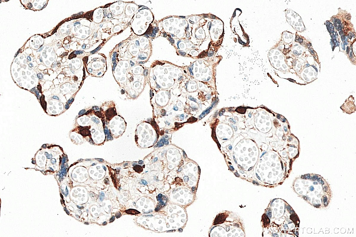
| Positive IHC detected in | mouse skin tissue, human placenta tissue Note: suggested antigen retrieval with TE buffer pH 9.0; (*) Alternatively, antigen retrieval may be performed with citrate buffer pH 6.0 |
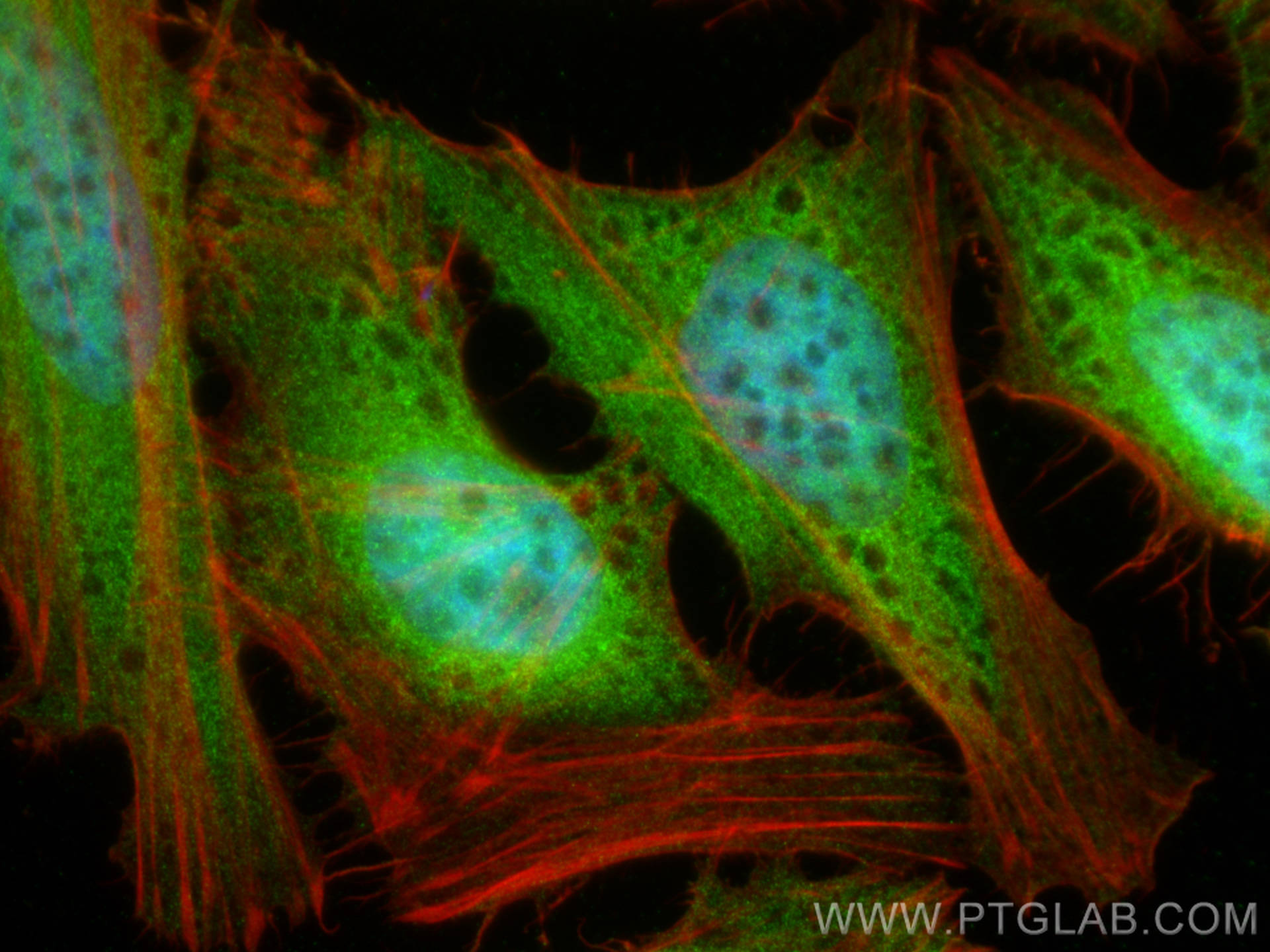
| Positive IF/ICC detected in | HeLa cells |
Recommended dilution
| Application | Dilution |
|---|---|
| Western Blot (WB) | WB : 1:2000-1:12000 |
| Immunohistochemistry (IHC) | IHC : 1:500-1:2000 |
| Immunofluorescence (IF)/ICC | IF/ICC : 1:200-1:800 |
| It is recommended that this reagent should be titrated in each testing system to obtain optimal results. | |
| Sample-dependent, Check data in validation data gallery. | |
Product Information
14412-1-AP targets PEG10 in WB, IHC, IF/ICC, IP, CoIP, ELISA applications and shows reactivity with human, mouse samples.
| Tested Reactivity | human, mouse |
| Cited Reactivity | human |
| Host / Isotype | Rabbit / IgG |
| Class | Polyclonal |
| Type | Antibody |
| Immunogen |
CatNo: Ag5754 Product name: Recombinant human PEG10 protein Source: e coli.-derived, PGEX-4T Tag: GST Domain: 1-325 aa of BC050659 Sequence: MTERRRDELSEEINNLREKVMKQSEENNNLQSQVQKLTEENTTLREQVEPTPEDEDDDIELRGAAAAAAPPPPIEEECPEDLPEKFDGNPDMLAPFMAQCQIFMEKSTRDFSVDRVRVCFVTSMMTGRAARWASAKLERSHYLMHNYPAFMMEMKHVFEDPQRREVAKRKIRRLRQGMGSVIDYSNAFQMIAQDLDWNEPALIDQYHEGLSDHIQEELSHLEVAKSLSALIGQCIHIERRLARAAAARKPRSPPRALVLPHIASHHQVDPTEPVGGARMRLTQEEKERRRKLNLCLYCGTGGHYADNCPAKASKSSPAGNSPAPL Predict reactive species |
| Full Name | paternally expressed 10 |
| Calculated Molecular Weight | 80 kDa |
| Observed Molecular Weight | 50 kDa, 100 kDa |
| GenBank Accession Number | BC050659 |
| Gene Symbol | PEG10 |
| Gene ID (NCBI) | 23089 |
| RRID | AB_10694427 |
| Conjugate | Unconjugated |
| Form | Liquid |
| Purification Method | Antigen affinity purification |
| UNIPROT ID | Q86TG7 |
| Storage Buffer | PBS with 0.02% sodium azide and 50% glycerol, pH 7.3. |
| Storage Conditions | Store at -20°C. Stable for one year after shipment. Aliquoting is unnecessary for -20oC storage. 20ul sizes contain 0.1% BSA. |
Background Information
Paternally expressed imprinted gene 10 (PEG10) is an evolutionarily conserved HERV gene belonging to the Ty3/Gypsy family of retrotransposons. The mRNA of PEG10 encodes at least two protein isoforms: the major PEG10 protein product reading frame 1 (RF1-PEG10, ~50 kDa) and reading frames 1 and 2 (RF1/RF2-PEG10, ~100 kDa), which are translated by a typical retroviral-1 ribosomal frameshift mechanism. PEG10 is strongly expressed in placenta, ovary and testis as well as somatic adult organs including brain, kidney and lung . Researches on mice suggest that PEG10 plays an important role in placenta formation and adipocyte differentiation, and its knockout can cause embryonic lethality.(PMID: 30123090, PMID: 32244497)
Protocols
| Product Specific Protocols | |
|---|---|
| IF protocol for PEG10 antibody 14412-1-AP | Download protocol |
| IHC protocol for PEG10 antibody 14412-1-AP | Download protocol |
| WB protocol for PEG10 antibody 14412-1-AP | Download protocol |
| Standard Protocols | |
|---|---|
| Click here to view our Standard Protocols |
Publications
What published studies show
Able is busy answering your previous question. Please try again later.
| Species | Application | Title |
|---|---|---|
J Exp Clin Cancer Res Targeting PEG10 as a novel therapeutic approach to overcome CDK4/6 inhibitor resistance in breast cancer | ||
Sci Adv Endogenous retrovirus-like proteins recruit UBQLN2 to stress granules and shape their functional biology | ||
Cancer Lett Membrane bionic supramolecular nanomaterials for co-delivered of ribociclib and PEG10 siRNA for targeted therapy in breast cancer. | ||
Acta Pharmacol Sin Transcriptional regulator CTR9 promotes hepatocellular carcinoma progression and metastasis via increasing PEG10 transcriptional activity. | ||
Genome Biol Disruption of maternal vascular remodeling by a fetal endoretrovirus-derived gene in preeclampsia |
Reviews
What customers say
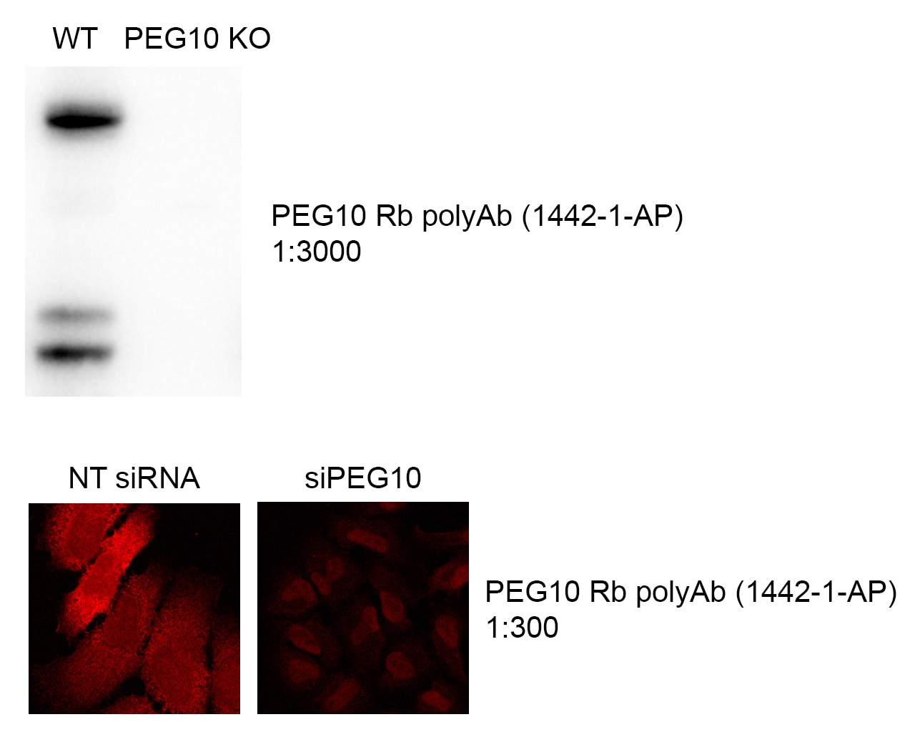
Both reviewers rated the antibody 5 stars. One user found it excellent for Western blotting at 1:3000, though noted some non-specific nuclear staining in immunofluorescence at 1:300. The other confirmed reliable WB performance in MCF-7 cells at 1:1000 in TBS-T with 5% BSA. Overall, users report strong WB results with clear specificity, while IF applications may require optimization to minimize background.
The reviews below have been submitted by verified Proteintech customers who received an incentive for providing their feedback.
Harihar (Verified Customer) (01-28-2024) | The antibody is excellent for Western blotting. There is some non-specific nuclear staining in case of IF at the dilutions we have tested at.
 |
Jordan (Verified Customer) (09-03-2019) | The antibody works well in our condition. MCF-7 cell lines. 1/1000 in TBS-T 5%BSA.
|





