Filter:


Tested Applications
| Positive WB detected in | A375 cells, A549 cells, HL-60 cells, K-562 cells |
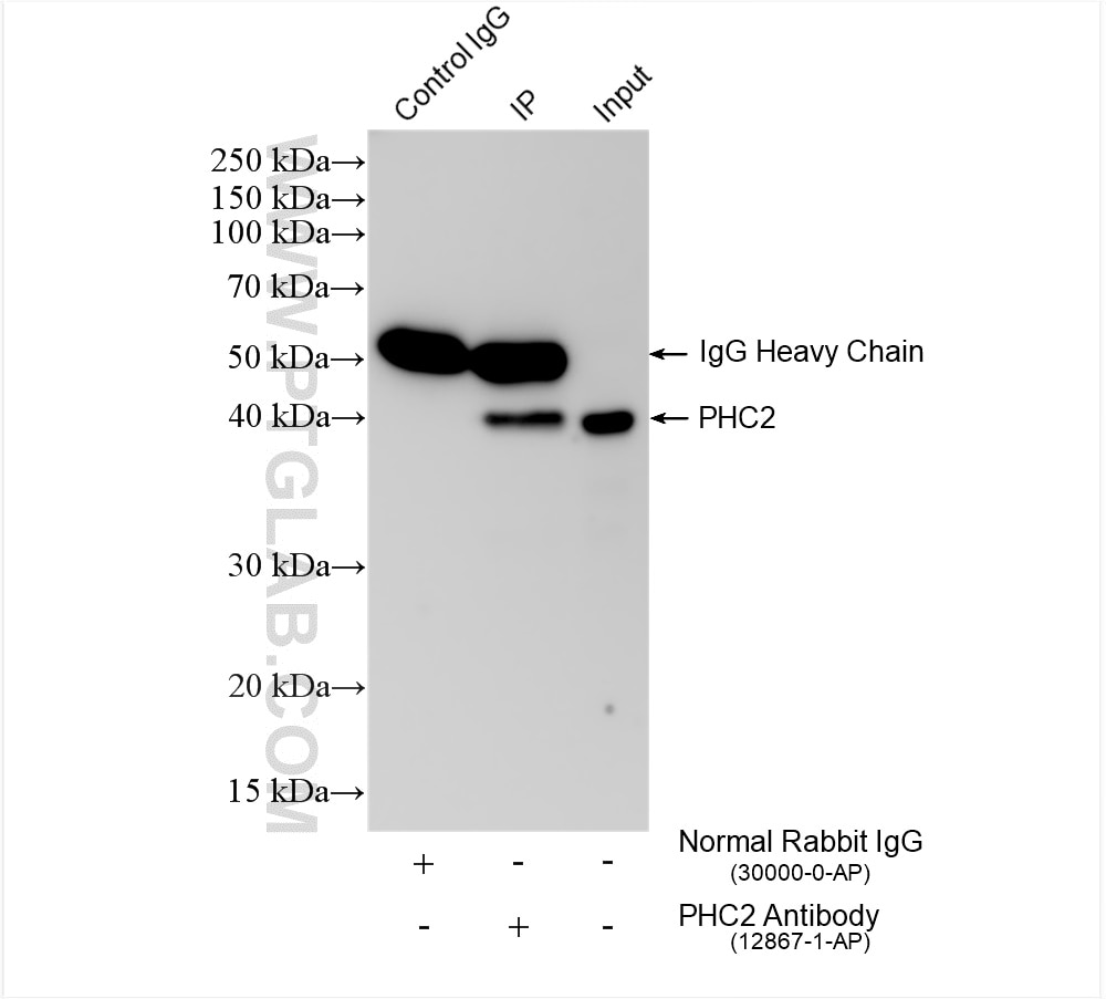
| Positive IP detected in | HL-60 cells |
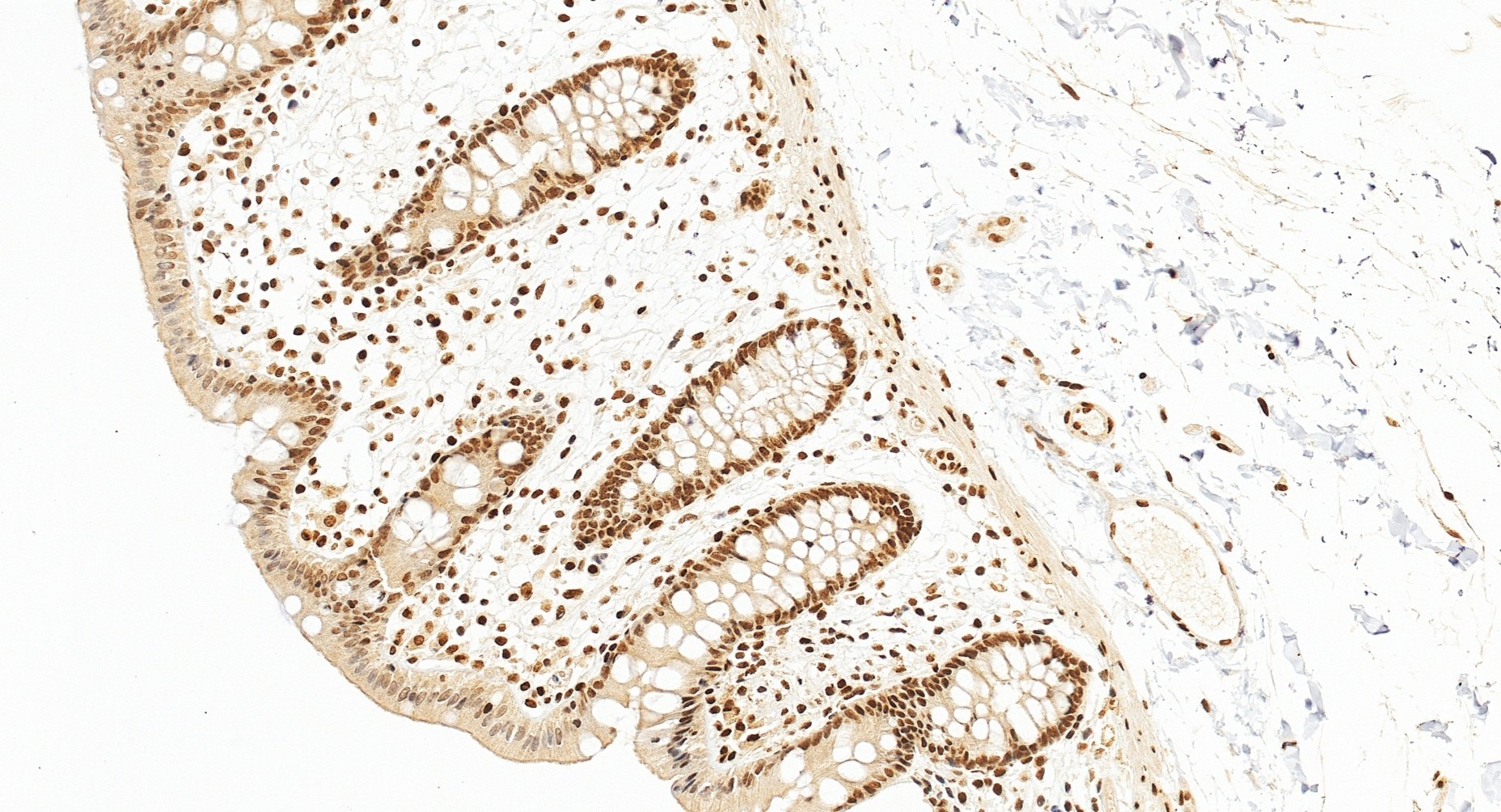
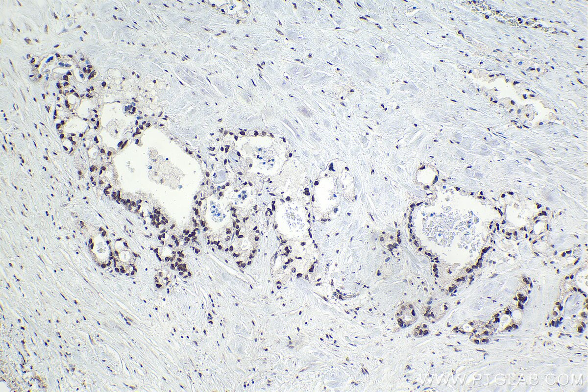
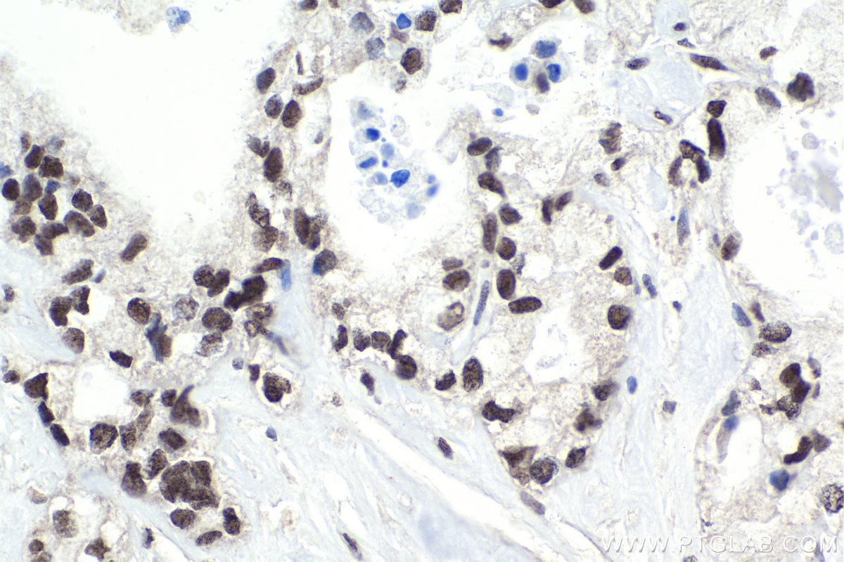
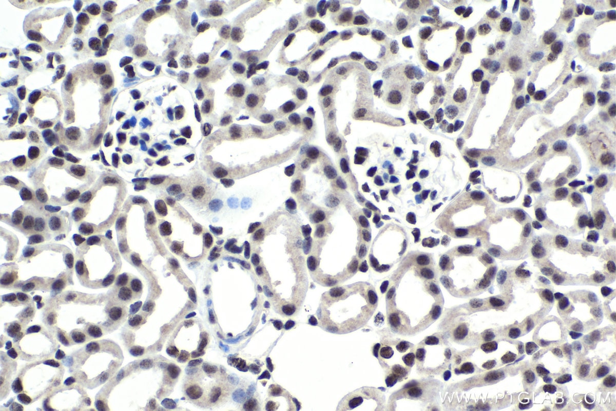
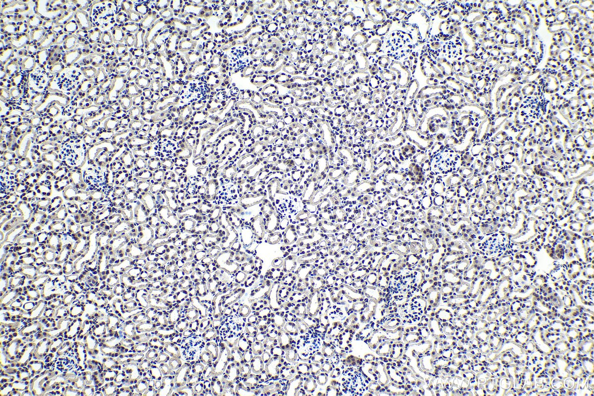
| Positive IHC detected in | human pancreas cancer tissue, human colon tissue, mouse kidney tissue Note: suggested antigen retrieval with TE buffer pH 9.0; (*) Alternatively, antigen retrieval may be performed with citrate buffer pH 6.0 |
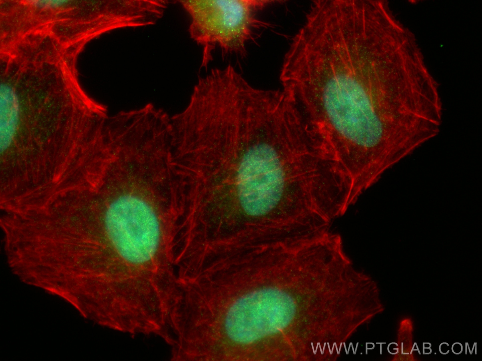
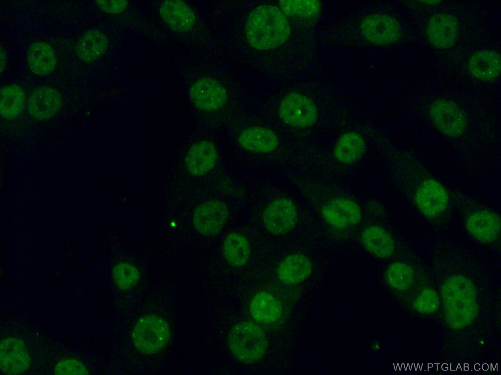
| Positive IF/ICC detected in | A549 cells, A375 cells |
Recommended dilution
| Application | Dilution |
|---|---|
| Western Blot (WB) | WB : 1:1000-1:4000 |
| Immunoprecipitation (IP) | IP : 0.5-4.0 ug for 1.0-3.0 mg of total protein lysate |
| Immunohistochemistry (IHC) | IHC : 1:50-1:500 |
| Immunofluorescence (IF)/ICC | IF/ICC : 1:500-1:2000 |
| It is recommended that this reagent should be titrated in each testing system to obtain optimal results. | |
| Sample-dependent, Check data in validation data gallery. | |
Published Applications
| IHC | See 1 publications below |
| IF | See 1 publications below |
Product Information
12867-1-AP targets PHC2 in WB, IHC, IF/ICC, IP, ELISA applications and shows reactivity with human, mouse, rat samples.
| Tested Reactivity | human, mouse, rat |
| Cited Reactivity | human, mouse |
| Host / Isotype | Rabbit / IgG |
| Class | Polyclonal |
| Type | Antibody |
| Immunogen |
CatNo: Ag3507 Product name: Recombinant human PHC2 protein Source: e coli.-derived, PGEX-4T Tag: GST Domain: 1-323 aa of BC028396 Sequence: MTSGNGNSASSIAGTAPQNGENKPPQAIVKPQILTHVIEGFVIQEGAEPFPVGRSSLLVGNLKKKYAQGFLPEKLPQQDHTTTTDSEMEEPYLQESKEEGAPLKLKCELCGRVDFAYKFKRSKRFCSMACAKRYNVGCTKRVGLFHSDRSKLQKAGAATHNRRRASKASLPPLTKDTKKQPTGTVPLSVTAALQLTHSQEDSSRCSDNSSYEEPLSPISASSSTSRRRQGQRDLELPDMHMRDLVGMGHHFLPSEPTKWNVEDVYEFIRSLPGCQEIEEEFRAQEIDGQALLLLKEDHLMSAMNIKLGPALKIYARISMLKDS Predict reactive species |
| Full Name | polyhomeotic homolog 2 (Drosophila) |
| Calculated Molecular Weight | 858 aa, 91 kDa |
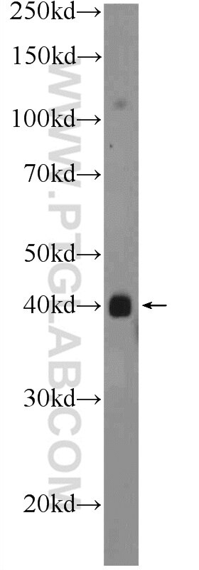
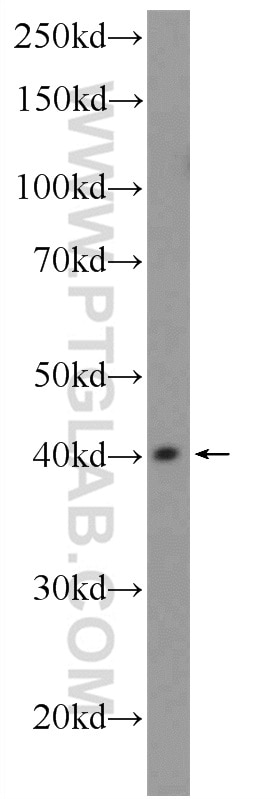
| Observed Molecular Weight | 40 kDa |
| GenBank Accession Number | BC028396 |
| Gene Symbol | PHC2 |
| Gene ID (NCBI) | 1912 |
| RRID | AB_2877890 |
| Conjugate | Unconjugated |
| Form | Liquid |
| Purification Method | Antigen affinity purification |
| UNIPROT ID | Q8IXK0 |
| Storage Buffer | PBS with 0.02% sodium azide and 50% glycerol, pH 7.3. |
| Storage Conditions | Store at -20°C. Stable for one year after shipment. Aliquoting is unnecessary for -20oC storage. 20ul sizes contain 0.1% BSA. |
Protocols
| Product Specific Protocols | |
|---|---|
| IF protocol for PHC2 antibody 12867-1-AP | Download protocol |
| IHC protocol for PHC2 antibody 12867-1-AP | Download protocol |
| IP protocol for PHC2 antibody 12867-1-AP | Download protocol |
| WB protocol for PHC2 antibody 12867-1-AP | Download protocol |
| Standard Protocols | |
|---|---|
| Click here to view our Standard Protocols |
Publications
| Species | Application | Title |
|---|---|---|
Cell Signal PHC2 promotes hepatocellular carcinoma progression and serves as a robust prognostic biomarker: A pancancer multiomics and clinical validation study. | ||
Mol Cell Modularity of PRC1 composition and chromatin interaction define condensate properties |