Tested Applications
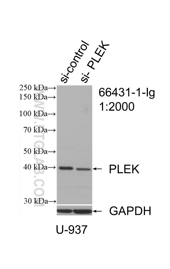
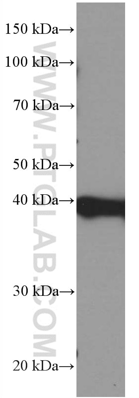
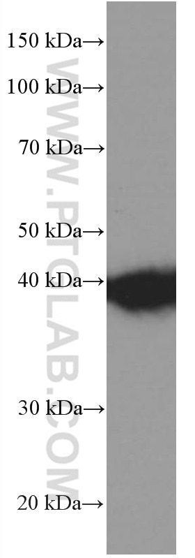
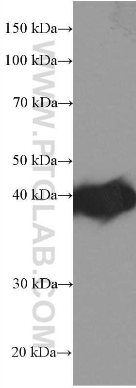
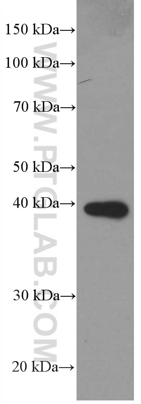
| Positive WB detected in | HL-60 cells, pig spleen tissue, THP-1 cells, U-937 cells |
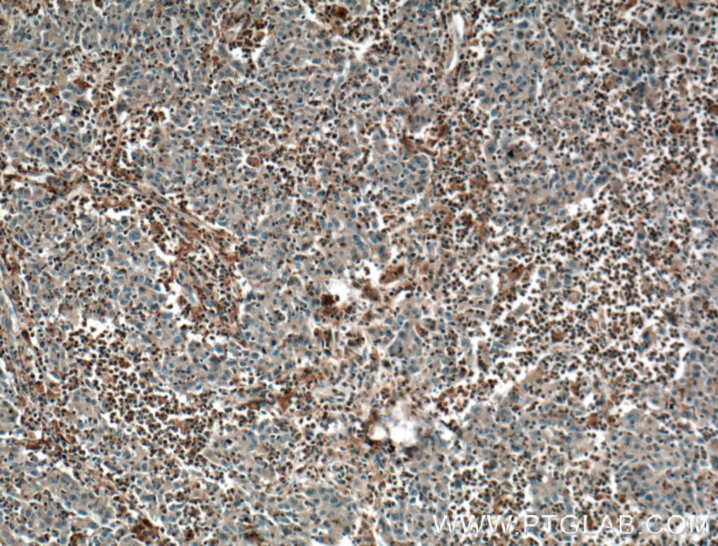
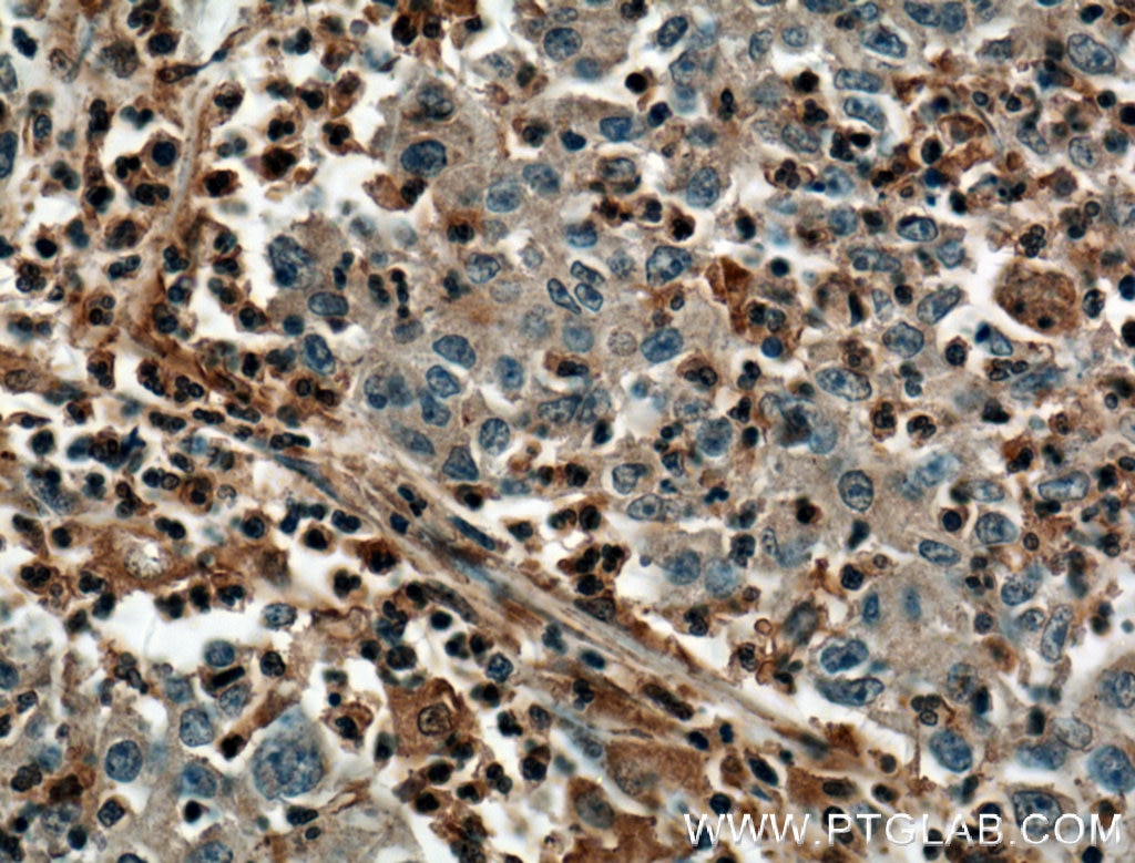
| Positive IHC detected in | human colon cancer tissue Note: suggested antigen retrieval with TE buffer pH 9.0; (*) Alternatively, antigen retrieval may be performed with citrate buffer pH 6.0 |
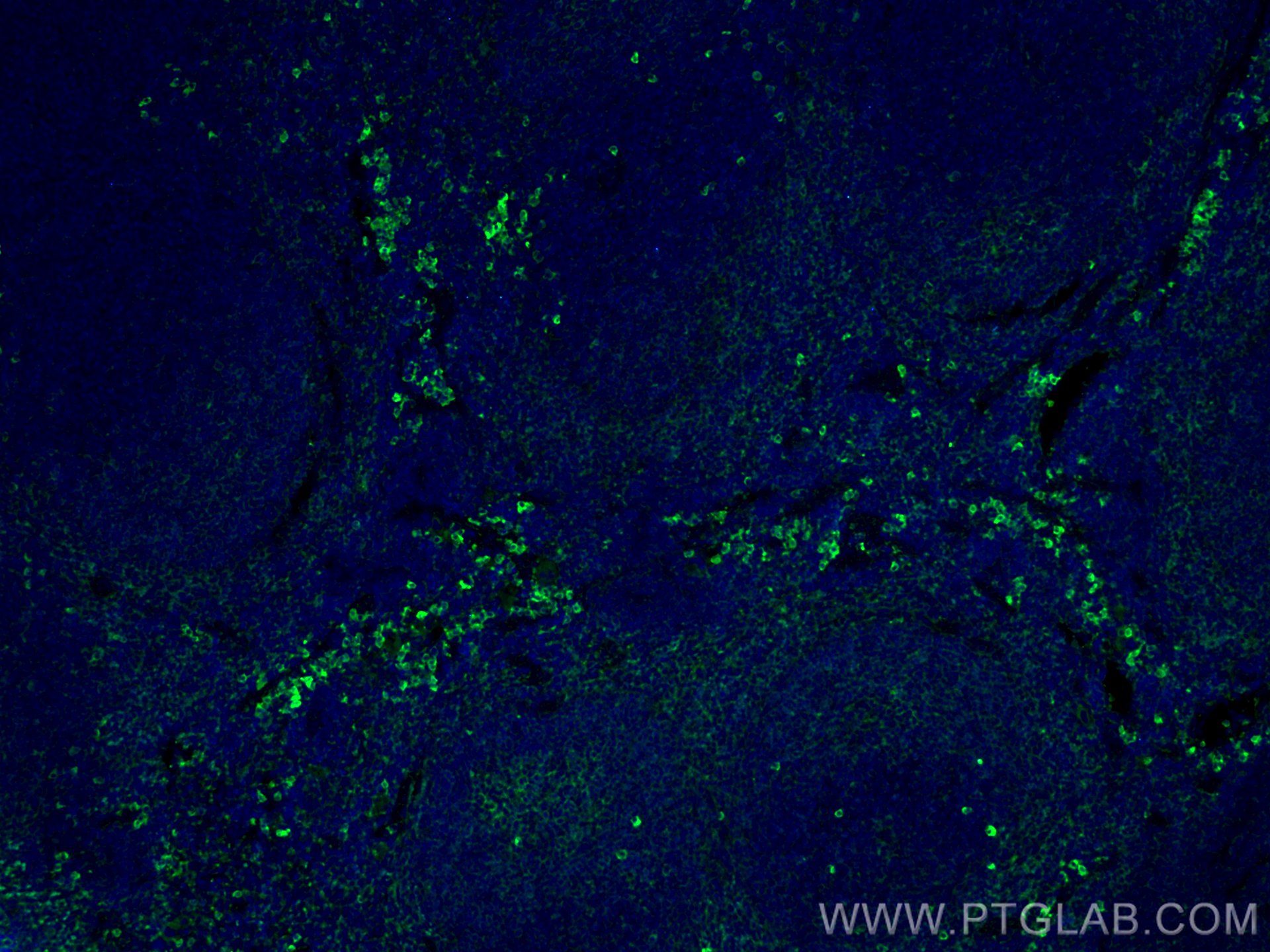
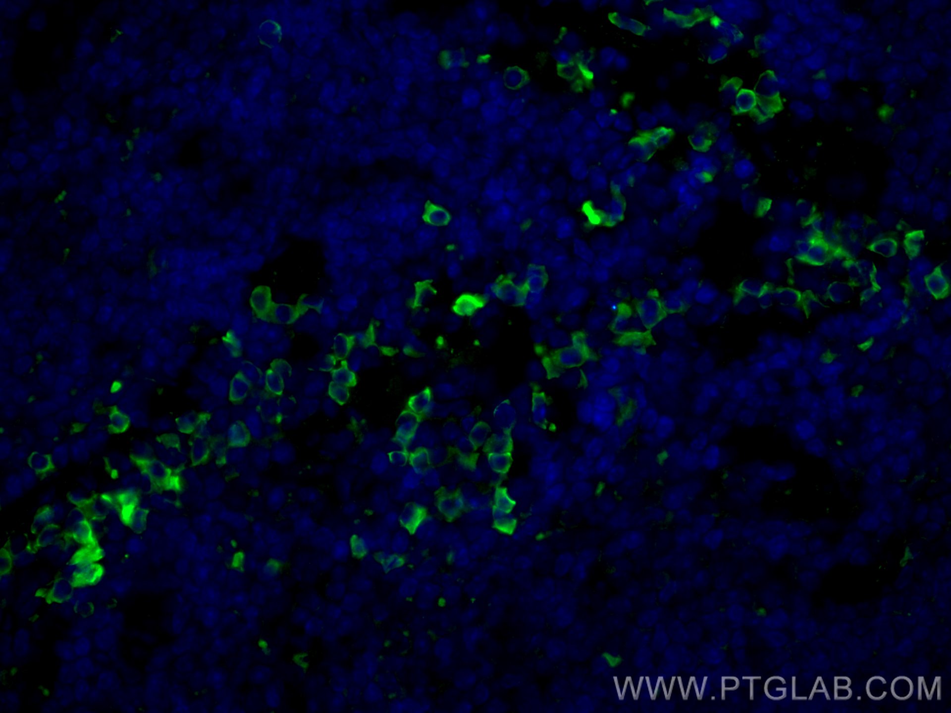
| Positive IF-P detected in | mouse spleen tissue |
Recommended dilution
| Application | Dilution |
|---|---|
| Western Blot (WB) | WB : 1:2000-1:20000 |
| Immunohistochemistry (IHC) | IHC : 1:50-1:500 |
| Immunofluorescence (IF)-P | IF-P : 1:200-1:800 |
| It is recommended that this reagent should be titrated in each testing system to obtain optimal results. | |
| Sample-dependent, Check data in validation data gallery. | |
Published Applications
| IHC | See 1 publications below |
| IF | See 1 publications below |
Product Information
66431-1-Ig targets PLEK in WB, IHC, IF-P, ELISA applications and shows reactivity with human, pig, mouse samples.
| Tested Reactivity | human, pig, mouse |
| Cited Reactivity | human |
| Host / Isotype | Mouse / IgG1 |
| Class | Monoclonal |
| Type | Antibody |
| Immunogen |
CatNo: Ag3187 Product name: Recombinant human PLEK protein Source: e coli.-derived, PGEX-4T Tag: GST Domain: 1-350 aa of BC018549 Sequence: MEPKRIREGYLVKKGSVFNTWKPMWVVLLEDGIEFYKKKSDNSPKGMIPLKGSTLTSPCQDFGKRMFVFKITTTKQQDHFFQAAFLEERDAWVRDIKKAIKCIEGGQKFARKSTRRSIRLPETIDLGALYLSMKDTEKGIKELNLEKDKKIFNHCFTGNCVIDWLVSNQSVRNRQEGLMIASSLLNEGYLQPAGDMSKSAVDGTAENPFLDNPDAFYYFPDSGFFCEENSSDDDVILKEEFRGVIIKQGCLLKQGHRRKNWKVRKFILREDPAYLHYYDPAGAEDPLGAIHLRGCVVTSVESNSNGRKSEEENLFEIITADEVHYFLQAATPKERTEWIKAIQMASRTGK Predict reactive species |
| Full Name | pleckstrin |
| Calculated Molecular Weight | 350 aa, 40 kDa |
| Observed Molecular Weight | 40-47 kDa |
| GenBank Accession Number | BC018549 |
| Gene Symbol | PLEK |
| Gene ID (NCBI) | 5341 |
| RRID | AB_2881802 |
| Conjugate | Unconjugated |
| Form | Liquid |
| Purification Method | Protein G purification |
| UNIPROT ID | P08567 |
| Storage Buffer | PBS with 0.02% sodium azide and 50% glycerol, pH 7.3. |
| Storage Conditions | Store at -20°C. Stable for one year after shipment. Aliquoting is unnecessary for -20oC storage. 20ul sizes contain 0.1% BSA. |
Background Information
PLEK (pleckstrin; also known as p47) was originally identified as the major PKC substrate in platelets and later is found to be expressed in all cells of the hemopoietic system. Through two pleckstrin homology (PH) domains at its N and C termini, PLEK may interact with various protein and/or lipid ligands and serve as an intracellular adaptor/targeting protein. Predominantly cytosolic in unstimulated cells, PLEK would undergo transient redistribution to phagosomal membrane in response to stimuli. PLEK has been found to be hyperphosphorylated in diabetic mononuclear phagocytes and promote proinflammatory cytokine secretion in diabetes. (10477609, 17579087)
Protocols
| Product Specific Protocols | |
|---|---|
| IF protocol for PLEK antibody 66431-1-Ig | Download protocol |
| IHC protocol for PLEK antibody 66431-1-Ig | Download protocol |
| WB protocol for PLEK antibody 66431-1-Ig | Download protocol |
| Standard Protocols | |
|---|---|
| Click here to view our Standard Protocols |