Tested Applications
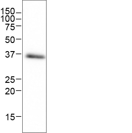
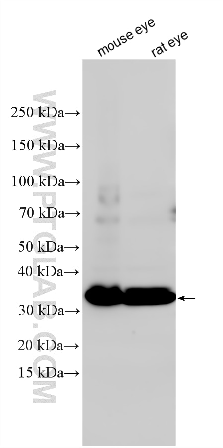
| Positive WB detected in | mouse eye tissue, rat eye tissue |
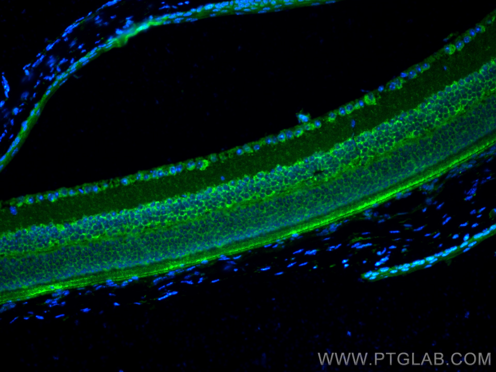
| Positive IF-P detected in | mouse eye tissue |
Recommended dilution
| Application | Dilution |
|---|---|
| Western Blot (WB) | WB : 1:1000-1:8000 |
| Immunofluorescence (IF)-P | IF-P : 1:200-1:800 |
| It is recommended that this reagent should be titrated in each testing system to obtain optimal results. | |
| Sample-dependent, Check data in validation data gallery. | |
Published Applications
| WB | See 10 publications below |
| IHC | See 5 publications below |
| IF | See 12 publications below |
| IP | See 1 publications below |
Product Information
18109-1-AP targets PRPH2 in WB, IHC, IF-P, IP, ELISA applications and shows reactivity with human, mouse, rat samples.
| Tested Reactivity | human, mouse, rat |
| Cited Reactivity | human, mouse, rat, zebrafish |
| Host / Isotype | Rabbit / IgG |
| Class | Polyclonal |
| Type | Antibody |
| Immunogen |
CatNo: Ag12555 Product name: Recombinant human PRPH2 protein Source: e coli.-derived, PET28a Tag: 6*His Domain: 124-346 aa of BC074720 Sequence: GSLENTLGQGLKNGMKYYRDTDTPGRCFMKKTIDMLQIEFKCCGNNGFRDWFEIQWISNRYLDFSSKEVKDRIKSNVDGRYLVDGVPFSCCNPSSPRPCIQYQITNNSAHYSYDHQTEELNLWVRGCRAALLSYYSSLMNSMGVVTLLIWLFEVTITIGLRYLQTSLDGVSNPEESESESEGWLLEKSVPETWKAFLESVKKLGKGNQVEAEGAGAGQAPEAG Predict reactive species |
| Full Name | peripherin 2 (retinal degeneration, slow) |
| Calculated Molecular Weight | 346 aa, 39 kDa |
| Observed Molecular Weight | 35 kDa |
| GenBank Accession Number | BC074720 |
| Gene Symbol | PRPH2 |
| Gene ID (NCBI) | 5961 |
| RRID | AB_10665364 |
| Conjugate | Unconjugated |
| Form | Liquid |
| Purification Method | Antigen affinity purification |
| UNIPROT ID | P23942 |
| Storage Buffer | PBS with 0.02% sodium azide and 50% glycerol, pH 7.3. |
| Storage Conditions | Store at -20°C. Stable for one year after shipment. Aliquoting is unnecessary for -20oC storage. 20ul sizes contain 0.1% BSA. |
Background Information
Peripherin-2 (PRPH2), also known as retinal degeneration slow protein (RDS), is a photoreceptor-specific tetraspanin protein implicated in outer segment disk morphogenesis. It may function as an adhesion molecule involved in stabilization and compaction of outer segment disks or in the maintenance of the curvature of the rim. Mutations in peripherin-2 are responsible for various retinal degenerative diseases including autosomal dominant retinitis pigmentosa (ADRP).
Protocols
| Product Specific Protocols | |
|---|---|
| IF protocol for PRPH2 antibody 18109-1-AP | Download protocol |
| WB protocol for PRPH2 antibody 18109-1-AP | Download protocol |
| Standard Protocols | |
|---|---|
| Click here to view our Standard Protocols |
Publications
| Species | Application | Title |
|---|---|---|
Biomaterials Hydrogel-mediated co-transplantation of retinal pigmented epithelium and photoreceptors restores vision in an animal model of advanced retinal degeneration. | ||
Proc Natl Acad Sci U S A Reversal of end-stage retinal degeneration and restoration of visual function by photoreceptor transplantation. | ||
Proc Natl Acad Sci U S A Accumulation of non-outer segment proteins in the outer segment underlies photoreceptor degeneration in Bardet-Biedl syndrome. | ||
Proc Natl Acad Sci U S A Transplantation of human embryonic stem cell-derived retinal tissue in two primate models of retinal degeneration. | ||
Cell Death Dis Prph2 knock-in mice recapitulate human central areolar choroidal dystrophy retinal degeneration and exhibit aberrant synaptic remodeling and microglial activation | ||
Reviews
The reviews below have been submitted by verified Proteintech customers who received an incentive for providing their feedback.
FH XINYU (Verified Customer) (10-09-2025) | Nice results for western blot application!
|
FH Melina (Verified Customer) (05-13-2024) | Worked very well in western blots of whole retina lysate
 |


