Tested Applications
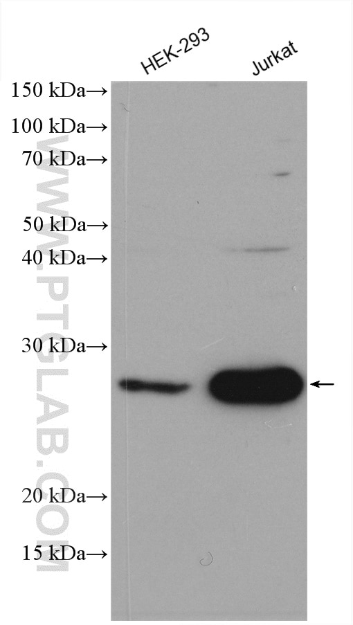
| Positive WB detected in | HEK-293 cells, K-562 cells, Jurkat cells |
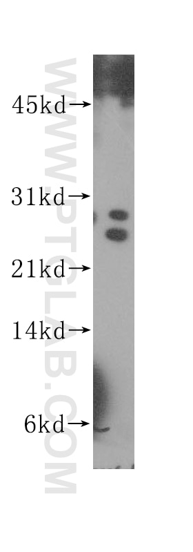
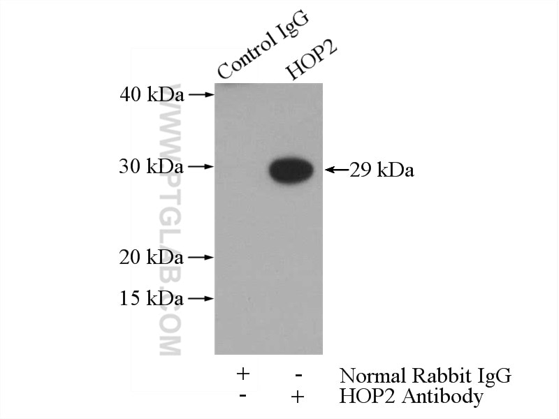
| Positive IP detected in | Jurkat cells |
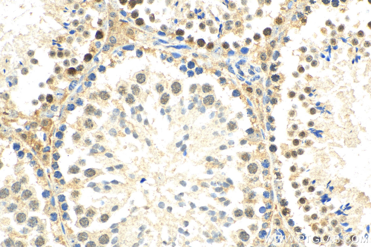
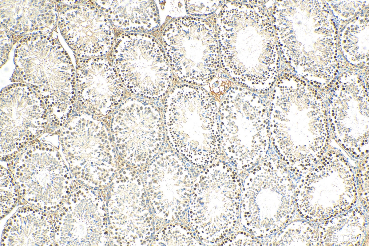
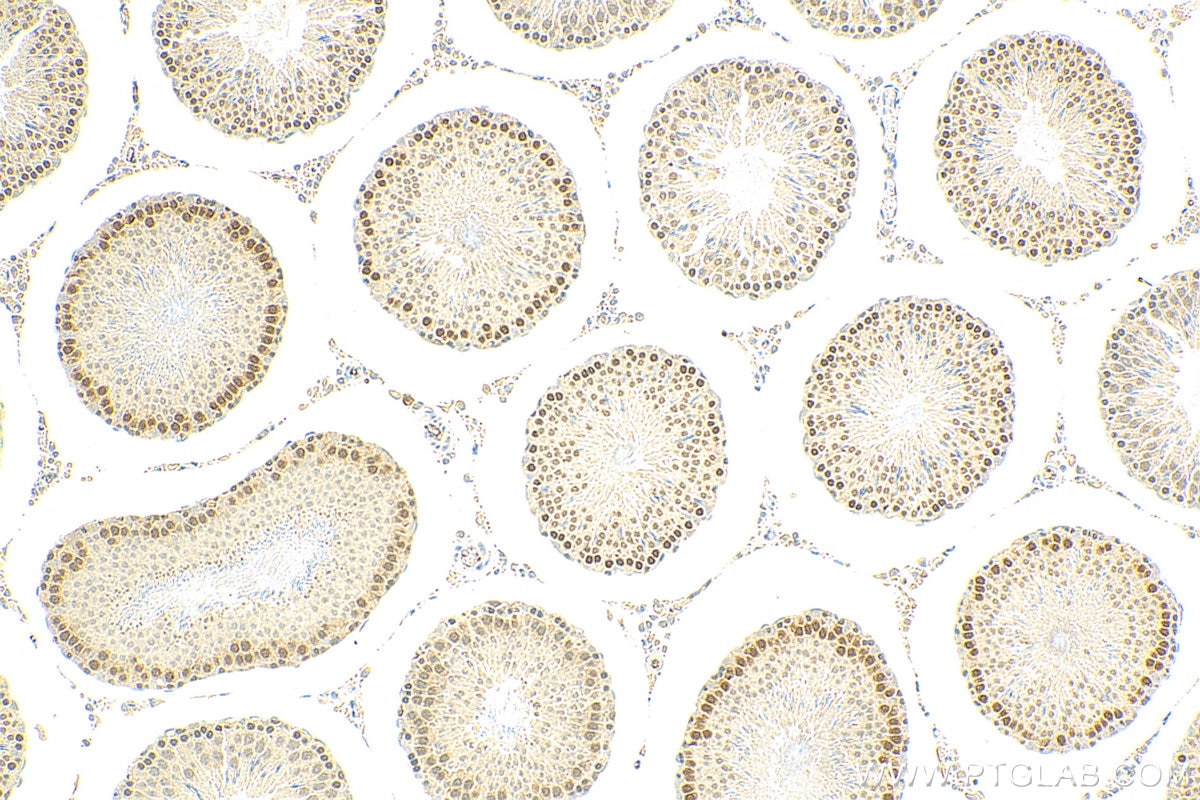
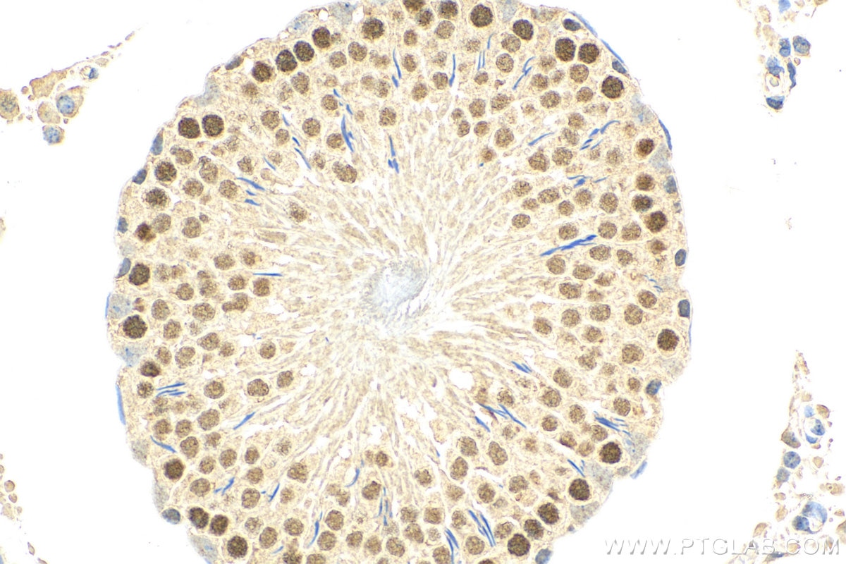
| Positive IHC detected in | mouse testis tissue, rat testis tissue Note: suggested antigen retrieval with TE buffer pH 9.0; (*) Alternatively, antigen retrieval may be performed with citrate buffer pH 6.0 |
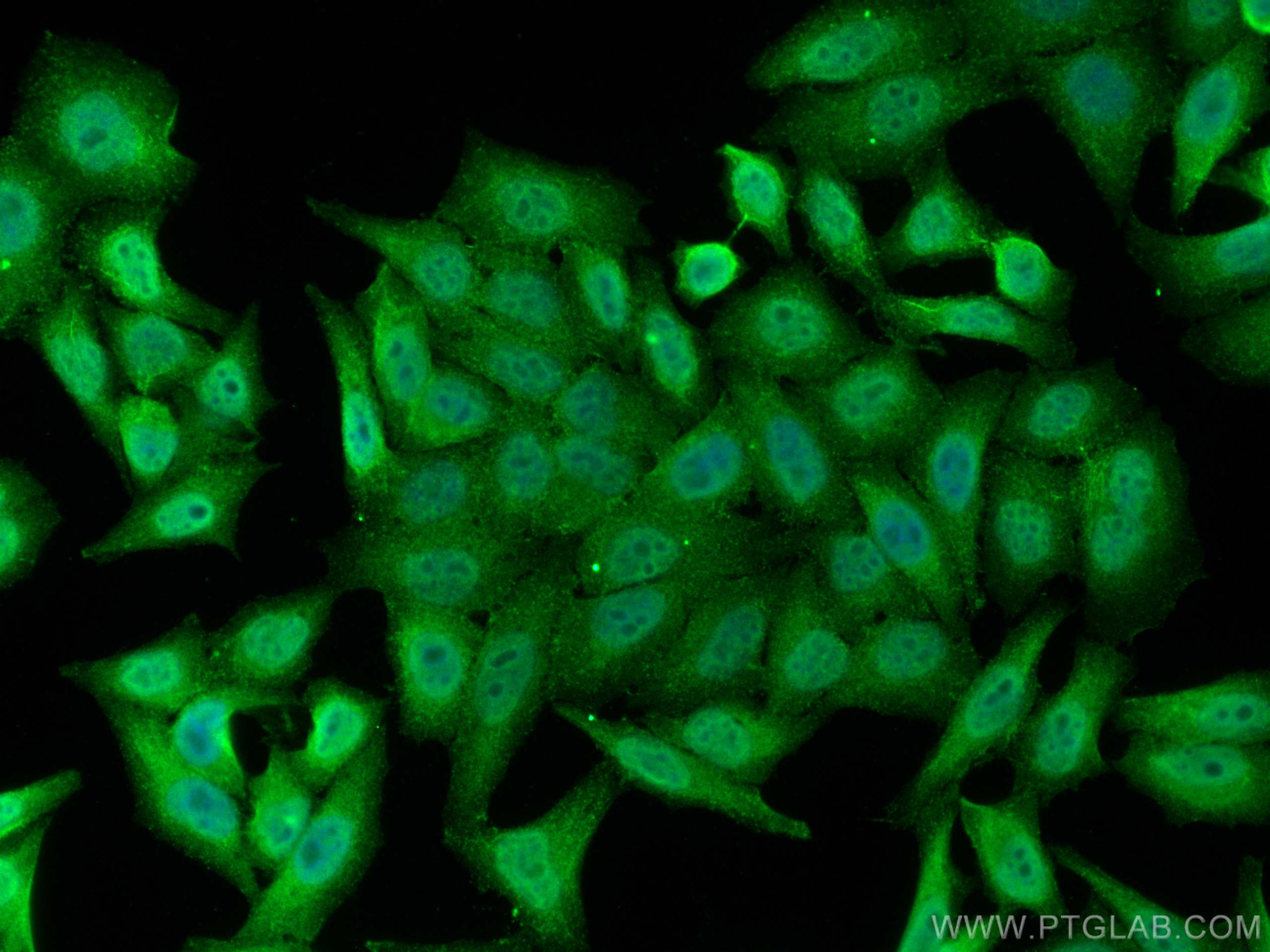
| Positive IF/ICC detected in | HepG2 cells |
Recommended dilution
| Application | Dilution |
|---|---|
| Western Blot (WB) | WB : 1:500-1:2000 |
| Immunoprecipitation (IP) | IP : 0.5-4.0 ug for 1.0-3.0 mg of total protein lysate |
| Immunohistochemistry (IHC) | IHC : 1:400-1:1600 |
| Immunofluorescence (IF)/ICC | IF/ICC : 1:200-1:800 |
| It is recommended that this reagent should be titrated in each testing system to obtain optimal results. | |
| Sample-dependent, Check data in validation data gallery. | |
Published Applications
| KD/KO | See 1 publications below |
| WB | See 3 publications below |
| IF | See 2 publications below |
Product Information
11339-1-AP targets PSMC3IP in WB, IHC, IF/ICC, IP, ELISA applications and shows reactivity with human, mouse, rat samples.
| Tested Reactivity | human, mouse, rat |
| Cited Reactivity | human, mouse |
| Host / Isotype | Rabbit / IgG |
| Class | Polyclonal |
| Type | Antibody |
| Immunogen |
CatNo: Ag1883 Product name: Recombinant human PSMC3IP protein Source: e coli.-derived, PGEX-4T Tag: GST Domain: 1-205 aa of BC008792 Sequence: MSKGRAEAAAGAAGILLRYLQEQNRPYSSQDVFGNLQREHGLGKAVVVKTLEQLAQQGKIKEKMYGKQKIYFADQDQFDMVSDADLQVLDGKIVALTAKVQSLQQSCRYMEAEMQKEIQELKKECAGYRERLKNIKAATNHVTPEEKEQVYRERQKYCKEWRKRKRMATELSDAILEGYPKSKKQFFEEVGIETDEDYNVTLPDP Predict reactive species |
| Full Name | PSMC3 interacting protein |
| Calculated Molecular Weight | 25 kDa |
| Observed Molecular Weight | 24-29 kDa |
| GenBank Accession Number | BC008792 |
| Gene Symbol | PSMC3IP |
| Gene ID (NCBI) | 29893 |
| RRID | AB_2172642 |
| Conjugate | Unconjugated |
| Form | Liquid |
| Purification Method | Antigen affinity purification |
| UNIPROT ID | Q9P2W1 |
| Storage Buffer | PBS with 0.02% sodium azide and 50% glycerol, pH 7.3. |
| Storage Conditions | Store at -20°C. Stable for one year after shipment. Aliquoting is unnecessary for -20oC storage. 20ul sizes contain 0.1% BSA. |
Background Information
PSMC3IP, also called HOP2, encodes a protein that functions in meiotic recombination. It is a subunit of the PSMC3IP/MND1 complex, which interacts with PSMC3/TBP1 to stimulate DMC1- and RAD51-mediated strand exchange during meiosis. The protein encoded by this gene can also co-activate ligand-driven transcription mediated by estrogen, androgen, glucocorticoid, progesterone, and thyroid nuclear receptors. Mutations in this gene cause XX female gonadal dysgenesis.
Protocols
| Product Specific Protocols | |
|---|---|
| IF protocol for PSMC3IP antibody 11339-1-AP | Download protocol |
| IHC protocol for PSMC3IP antibody 11339-1-AP | Download protocol |
| IP protocol for PSMC3IP antibody 11339-1-AP | Download protocol |
| WB protocol for PSMC3IP antibody 11339-1-AP | Download protocol |
| Standard Protocols | |
|---|---|
| Click here to view our Standard Protocols |
Publications
| Species | Application | Title |
|---|---|---|
Cell Interchromosomal Homology Searches Drive Directional ALT Telomere Movement and Synapsis.
| ||
Nucleic Acids Res RNase H1 facilitates recombinase recruitment by degrading DNA-RNA hybrids during meiosis | ||
Oncotarget A study of meiomitosis and novel pathways of genomic instability in cutaneous T-cell lymphomas (CTCL). |