Filter:


Tested Applications
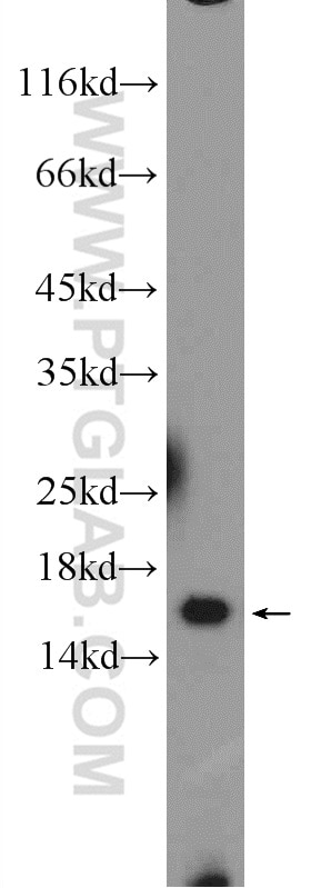
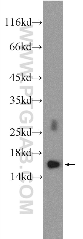
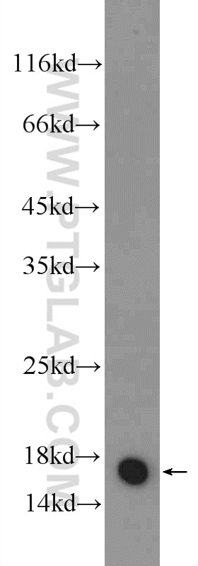
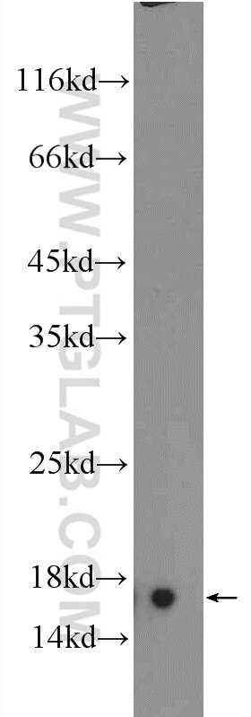
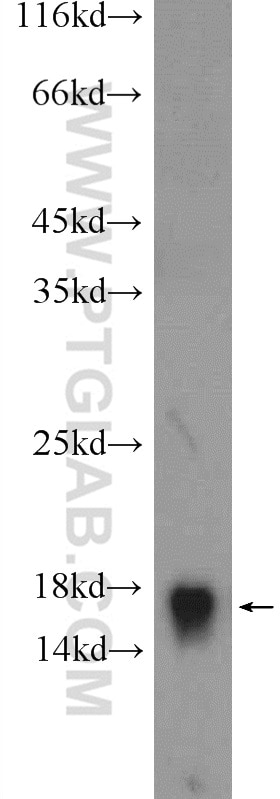
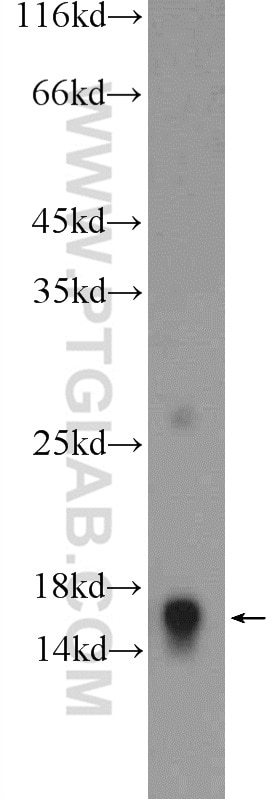
| Positive WB detected in | mouse heart tissue, HeLa cells, mouse skeletal muscle tissue |
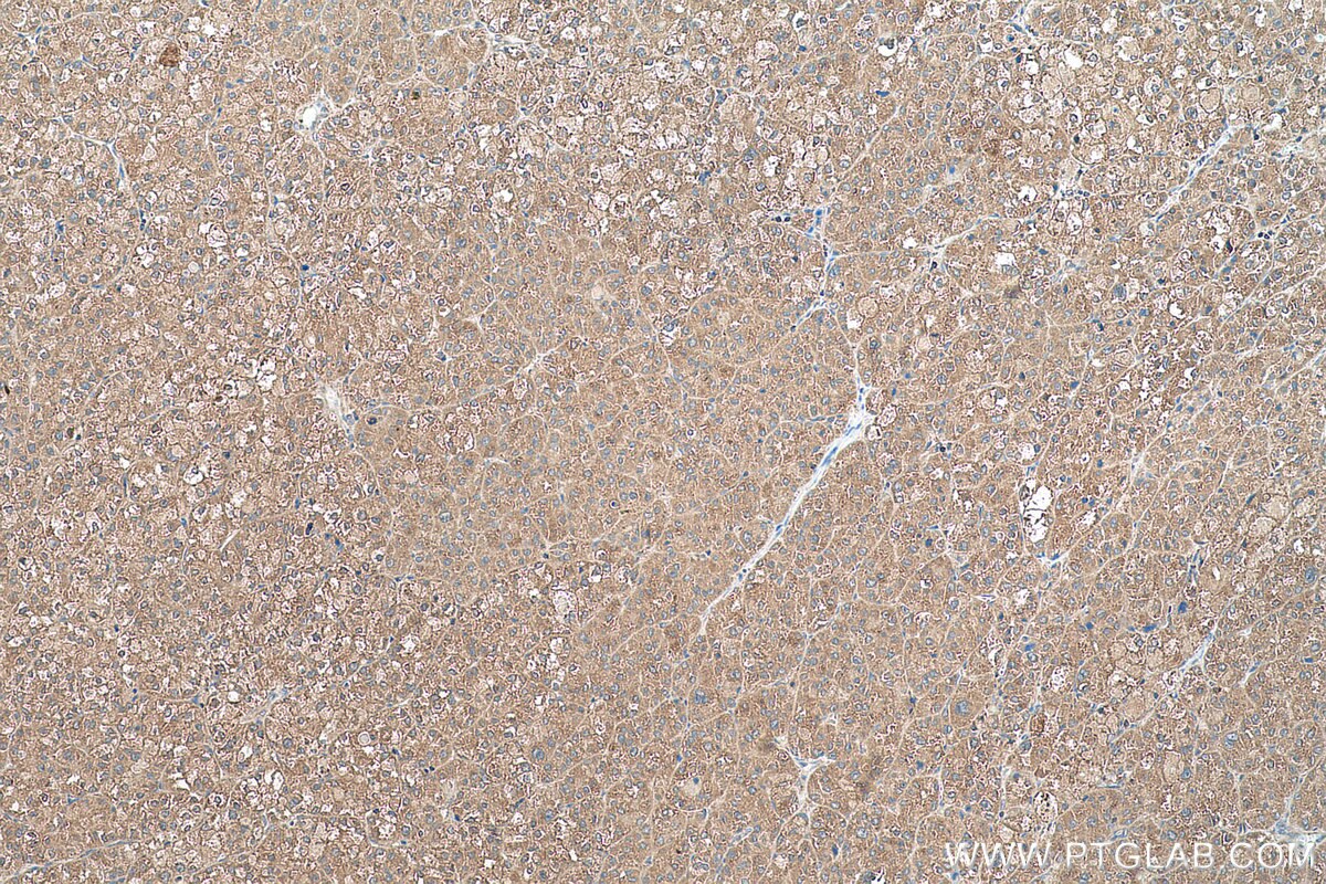
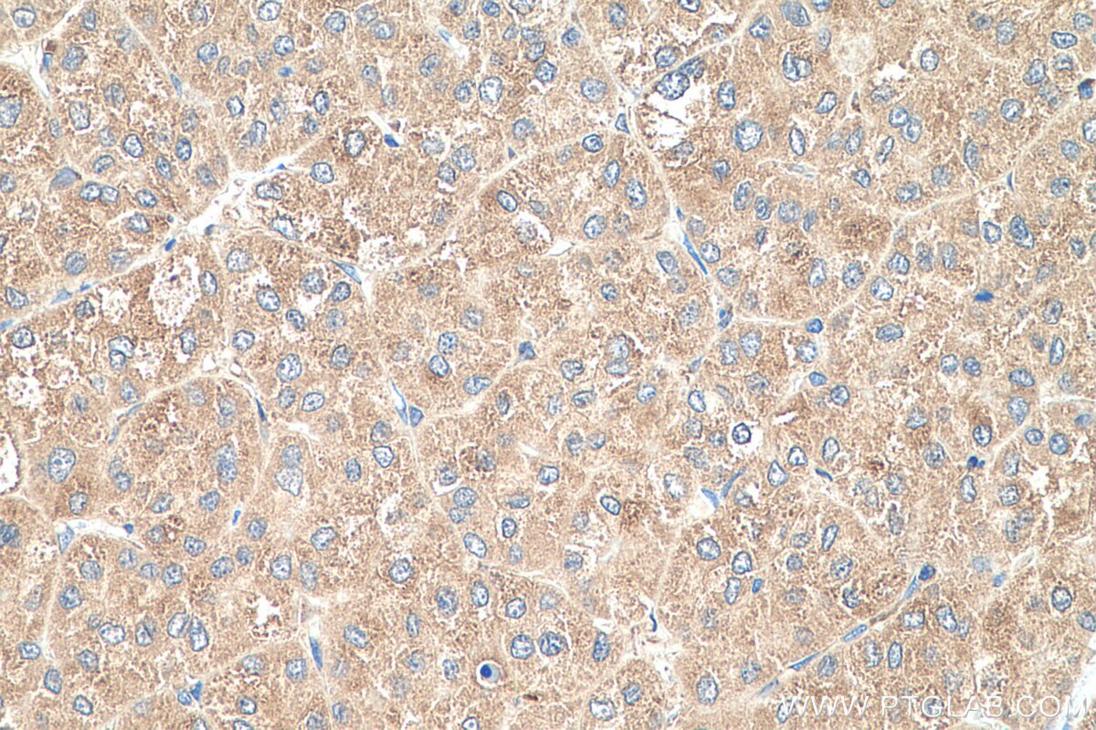
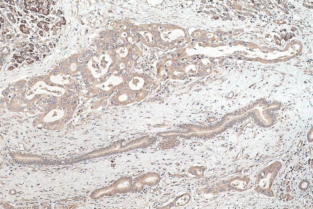
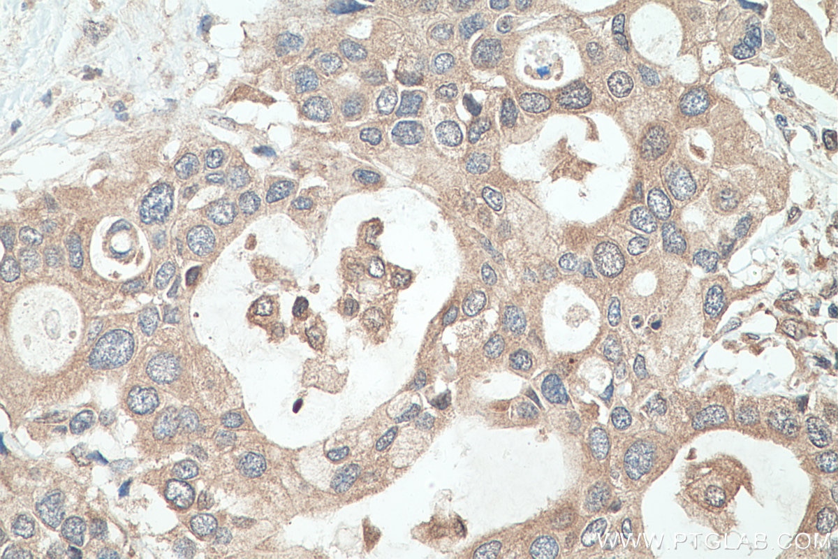
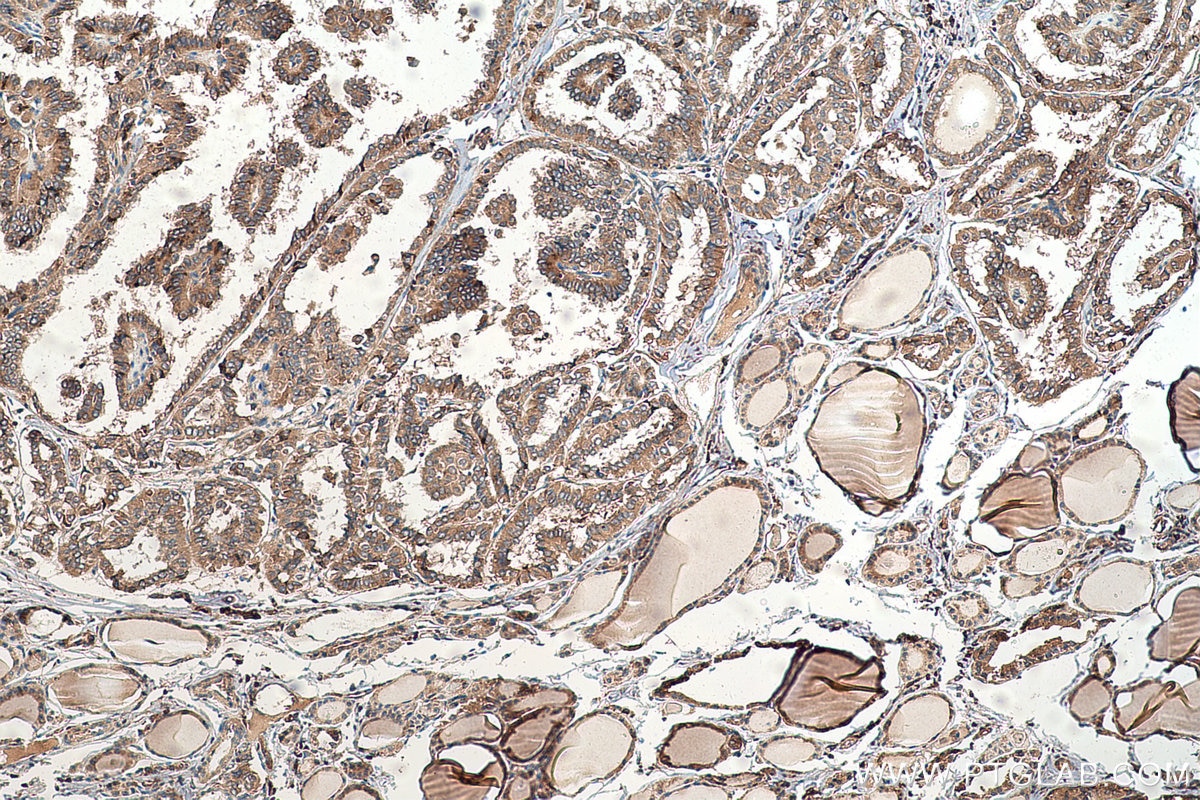
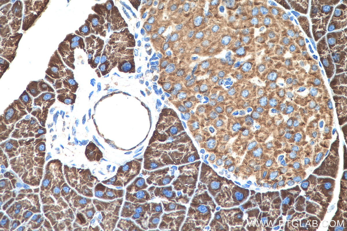
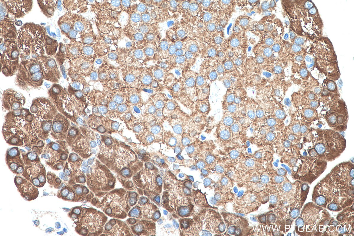
| Positive IHC detected in | rat pancreas tissue, human liver cancer tissue, human pancreas cancer tissue, human stomach cancer tissue, human thyroid cancer tissue, mouse pancreas tissue Note: suggested antigen retrieval with TE buffer pH 9.0; (*) Alternatively, antigen retrieval may be performed with citrate buffer pH 6.0 |
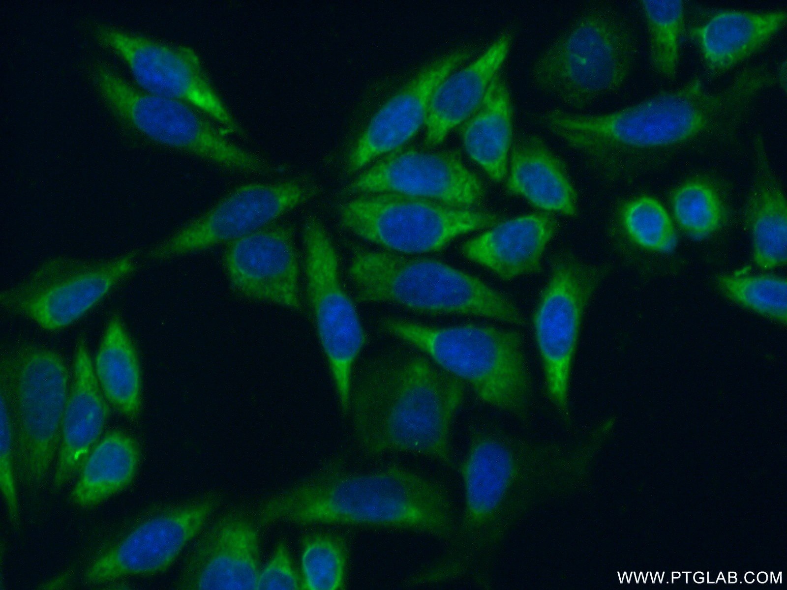
| Positive IF/ICC detected in | HeLa cells |
Recommended dilution
| Application | Dilution |
|---|---|
| Western Blot (WB) | WB : 1:200-1:1000 |
| Immunohistochemistry (IHC) | IHC : 1:500-1:2000 |
| Immunofluorescence (IF)/ICC | IF/ICC : 1:50-1:500 |
| It is recommended that this reagent should be titrated in each testing system to obtain optimal results. | |
| Sample-dependent, Check data in validation data gallery. | |
Published Applications
| KD/KO | See 1 publications below |
| WB | See 4 publications below |
| IF | See 1 publications below |
| IP | See 1 publications below |
Product Information
14826-1-AP targets RPL35 in WB, IHC, IF/ICC, IP, ELISA applications and shows reactivity with human, mouse, rat samples.
| Tested Reactivity | human, mouse, rat |
| Cited Reactivity | human, mouse, monkey |
| Host / Isotype | Rabbit / IgG |
| Class | Polyclonal |
| Type | Antibody |
| Immunogen |
CatNo: Ag6615 Product name: Recombinant human RPL35 protein Source: e coli.-derived, PGEX-4T Tag: GST Domain: 1-123 aa of BC000348 Sequence: MAKIKARDLRGKKKEELLKQLDDLKVELSQLRVAKVTGGAASKLSKIRVVRKSIARVLTVINQTQKENLRKFYKGKKYKPLDLRPKKTRAMRRRLNKHEENLKTKKQQRKERLYPLRKYAVKA Predict reactive species |
| Full Name | ribosomal protein L35 |
| Calculated Molecular Weight | 15 kDa |
| Observed Molecular Weight | 15 kDa |
| GenBank Accession Number | BC000348 |
| Gene Symbol | RPL35 |
| Gene ID (NCBI) | 11224 |
| RRID | AB_2878083 |
| Conjugate | Unconjugated |
| Form | Liquid |
| Purification Method | Antigen affinity purification |
| UNIPROT ID | P42766 |
| Storage Buffer | PBS with 0.02% sodium azide and 50% glycerol, pH 7.3. |
| Storage Conditions | Store at -20°C. Stable for one year after shipment. Aliquoting is unnecessary for -20oC storage. 20ul sizes contain 0.1% BSA. |
Protocols
| Product Specific Protocols | |
|---|---|
| IF protocol for RPL35 antibody 14826-1-AP | Download protocol |
| IHC protocol for RPL35 antibody 14826-1-AP | Download protocol |
| WB protocol for RPL35 antibody 14826-1-AP | Download protocol |
| Standard Protocols | |
|---|---|
| Click here to view our Standard Protocols |
Publications
| Species | Application | Title |
|---|---|---|
Cancer Cell Int GADD45GIP1 promotes osteosarcoma progression by modulating RPL35 ubiquitination and alleviating endoplasmic reticulum stress via the PERK/eIF2α pathway | ||
Zool Res Ribosome-associated pathological TDP-43 alters the expression of multiple mRNAs in the monkey brain | ||
J Virol Ribosomal protein L35 negatively regulates FMDV replication by recruiting AMFR to promote the ubiquitination and degradation of VP2.
|