Tested Applications
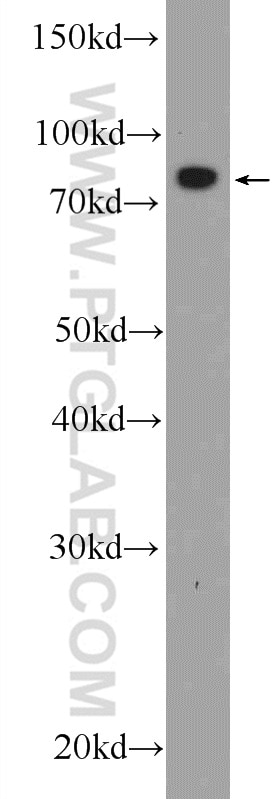
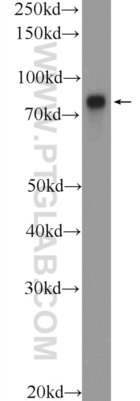
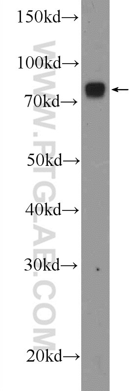
| Positive WB detected in | HepG2 cells, HeLa cells, mouse liver tissue |
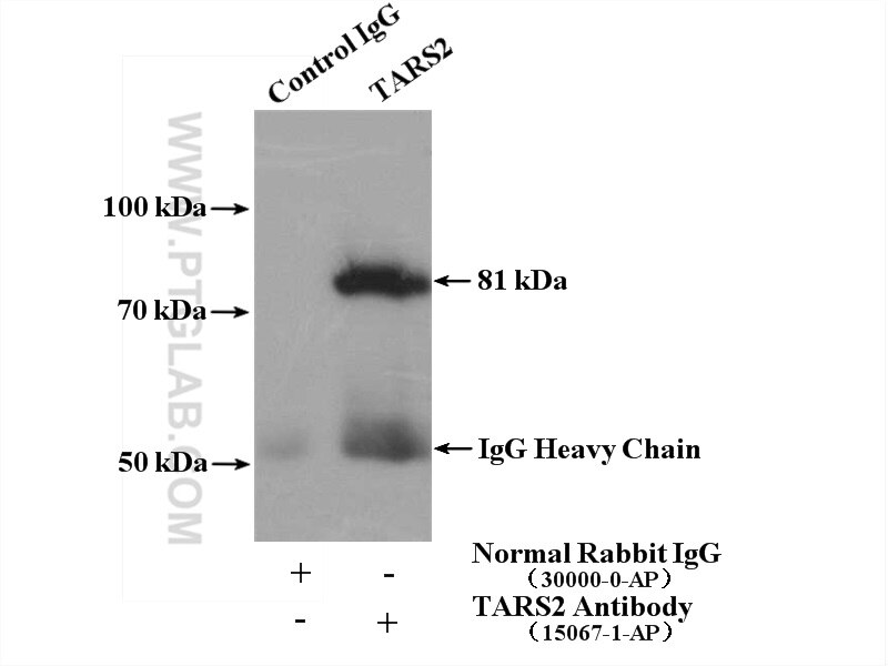
| Positive IP detected in | mouse liver tissue |
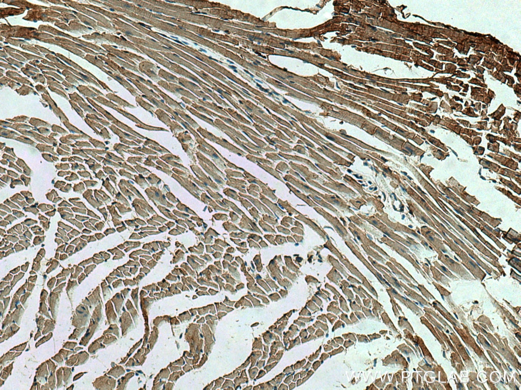
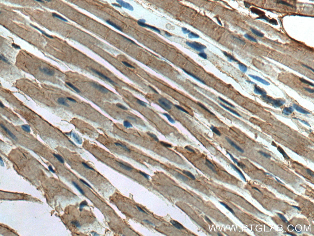
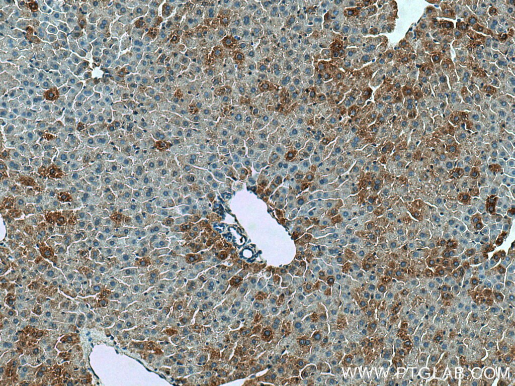
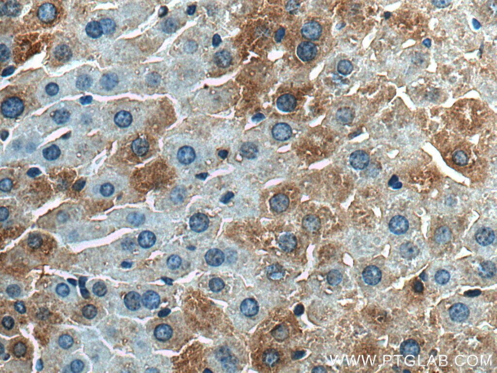
| Positive IHC detected in | mouse heart tissue, mouse liver tissue Note: suggested antigen retrieval with TE buffer pH 9.0; (*) Alternatively, antigen retrieval may be performed with citrate buffer pH 6.0 |
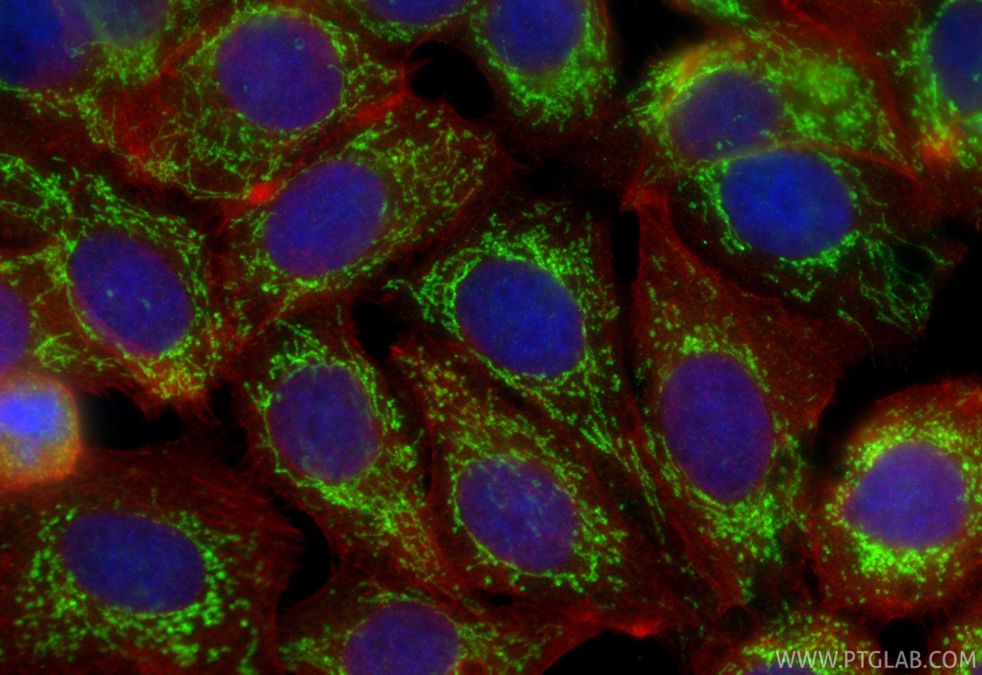
| Positive IF/ICC detected in | MCF-7 cells |
Recommended dilution
| Application | Dilution |
|---|---|
| Western Blot (WB) | WB : 1:500-1:1000 |
| Immunoprecipitation (IP) | IP : 0.5-4.0 ug for 1.0-3.0 mg of total protein lysate |
| Immunohistochemistry (IHC) | IHC : 1:50-1:500 |
| Immunofluorescence (IF)/ICC | IF/ICC : 1:200-1:800 |
| It is recommended that this reagent should be titrated in each testing system to obtain optimal results. | |
| Sample-dependent, Check data in validation data gallery. | |
Published Applications
| KD/KO | See 2 publications below |
| WB | See 4 publications below |
| IHC | See 1 publications below |
| IF | See 1 publications below |
| IP | See 2 publications below |
Product Information
15067-1-AP targets TARS2 in WB, IHC, IF/ICC, IP, ELISA applications and shows reactivity with human, mouse, rat samples.
| Tested Reactivity | human, mouse, rat |
| Cited Reactivity | human, mouse, pig |
| Host / Isotype | Rabbit / IgG |
| Class | Polyclonal |
| Type | Antibody |
| Immunogen |
CatNo: Ag7074 Product name: Recombinant human TARS2 protein Source: e coli.-derived, PGEX-4T Tag: GST Domain: 369-718 aa of BC000541 Sequence: EHYQEDMFAVQPPGSDRPPSSQSDDSTRHITDTLALKPMNCPAHCLMFAHRPRSWRELPLRLADFGALHRAEASGGLGGLTRLRCFQQDDAHIFCTTDQLEAEIQSCLDFLRSVYAVLGFSFRLALSTRPSGFLGDPCLWDQAEQVLKQALKEFGEPWDLNSGDGAFYGPKIDVHLHDALGRPHQCGTIQLDFQLPLRFDLQYKGQAGALERPVLIHRAVLGSVERLLGVLAESCGGKWPLWLSPFQVVVIPVGSEQEEYAKEAQQSLRAAGLVSDLDADSGLTLSRRIRRAQLAHYNFQFVVGQKEQSKRTVNIRTRDNRRLGEWDLPEAVQRLVELQNTRVPNAEEIF Predict reactive species |
| Full Name | threonyl-tRNA synthetase 2, mitochondrial (putative) |
| Calculated Molecular Weight | 81 kDa |
| Observed Molecular Weight | 81 kDa |
| GenBank Accession Number | BC000541 |
| Gene Symbol | TARS2 |
| Gene ID (NCBI) | 80222 |
| RRID | AB_2200668 |
| Conjugate | Unconjugated |
| Form | Liquid |
| Purification Method | Antigen affinity purification |
| UNIPROT ID | Q9BW92 |
| Storage Buffer | PBS with 0.02% sodium azide and 50% glycerol, pH 7.3. |
| Storage Conditions | Store at -20°C. Stable for one year after shipment. Aliquoting is unnecessary for -20oC storage. 20ul sizes contain 0.1% BSA. |
Background Information
TARS2 (mitochondrial (mt) threonyl-tRNA synthetase 2) is a class-II aminoacyl tRNA synthetase. Its canonical role is to ligate threonine to its cognate tRNA molecule during mitochondrial protein translation. TARS2 is required for the activation of mTORC1, a master regulator of cell growth and proliferation (PMID: 39509107, 37454282). TARS2 mainly localizes to mitochondria because of its N-terminal mitochondrial targeting sequence (MTS) (PMID: 33340489).
Protocols
| Product Specific Protocols | |
|---|---|
| IF protocol for TARS2 antibody 15067-1-AP | Download protocol |
| IHC protocol for TARS2 antibody 15067-1-AP | Download protocol |
| IP protocol for TARS2 antibody 15067-1-AP | Download protocol |
| WB protocol for TARS2 antibody 15067-1-AP | Download protocol |
| Standard Protocols | |
|---|---|
| Click here to view our Standard Protocols |
Publications
| Species | Application | Title |
|---|---|---|
Mol Cell Mitochondrial Threonyl-tRNA Synthetase TARS2 Is Required for Threonine-Sensitive mTORC1 Activation.
| ||
Bioengineered Knockdown of mitochondrial threonyl-tRNA synthetase 2 inhibits lung adenocarcinoma cell proliferation and induces apoptosis.
| ||
Int J Biol Markers Potential prognostic biomarkers of hepatocellular carcinoma based on 4D label-free quantitative proteomics analysis pilot investigation | ||
Redox Biol Decreased expression of mitochondrial aminoacyl-tRNA synthetases causes downregulation of OXPHOS subunits in type 2 diabetic muscle | ||
J Nutr Amino Acid Signaling in Skeletal Muscle Is Blunted by Prematurity in a Piglet Model. |