





Tested Applications
| Positive WB detected in | A431 cells, PC-3 cells, HEK-293T cells, Jurkat cells |
| Positive IP detected in | Jurkat cells |
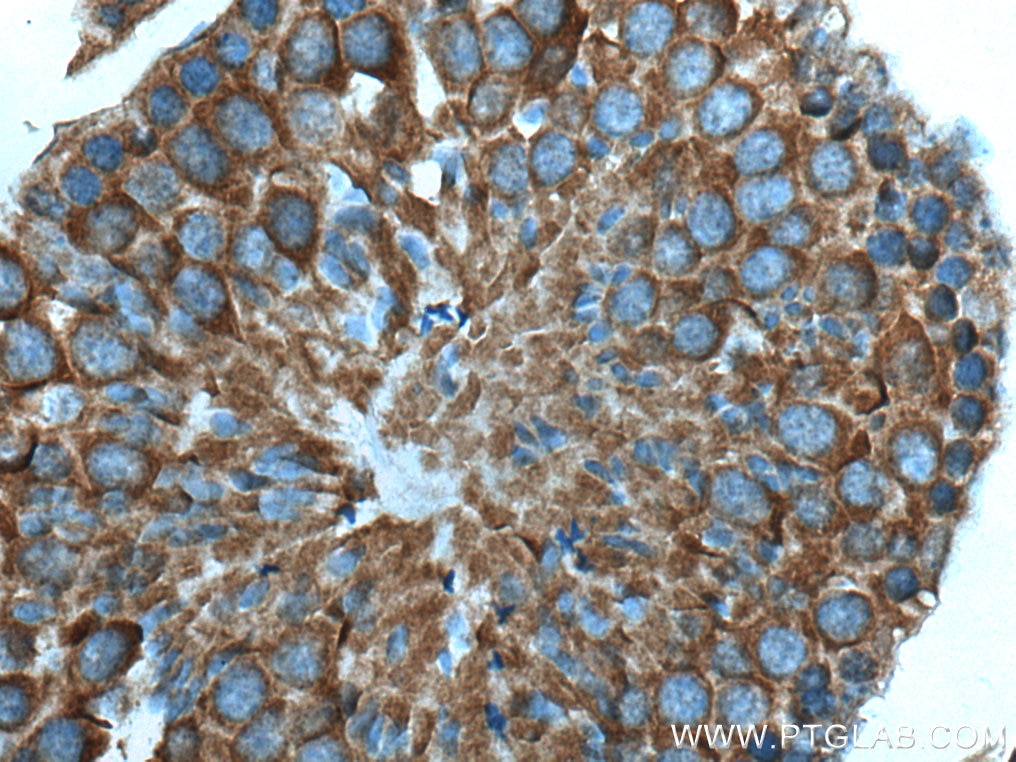
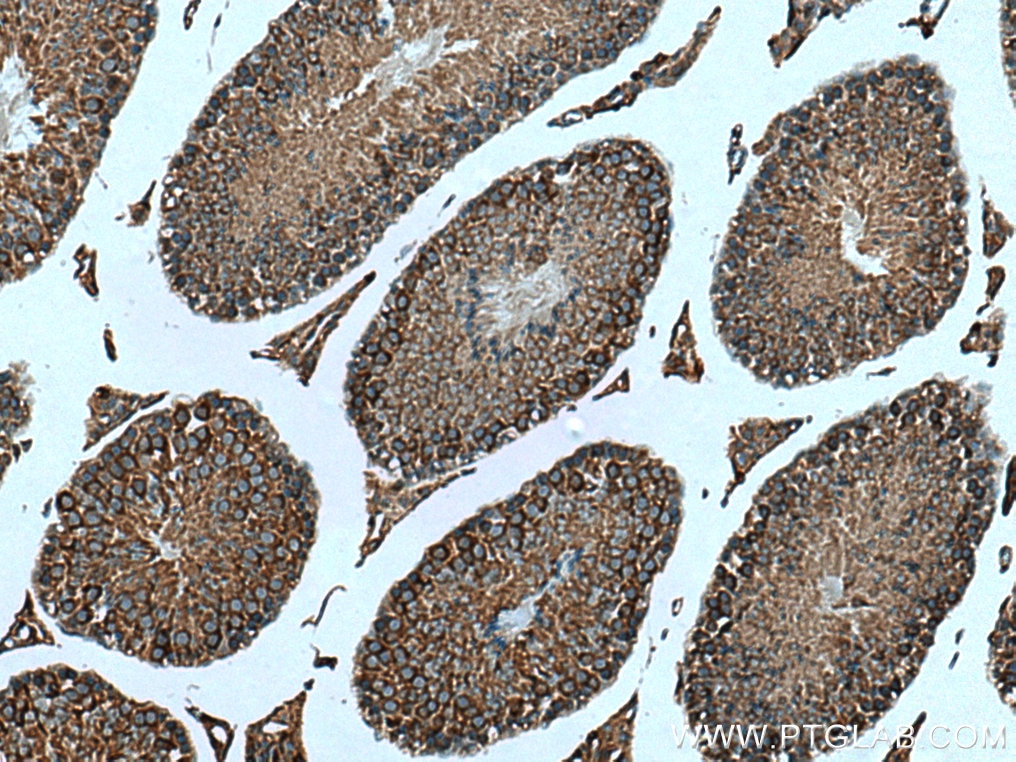
| Positive IHC detected in | human stomach cancer tissue, rat stomach tissue, mouse lung tissue, mouse stomach tissue, mouse testis tissue, rat kidney tissue, rat lung tissue Note: suggested antigen retrieval with TE buffer pH 9.0; (*) Alternatively, antigen retrieval may be performed with citrate buffer pH 6.0 |
Recommended dilution
| Application | Dilution |
|---|---|
| Western Blot (WB) | WB : 1:2000-1:10000 |
| Immunoprecipitation (IP) | IP : 0.5-4.0 ug for 1.0-3.0 mg of total protein lysate |
| Immunohistochemistry (IHC) | IHC : 1:500-1:2000 |
| It is recommended that this reagent should be titrated in each testing system to obtain optimal results. | |
| Sample-dependent, Check data in validation data gallery. | |
Product Information
17078-1-AP targets TBC1D5 in WB, IHC, IF, IP, ELISA applications and shows reactivity with human, mouse, rat samples.
| Tested Reactivity | human, mouse, rat |
| Cited Reactivity | human, mouse |
| Host / Isotype | Rabbit / IgG |
| Class | Polyclonal |
| Type | Antibody |
| Immunogen |
CatNo: Ag10991 Product name: Recombinant human TBC1D5 protein Source: e coli.-derived, PGEX-4T Tag: GST Domain: 445-795 aa of BC013145 Sequence: TNAKGAPLNINKVSNSLINFGRKLISPAMAPGSAGGPVPGGNSSSSSSVVIPTRTSAEAPSHHLQQQQQQQRLMKSESMPVQLNKGLSSKNISSSPSVESLPGGREFTGSPPSSATKKDSFFSNISRSRSHSKTMGRKESEEELEAQISFLQGQLNDLDAMCKYCAKVMDTHLVNIQDVILQENLEKEDQILVSLAGLKQIKDILKGSLRFNQSQLEAEENEQITIADNHYCSSGQGQGRGQGQSVQMSGAIKQASSETPGCTDRGNSDDFILISKDDDGSSARGSFSGQAQPLRTLRSTSGKSQAPVCSPLVFSDPLMGPASASSSNPSSSPDDDSSKDSGFTIVSPLDI Predict reactive species |
| Full Name | TBC1 domain family, member 5 |
| Calculated Molecular Weight | 795 aa, 89 kDa |
| Observed Molecular Weight | 89 kDa |
| GenBank Accession Number | BC013145 |
| Gene Symbol | TBC1D5 |
| Gene ID (NCBI) | 9779 |
| RRID | AB_2199389 |
| Conjugate | Unconjugated |
| Form | Liquid |
| Purification Method | Antigen affinity purification |
| UNIPROT ID | Q92609 |
| Storage Buffer | PBS with 0.02% sodium azide and 50% glycerol, pH 7.3. |
| Storage Conditions | Store at -20°C. Stable for one year after shipment. Aliquoting is unnecessary for -20oC storage. 20ul sizes contain 0.1% BSA. |
Background Information
TBC1D5, a member of TBC (Tre2/Bub2/Cdc16)1 domain family, is a novel retromer-interacting protein. TBC1D5 is a Rab GTPase-activating proteins (GAPs) proteins that negatively regulates VPS35/29/26 recruitment and causes Rab7 to dissociate from the membrane. TBC1D5 could bridge the endosome and autophagosome via its C-terminal LIR motif, and Rab GAPs are implicated in the reprogramming of the endocytic trafficking events under starvation-induced autophagy. The TBC1D5 protein exists 89 kDa and 91 kDa isoforms.
Protocols
| Product Specific Protocols | |
|---|---|
| IHC protocol for TBC1D5 antibody 17078-1-AP | Download protocol |
| IP protocol for TBC1D5 antibody 17078-1-AP | Download protocol |
| WB protocol for TBC1D5 antibody 17078-1-AP | Download protocol |
| Standard Protocols | |
|---|---|
| Click here to view our Standard Protocols |
Publications
What published studies show
The TBC1D5 Polyclonal antibody (Cat# 17078-1-AP) has 16 citations. These appear in journals including Nature Communications, Science Bulletin, Autophagy, Advanced Science, Science Advances, EMBO Journal, EMBO Reports, Journal of Cell Biology, Journal of Cell Science, International Journal of Biological Sciences, Journal of Translational Medicine, Journal of Alzheimer's Disease, Molecular Biology of the Cell, eLife, bioRxiv, and Research Square. Research fields span autophagy, mitophagy, endosomal/retromer trafficking, mTORC1 signaling, cancer, and Alzheimer's disease.
| Species | Application | Title |
|---|---|---|
Sci Bull (Beijing) Hepatitis B virus core protein as a Rab-GAP suppressor driving liver disease progression
| ||
Nat Commun Endosome maturation is orchestrated by inside-out proton signaling through a Na(+)/H(+) exchanger and pH-dependent Rab GTPase cycling. | ||
Adv Sci (Weinh) VPS34 Governs Oocyte Developmental Competence by Regulating Mito/Autophagy: A Novel Insight into the Significance of RAB7 Activity and Its Subcellular Location | ||
Sci Adv De novo macrocyclic peptides for inhibiting, stabilizing, and probing the function of the retromer endosomal trafficking complex. | ||
Int J Biol Sci TBK1 Facilitates GLUT1-Dependent Glucose Consumption by suppressing mTORC1 Signaling in Colorectal Cancer Progression.
|
Reviews
What customers say
Reviewers rate this TBC1D5 antibody 3–4 stars. It is described as a strong, reasonably specific antibody for Western blot, detecting the expected ~100 kD band in HeLa cells, mouse brain, and postmortem brain lysates. Specificity was confirmed via CRISPR/Cas9 knockout. Several weaker non-specific bands appear with 5% milk blocking, which may be reduced at lower dilutions (e.g., 1:3000). One user also validated it for immunofluorescence in HeLa cells at 1:200. Overall, it is considered a reliable reagent with minor background issues.
The reviews below have been submitted by verified Proteintech customers who received an incentive for providing their feedback.
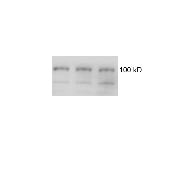
q (Verified Customer) (06-01-2021) | It is OK to detect TBC1D5 (100 kD) in both mouse and postmortem brain lysates, although there are some non specific bands.
 |
Florian (Verified Customer) (04-09-2019) | This is a very strong antibody that is reasonably specific in western blot applications. We have validated it through Crispr/Cas9 mediated knockout of TBC1D5 in HeLa cells (Jimenez-Orgaz et al, EMBOJ, 2018). While the strongest band is indeed TBC1D5, as our knockout confirms, it also produces several weaker, unspecific bands on membranes blocked with 5% milk. Because of the unspecific bands, I rate it with four stars instead of five. We have not tested it for immunofluorescence. Overall, it is a very good antibody that will probably also work at much lower dilutions than 1:1000. Diluting it further may also reduce the unspecific signal.
|
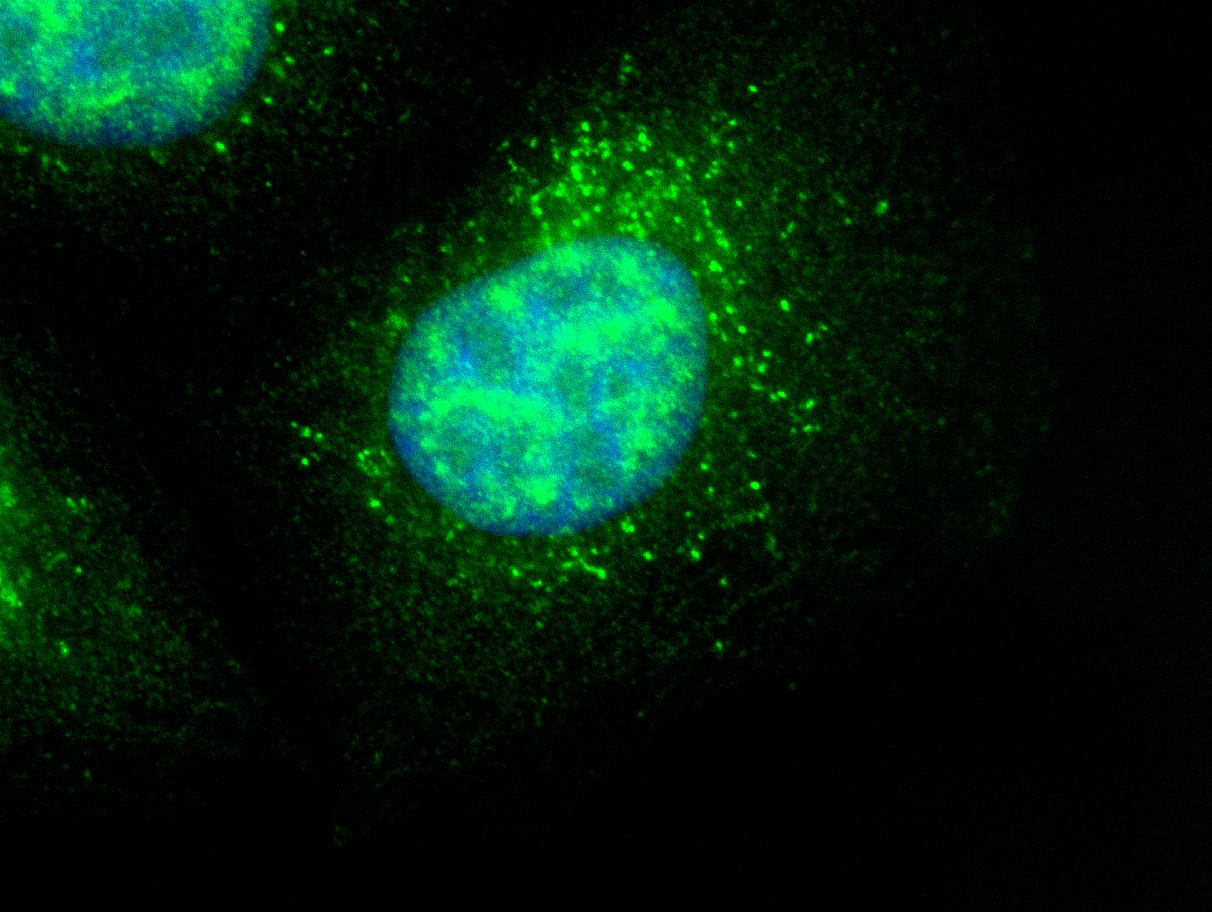
David (Verified Customer) (02-28-2019) | Hela cells stained with anti-TBC1D5 (green) and Hoechst (blue)
 |






