Tested Applications
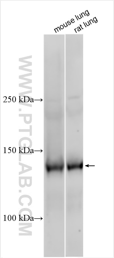
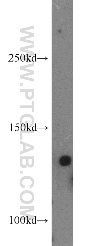
| Positive WB detected in | mouse lung tissue, mouse liver tissue, Rat lung tissue |
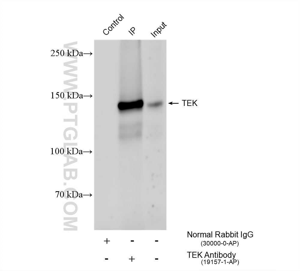
| Positive IP detected in | mouse lung tissue |
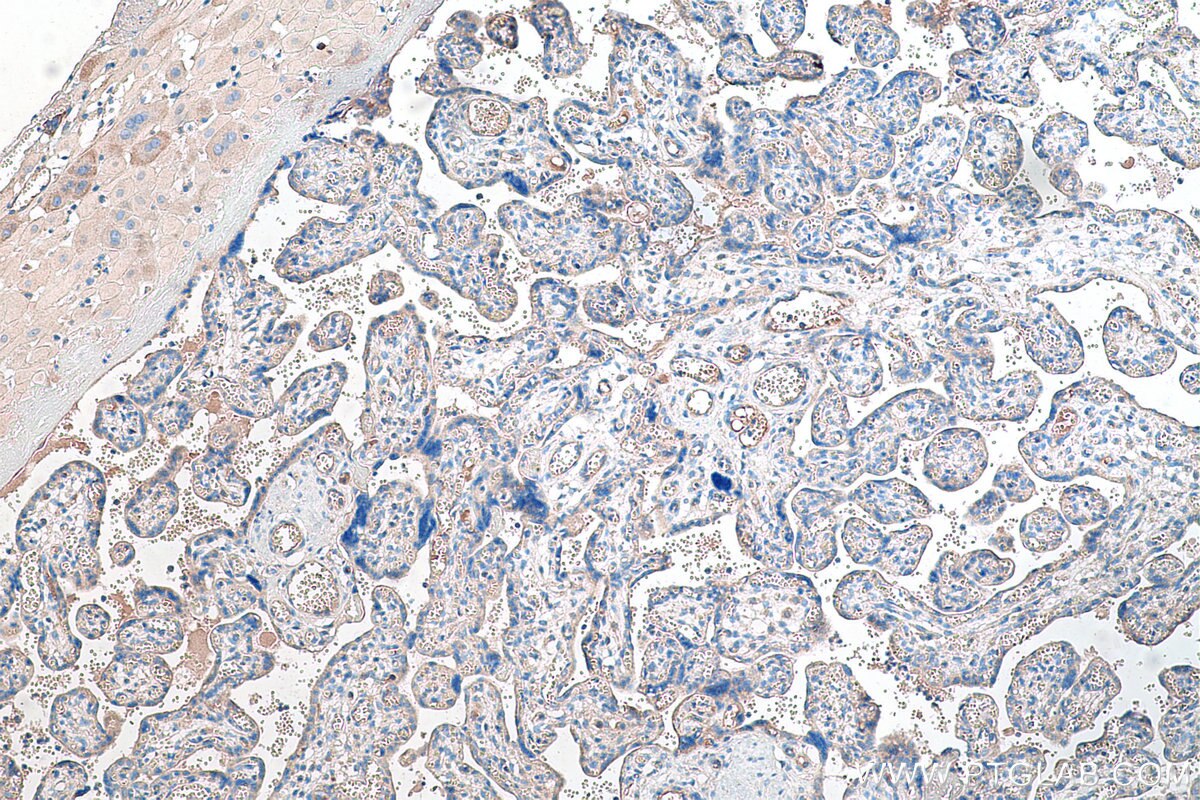
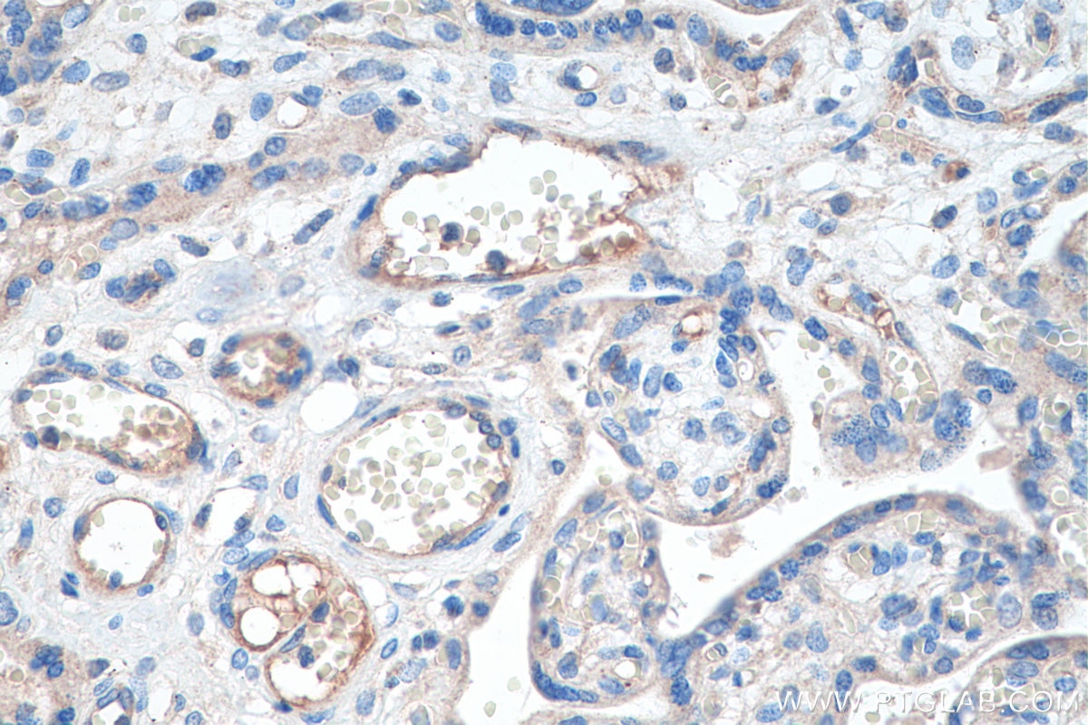
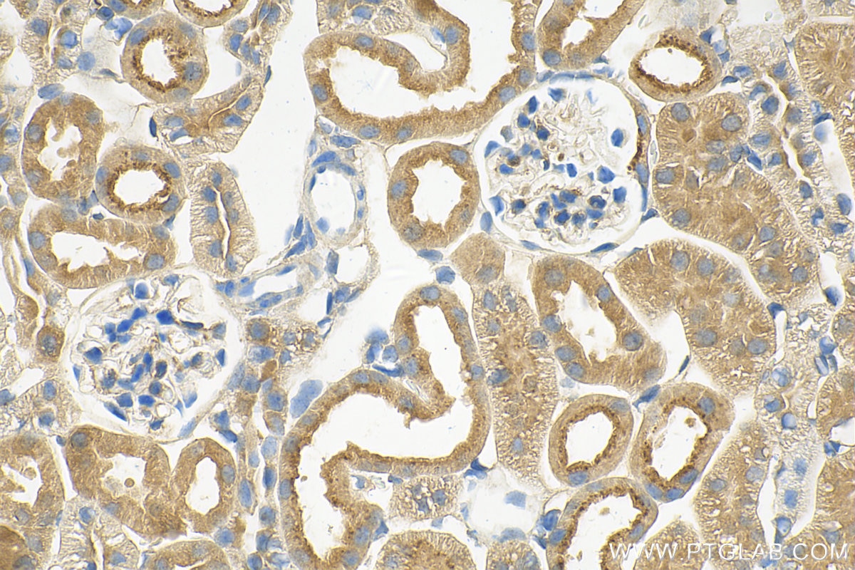
| Positive IHC detected in | human placenta tissue, mouse kidney tissue Note: suggested antigen retrieval with TE buffer pH 9.0; (*) Alternatively, antigen retrieval may be performed with citrate buffer pH 6.0 |
Recommended dilution
| Application | Dilution |
|---|---|
| Western Blot (WB) | WB : 1:500-1:2000 |
| Immunoprecipitation (IP) | IP : 0.5-4.0 ug for 1.0-3.0 mg of total protein lysate |
| Immunohistochemistry (IHC) | IHC : 1:1000-1:4000 |
| It is recommended that this reagent should be titrated in each testing system to obtain optimal results. | |
| Sample-dependent, Check data in validation data gallery. | |
Published Applications
| WB | See 21 publications below |
| IHC | See 3 publications below |
| IF | See 5 publications below |
Product Information
19157-1-AP targets Tie-2/CD202b in WB, IHC, IF, IP, ELISA applications and shows reactivity with human, mouse, rat samples.
| Tested Reactivity | human, mouse, rat |
| Cited Reactivity | human, mouse, rat |
| Host / Isotype | Rabbit / IgG |
| Class | Polyclonal |
| Type | Antibody |
| Immunogen |
CatNo: Ag13523 Product name: Recombinant human TEK protein Source: e coli.-derived, PET28a Tag: 6*His Domain: 779-1124 aa of BC035514 Sequence: RRMAQAFQNVREEPAVQFNSGTLALNRKVKNNPDPTIYPVLDWNDIKFQDVIGEGNFGQVLKARIKKDGLRMDAAIKRMKEYASKDDHRDFAGELEVLCKLGHHPNIINLLGACEHRGYLYLAIEYAPHGNLLDFLRKSRVLETDPAFAIANSTASTLSSHHLLHFAADVARGMDYLSQKQFIHRDLAARNILVGENYVAKIADFGLSRGQEVYVKKTMGRLPVRWMAIESLNYSVYTTNSDVWSYGVLLWEIVSLGGTPYCGMTCAELYEKLPQGYRLEKPLNCDDEVYDLMRQCWREKPYERPSFAQILVSLNRMLEERKTYVNTTLYEKFTYAGIDCSAEEAA Predict reactive species |
| Full Name | TEK tyrosine kinase, endothelial |
| Calculated Molecular Weight | 1124 aa, 126 kDa |
| Observed Molecular Weight | 140 kDa |
| GenBank Accession Number | BC035514 |
| Gene Symbol | Tie2 |
| Gene ID (NCBI) | 7010 |
| RRID | AB_10644297 |
| Conjugate | Unconjugated |
| Form | Liquid |
| Purification Method | Antigen affinity purification |
| UNIPROT ID | Q02763 |
| Storage Buffer | PBS with 0.02% sodium azide and 50% glycerol, pH 7.3. |
| Storage Conditions | Store at -20°C. Stable for one year after shipment. Aliquoting is unnecessary for -20oC storage. 20ul sizes contain 0.1% BSA. |
Background Information
Tie2 (also known as TEK) is a tyrosine-protein kinase expressed almost exclusively on endothelial cells. It contains two immunoglobulin-like domains, three epidermal growth factor (EGF)--like domains and three fibronectin type III repeats. Tie2 acts as a cell-surface receptor for ANGPT1, ANGPT2, and ANGPT4 and regulates angiogenesis, endothelial cell survival, proliferation, migration, adhesion and cell spreading, reorganization of the actin cytoskeleton, but also maintenance of vascular quiescence. Mutations in the gene Tie2 are associated with inherited venous malformations of the skin and mucous membranes. Human Tie2 has a calculated molecular weight of 126 kDa. As a result of glycosylation, the apparent molecular mass of Tie2 is approximately 140-160 kDa.
Protocols
| Product Specific Protocols | |
|---|---|
| IHC protocol for Tie-2/CD202b antibody 19157-1-AP | Download protocol |
| IP protocol for Tie-2/CD202b antibody 19157-1-AP | Download protocol |
| WB protocol for Tie-2/CD202b antibody 19157-1-AP | Download protocol |
| Standard Protocols | |
|---|---|
| Click here to view our Standard Protocols |
Publications
| Species | Application | Title |
|---|---|---|
Ther Adv Med Oncol Development and external validation of a composite immune-clinical prognostic model associated with EGFR mutation in East-Asian patients with lung adenocarcinoma. | ||
Mol Cancer Ther JQ1 induces DNA damage and apoptosis, and inhibits tumor growth in a patient-derived xenograft model of cholangiocarcinoma. | ||
Front Cell Dev Biol Inhibiting Heat Shock Protein 90 Protects Nucleus Pulposus-Derived Stem/Progenitor Cells From Compression-Induced Necroptosis and Apoptosis. | ||
Stem Cell Res Ther Human endometrium-derived stem cell improves cardiac function after myocardial ischemic injury by enhancing angiogenesis and myocardial metabolism. | ||
Stem Cell Res Ther MicroRNA-9 modified bone marrow-derived mesenchymal stem cells (BMSCs) repair severe acute pancreatitis (SAP) via inducing angiogenesis in rats. | ||
J Mol Cell Cardiol BMP9 attenuates occurrence of venous malformation by maintaining endothelial quiescence and strengthening vessel walls via SMAD1/5/ID1/α-SMA pathway. |

